
Human Knee Pictures, Images and Stock Photos
Browse 23,600+ human knee stock photos and images available, or search for human knee anatomy or human knee bone to find more great stock photos and pictures.
Most popular
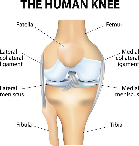
Human Knee joint anatomy. Vector

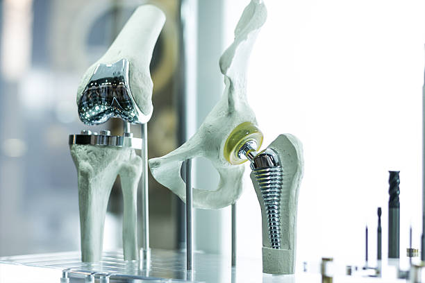
Modern knee and hip prosthesis made by cad engineer and manufactured by 3d printing
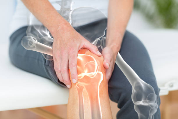
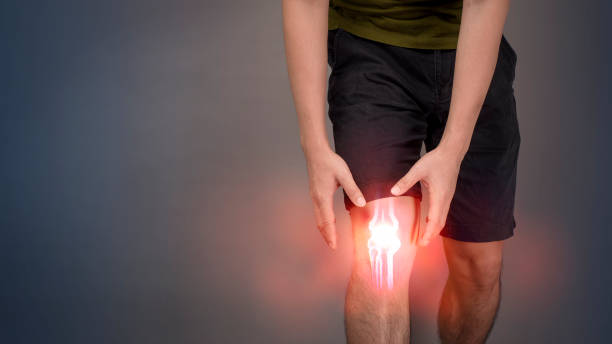
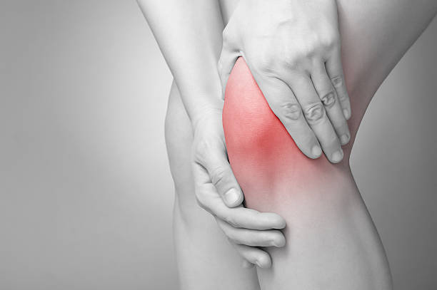
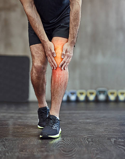
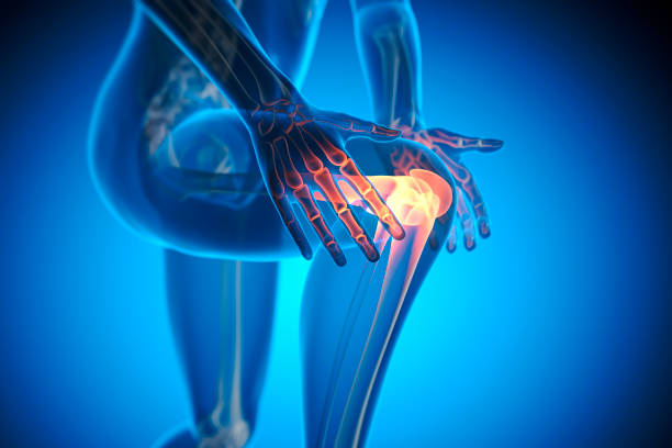
Digitally generated image of man suffering with knee pain

Portrait of sporty woman holding her injured knee after fitness workout. Red colouring spot has been added on the skin to demonstrate where the pain resides.
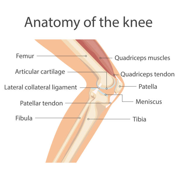
Anatomy of the knee joint, The main parts of the knee joint. Knee-joint For basic medical education. Vector illustration
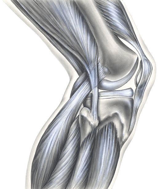
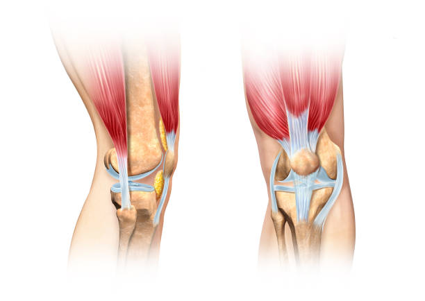
The bony view (left) shows the femur, cut edge of the synovial capsule, meniscus, fibula, tibia, patella, anterior cruciate ligament, posterior cruciate ligament, and medial collateral ligament. The muscular view (right) shows the hamstring muscles, fibula, gastrocnemius muscles, and quadriceps muscles.

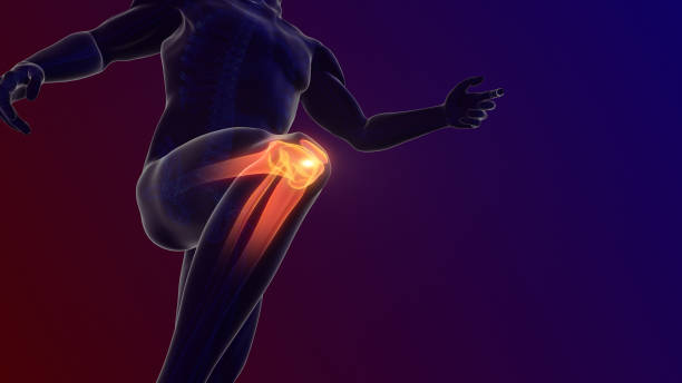
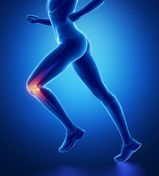
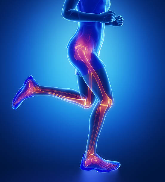
Knee painful - skeleton x-ray, 3D Illustration medical concept.


Pain in the knee joint. A polygonal model of lines and points. Blue background.

Unhappy young woman sitting on the mat, grabbing an ankle, unable to start yoga work out because of sport injury, feeling pain. Beginner doing wrong exercise without coacher

Cropped view of a hand gently caressing a perfectly smooth leg
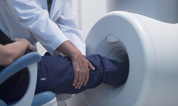
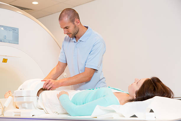
Patient and Doctor in fully open high field Magnetic Resonance Image MRI CAT Scan scanning knee and leg.
Human knee cutaway illustration. Side and front views detailed, scientifically correct cross section representation. On white background, with clipping path included. Anatomy image.

Shot of a mature doctor examining his patient who is concerned about his knee
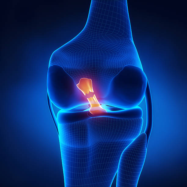
Torn Anterior Cruciate Ligament in x-ray view
An icon of a knee joint and knee with a starburst to indicate pain.
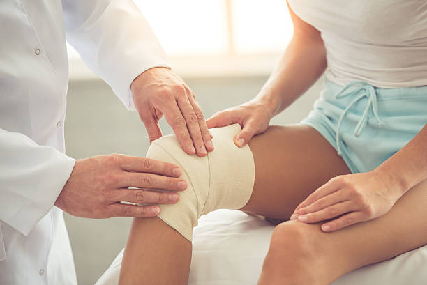
Cropped image of handsome doctor bandaging woman's injured knee while working in his office


The athlete takes a break from running due to knee pain

Young woman feeling pain in her knee

Human knee joint icon, emblem for orthopedic clinic


Cropped shot of a runner holding onto her injured kneehttp://195.154.178.81/DATA/i_collage/pi/shoots/805758.jpg
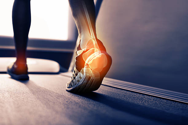
Rear view shot of the highlighted joints in a runner's foot
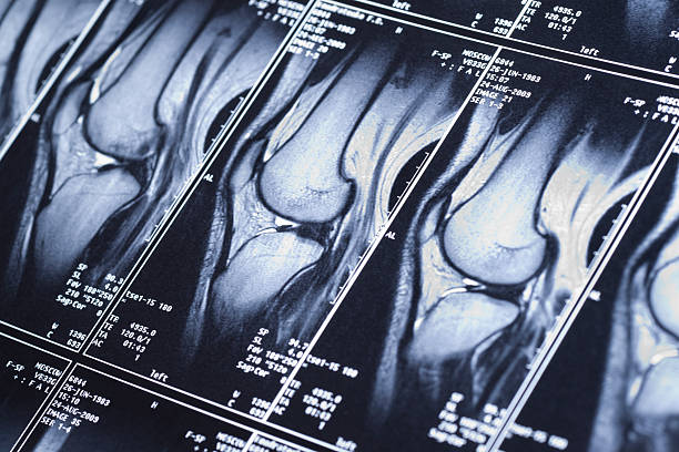
My knee MRI - sport trauma, damage of cross-shaped ligaments

Shot of a father applying antibacterial medical bandage on child's knee after falling down from a cycle
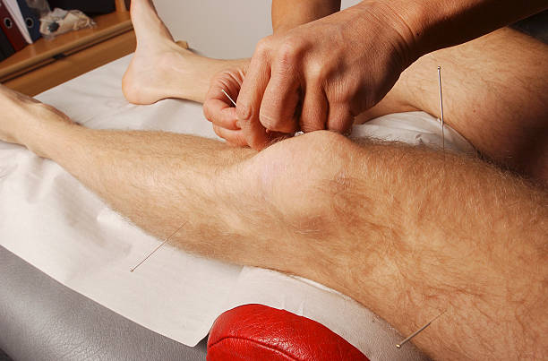
Man getting acupuncture at a clinic
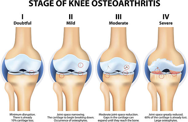
Stages of knee Osteoarthritis (OA). Kellgren and Lawrence criteria for assessment stage of osteoarthritis. The classifications are based on osteophyte formation and joint space narrowing.

Cropped shot of a runner's highlighted bones


Old age, health problem and people concept - senior woman suffering from pain in leg at home. Elderly woman suffering from pain in knee at home.

A young woman massaging her painful knee
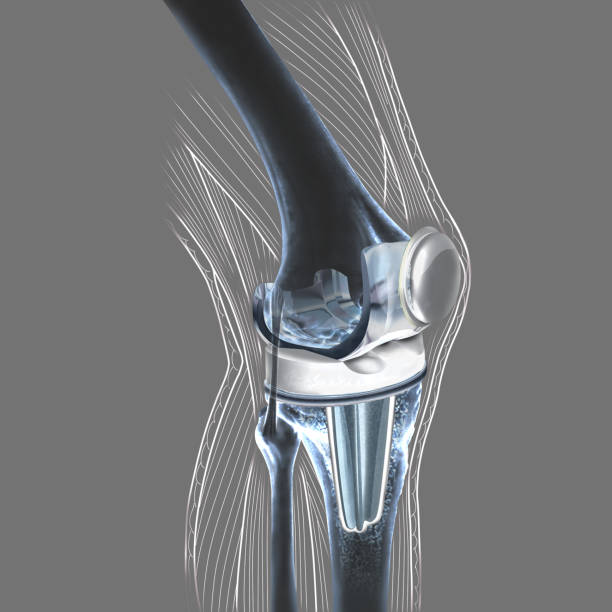
The knee prosthesis replaces the damaged joint of the patella. Knee prosthesis is offered to patients who suffer from very advanced osteoarthritis.

Doctors at the hospital looking at an x-ray - medical exam concepts
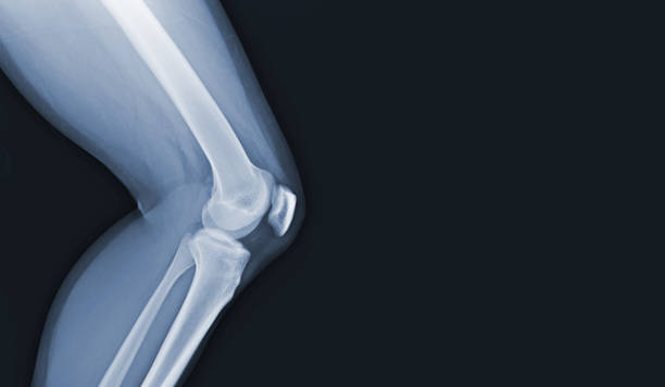
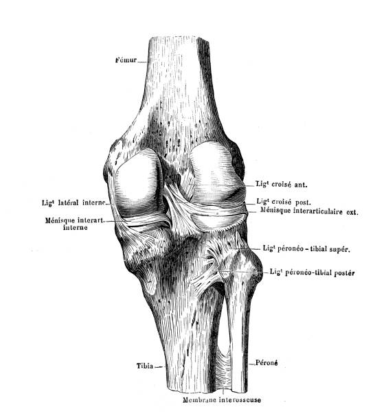
illustration of a knee

A cropped view of a female jogger on the road experiencing joint inflammation
Shot of an unrecognizable man holding his knee in pain

Cropped shot of a young woman out on a country road for a jog
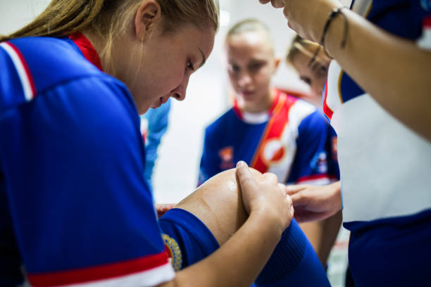
Teenage female soccer player holding her knee in pain at dressing room.

Always warm-up before jogging or injury occurs...
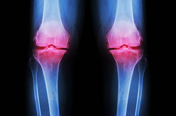
Osteoarthritis Knee ( OA Knee ). Film x-ray both knee ( front view ) show narrow joint space ( joint cartilage loss ) , osteophyte , subchondral sclerosis

Physiotherapist giving knee therapy to a woman in clinic
A set of orthopedics icons that include editable strokes or outlines using the EPS vector file. The icons include an orthopedist, orthopedist giving an exam, medicine, injured foot, injured shoulder, injured knee, injured back, broken arm, person doing rehabilitation, female nurse, person doing physical therapy, person with injury, human hip, surgery using a scalpel, person rehabbing using an exercise ball, injured wrist, knee x-ray, knee bone, patient in hospital bed, person slipping and falling, checklist, physical therapist, person doing water rehabilitation and other related icons.

Unrecognizable athletic woman feeling pain in her knee at the park. Copy space.

Medical X-Ray illustration of pain in knee joint - 3D illustration

Shot of a sportswoman with a knee injury. Front view of athletic woman holding her knee in pain on sports training in a health club. Cropped shot of a woman suffering from a knee injury.

X-Ray of human knee
Medical technical assistant councelling patient and preparing scan of the knee with magnetic resonance tomography MRI in radiology
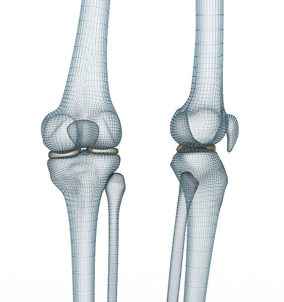
Knee joint anatomy. Medically accurate wire 3d illustration.
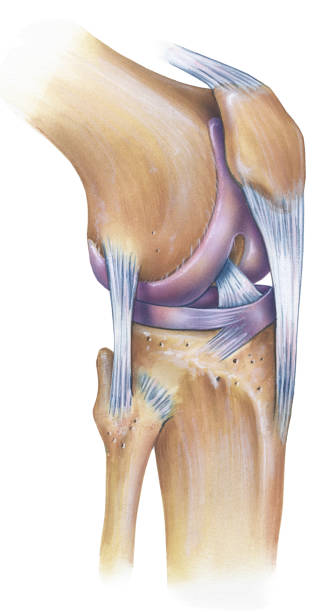
The normal human anatomy of a knee, anterolateral view. Shown are the patella, anterior cruciate ligament, patellar ligament, fibular collateral ligament, lateral meniscus, lateral femoral condyle, femur, tibia, and fibula.
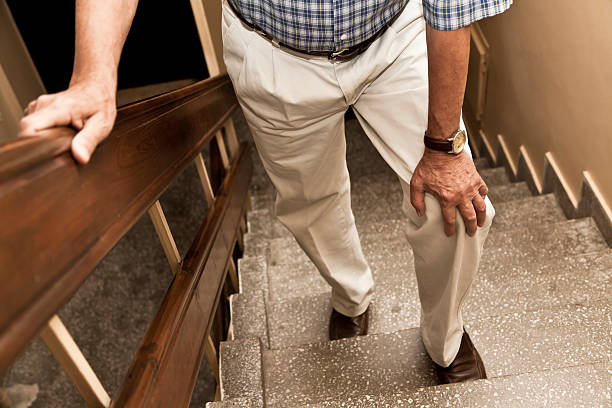
Senior adult having knee pain while climbing up stairs