quadriceps muscle anatomy 3d medical vector illustration eps 10
Browse 250+ vastus lateralis stock photos and images available, or search for vastus medialis or vastus intermedius to find more great stock photos and pictures.
quadriceps muscle anatomy 3d medical vector illustration eps 10

Vastus Lateralis Muscle - Anatomy Muscles isolated on white - 3D illustration
Frontal view of man's isolated leg muscles with labeled names
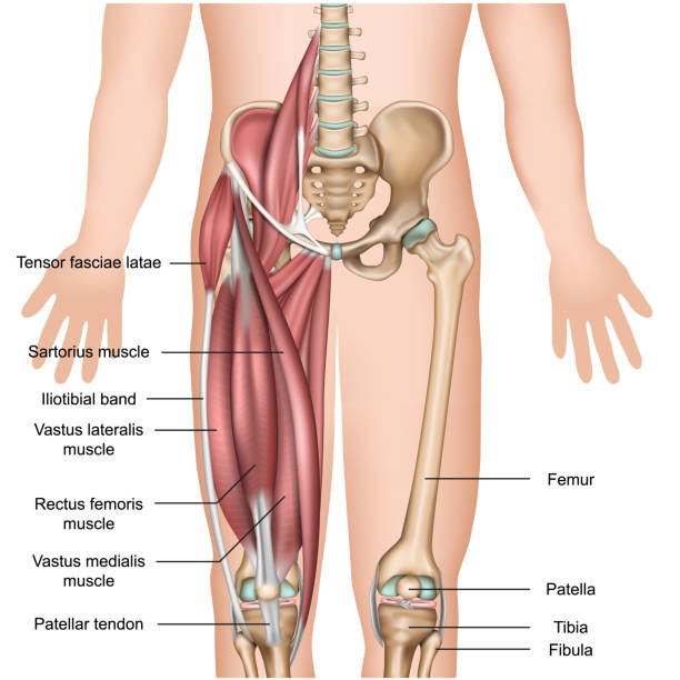
leg muscle anatomy 3d medical vector illustration quadriceps eps 10
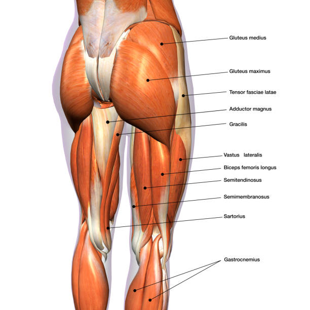
Rear view of woman's thigh and knee muscles with names

medical accurate illustration of the vastus lateralis

Frontal view of the vastus lateralis quadriceps thigh muscles isolated within the skeleton on a white background. 3D rendering.

3D Illustration, Muscle is a soft tissue, Muscle cells contain proteins , producing a contraction that changes both the length and the shape of the cell. Muscles function to produce force and motion.

Full body side view of strong young female in sportswear performing rear lunge exercise with raised arms during training against white background

medical accurate illustration of the vastus lateralis

This 3d illustration shows the vastus lateralis muscles anatomical position on xray body

Vastus Lateralis Muscle - Anatomy Muscles isolated on white - 3D illustration

3D Illustration, Muscle is a soft tissue, Muscle cells contain proteins , producing a contraction that changes both the length and the shape of the cell. Muscles function to produce force and motion.

Student with human knee. Anatomical model. White background.

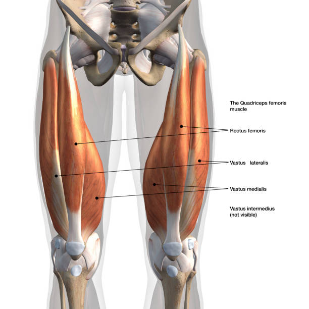
Frontal view of male quadriceps muscles of the thighs in isolation on white background with names.

3D Illustration, Muscle is a soft tissue, Muscle cells contain proteins , producing a contraction that changes both the length and the shape of the cell. Muscles function to produce force and motion.

3D Illustration, Muscle is a soft tissue, Muscle cells contain proteins , producing a contraction that changes both the length and the shape of the cell. Muscles function to produce force and motion.

3D Illustration, Muscle is a soft tissue, Muscle cells contain proteins , producing a contraction that changes both the length and the shape of the cell. Muscles function to produce force and motion.

3D Illustration, Muscle is a soft tissue, Muscle cells contain proteins , producing a contraction that changes both the length and the shape of the cell. Muscles function to produce force and motion.

3D Illustration, Muscle is a soft tissue, Muscle cells contain proteins , producing a contraction that changes both the length and the shape of the cell. Muscles function to produce force and motion.

3D Illustration, Muscle is a soft tissue, Muscle cells contain proteins , producing a contraction that changes both the length and the shape of the cell. Muscles function to produce force and motion.

Gluteus maximus male muscle anatomy posterior view isolated in 3D

medical accurate illustration of the vastus lateralis

3D Illustration, Muscle is a soft tissue, Muscle cells contain proteins , producing a contraction that changes both the length and the shape of the cell. Muscles function to produce force and motion.

Sartorius male muscles anatomy anterior view isolated in 3D

image showing gluteal muscles, hamstring muscles and the sciatic nerve passage

dissection picture of gluteal region showing sciatic nerve course, hamstring muscles and gluteal muscle

"Right Knee includes: rectus femoris, vastus lateralis and vastus medialis muscles, femur, fibula, patella and tibia bones; anterior cruciate ligament, quadriceps femoris tendon plus six more ligaments and tendons. Blue background."

The rectus femoris is the anterior thigh compartment's most superficial and nearly vertically oriented muscle. This bipennate structure is a component of the quadriceps muscle complex, one of the knee's most important dynamic stabilizers.

3D Illustration, Muscle is a soft tissue, Muscle cells contain proteins , producing a contraction that changes both the length and the shape of the cell. Muscles function to produce force and motion.

Computer generated 3D model camera positioned from all 6 directions white background with alpha channel for transparency

Full Body Anterior View of Male Muscle System with Connective Tissues, White Background, No Genitals, 3D Rendering.

3D Rendering of Male Back Power Muscles Isolated on Skeleton on White Background with Text Labeling. Posterior view.

3D Illustration, Muscle is a soft tissue, Muscle cells contain proteins , producing a contraction that changes both the length and the shape of the cell. Muscles function to produce force and motion.

vector illustration of diagram of Muscular System

Anatomically correct model of human knee. Side view. Isolated on white.

Anatomically correct model of right human knee with muscles and tendons. Blue background.

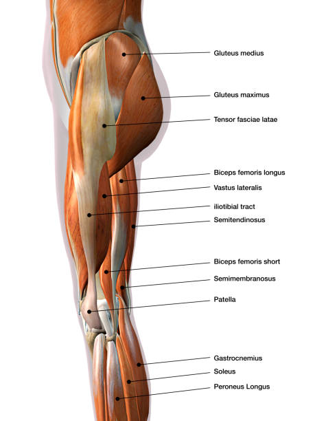
Shown are the biceps femoris muscle (long head), biceps femoris muscle (short head), plantaris muscle gastrocnemius muscle, lateral head, common fibular nerve, iliotibial tract, vastus lateralis muscle, tendon of rectus femoris muscle, fibular collateral ligament, patella, deep infrapatellar bursa patellar ligament, anterior ligament of head of fibula head of fibula, soleus muscle, tibialis anterior muscle, fibularis longus muscle, extensor digitorum longus muscle, fibuarlis (peroneus) longus muscle, fibularis (peroneus) brevis muscle, the lateral compartment (fibular muscles), fibularis brevis muscle, calcaneal tendon, lateral malleolus, superior fibular retinaculum, calcaneofibular ligament, inferior fibular retinaculum, tendons of the fibularis longus and brevis muscles, extensor hallucis longus muscle, tendon of tibialis anterior muscle (synovial sheath), tendon of extensor digitorum longus muscle (synovial sheath), inferior extensor retinaculum, tendon of extensor hallucis longus muscle (synovial sheath) extensor digitorum brevis muscle tendon of fibularis tertius muscle, tendon of extensor hallucis brevis muscle, tendons of extensor digitorum longus muscle opponens digiti minimi muscle.

image showing muscles of gluteal region and the posterior compartment of thigh with related nerves

The vastus lateralis is the largest muscle in the quadriceps group, which is located on the outside of the thigh. It works with the other quadriceps muscles to extend the knee joint, allowing the body to stand up from a squat.

"Male doctor holding anatomically correct model os right knee. The model shows rectus femoris, vastus lateralis and vastus medialis muscles, femur, fibula, patella and tibia bones; anterior cruciate ligament, quadriceps femoris tendon plus six more ligaments and tendons. White background."

The rectus femoris is the anterior thigh compartment's most superficial and nearly vertically oriented muscle. This bipennate structure is a component of the quadriceps muscle complex, one of the knee's most important dynamic stabilizers.

Right Knee shows rectus femoris, vastus lateralis and vastus medialis muscles, femur, fibula, patella and tibia bones; anterior cruciate ligament, quadriceps femoris tendon plus six more ligaments and tendons. White background.

The major muscle groups of the lower body are the quadriceps, hamstrings, gluteus, gastrocnemius, and soleus. The quadriceps, or thigh muscles, form the front part of your upper leg. These muscles are used for motions such as walking or kicking a ball.

3D Illustration Concept of Human Muscular System Leg Muscles Vastus Lateralis Muscle Anatomy

The gastrocnemius muscle is a complex muscle that is fundamentally involved in walking and posture. It affects the entire lower limb and the movement of the hip and the lumbar area. It is a muscular district called to work during daily and sports activities and maintain orthostatism.

"Right Knee shows rectus femoris, vastus lateralis and vastus medialis muscles, femur, fibula, patella and tibia bones; anterior cruciate ligament, quadriceps femoris tendon plus six more ligaments and tendons."

Smiling muscular built shirtless man - fitness instructor standing inside illuminated Gym, showing his Leg Muscles towards the camera. Smiling, looking confident and happy. Real People Body Building - Men's Fitness Studio Workout Portrait Series.

"Right Knee shows rectus femoris, vastus lateralis and vastus medialis muscles, femur, fibula, patella and tibia bones; anterior cruciate ligament, quadriceps femoris tendon plus six more ligaments and tendons."
© 2025 iStockphoto LP. The iStock design is a trademark of iStockphoto LP. Browse millions of high-quality stock photos, illustrations, and videos.
Do Not Sell or Share