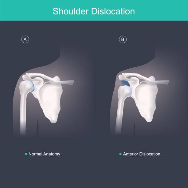
Shoulder dislocation types. Arm injury, upper arm bone pops out of the cup-shaped socket of shoulder blade. Glenohumeral joint dislocation. Flat vector illustration
Browse 1,100+ dislocation stock illustrations and vector graphics available royalty-free, or search for shoulder dislocation or hip dislocation to find more great stock images and vector art.
Shoulder dislocation types. Arm injury, upper arm bone pops out of the cup-shaped socket of shoulder blade. Glenohumeral joint dislocation. Flat vector illustration

Patellar dislocation. Normal position of kneecap and Patella displaced. Anatomy of the Knee

Shoulder dislocation. humerus bone trauma, Sports injuries, or Weak shoulder muscles. Human arm anatomy. Bones and joint of the Shoulder, and hand. Vector illustration
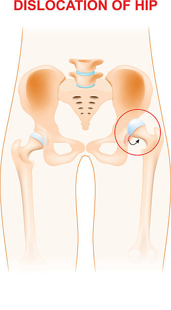
Dislocation of hip. A hip dislocation takes place when the head of the thigh bone is forced out of its socket in the pelvic bone.

Shoulder dislocation and humerus bone trauma explanation outline diagram. Labeled educational medical injury when skeletal part rotates out of scapula vector illustration. Painful arm vs healthy.

Healthy and unhealthy dog's knee joint, the medial luxating patella or knee cap dislocation illustration.

Hip joint anatomy. Ball-and-socket joint. Vector poster

Female character with different types of injuries. Vector medical illustration

Spondylolisthesis. Spinal disease when lower vertebrae slip forward. Birth defect or injury damage. Flat vector illustration

Human shoulder muscles and joints have a red signal. Illustration about chronic pain.
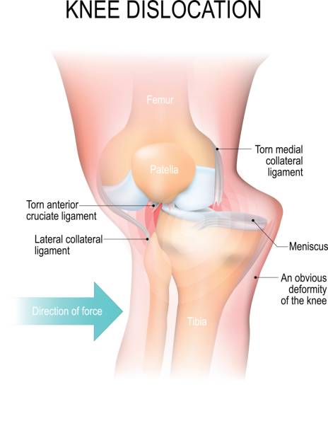
Knee dislocation. Lateral trauma to the knee, torn collateral ligaments, cruciate ligament injury and meniscus injury. An obvious deformity of the knee. Human anatomy

Normal position of kneecap and patella displaced. Illustration isolated on white background. Graphic concept for your design

Dislocation Shoulder symptoms or separated. Bone and joints to slipping out of place.
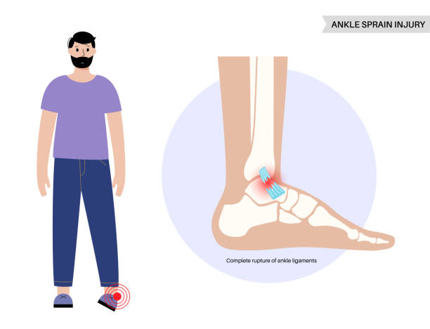
Sprained ankle injury. Twisted feet, pain and swelling. Tears, stretch or rupture of ligaments. Foot trauma anatomical poster, diagnosis and treatment in clinic. Leg problem, X ray vector illustration

Hip Labral Tears. Labrum torn from socket and Repaired labrum. Surgery for Repairing a Torn Hip Labral. Vector

Shoulder subluxation as partial dislocated arm joint problem outline diagram. Labeled educational medical scheme with body skeletal anatomy and dislocated bones vector illustration. Upper body trauma.

Bankart lesion as anterior part of the glenoid labrum trauma outline diagram. Labeled educational medical injury type with anterior and lateral view of xray with bones and tears vector illustration.

Radiogram of hand. Radiologist doctor watching an x-ray or roentgenogram. Vector illustration of radiograph
Poster illustrating the condition of Hernia
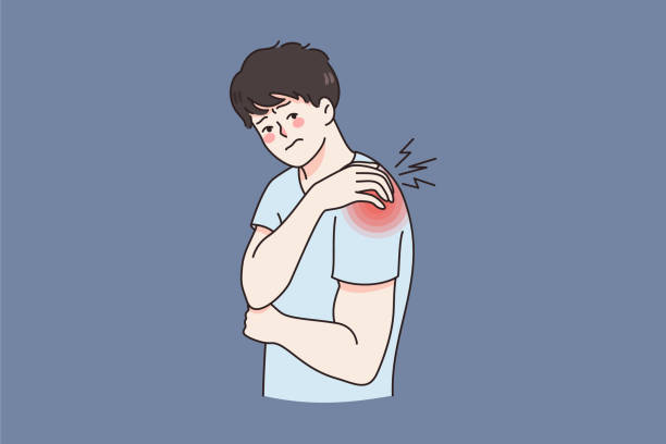
Unhealthy man touch shoulder suffer from back injury or trauma. Unwell sick guy struggle with acute pain or muscular spasm strain in arm. Healthcare and medicine. Flat vector illustration.

vector illustration of joint injuries and pain.
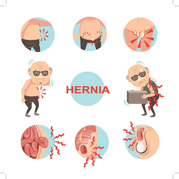
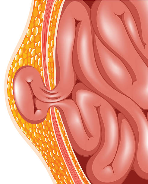
Diagram of inside umbilical and inguinal hernia, Men with hernia symptoms and signs that can be noticed.Cartoon vector illustration

Snapping hip syndrome. Coxa saltans. iliopsoas tendinitis. dancer's hip. Anatomy of a Human Hip. Vector illustration

Anatomy of a healthy human hip joint. Femoral head circulation system. Didactic scheme of structure of bone, joint and arteries with anatomical captions. Flat vector illustration
3D Isometric Flat Vector Conceptual Illustration of Knee Problems, Inflammatory Arthritis and Osteoporosis, Joint Inflammation
Vector illustrations of sling, bandage, and elevation techniques treatment for broken bones and pain.

Rheumatoid arthritis of the foot. Tiny doctors treat rheumatism, osteoarthritis, make ultrasound, x ray. World arthritis day in October. Flat concept vector for landing page, banner, app.

From "Treatise on External Pathology" (1864): Broken bone

Fracture and repair. Vector scheme

Illustration representing First Aid with ice compress on the injured arm. Ideal for catalogs of medical, institutional and educational

Back pain. Character having neck and shoulder muscle spasm. Trigger point in muscle tissue. Myositis, strained muscle inflamation. Flat vector illustration.

Inflammation and pain in the elbow joint. Polygonal design of interconnected lines and points. Blue background.
Broken bones thin line icons set isolated on white. Fracture, dislocation of leg, arm, skull, finger pictograms collection. Crutches, plaster of Paris, wheelchair vector elements for infographic, web.
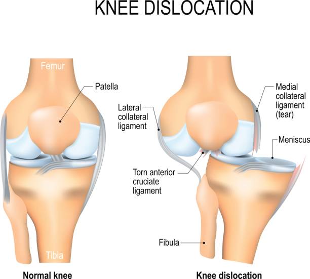
Knee dislocation and normal. Lateral trauma to the knee, torn collateral ligaments, cruciate ligament injury and meniscus injury. Human anatomy

Two male doctors examining knee joints, orthopedic health protection concept illustration.

Elbow pain location. Bursitis of arm joint, dislocated, sprain, ligaments rupture, arthritis, arthrosis, epicondylitis, injury. Vector medical minimal illustration isolated on white background.

Child with broken arm visits orthopedic doctor. Traumatologist treats injury of small patient. Characters in colored flat vector illustration isolated on white background.

Luxating patella in dogs, dislocated knee cap detailed info graphic poster, the severity of the condition, grades, diagnostic and treatment etc. beautiful aesthetic design.

Orthopedic traumatology. Doctor puts splint on child's broken arm. Characters in colored flat vector illustration isolated on white background.

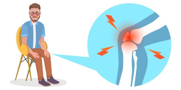
Young man with knee pain. Flat trendy hand drawn character vector illustration. Isolated on white background.

Medical illustration to explain Cubital tunnel syndrome. Ulnar nerve entrapment. Fascia involved in ulnar nerve compression. Vector
© 2025 iStockphoto LP. The iStock design is a trademark of iStockphoto LP. Browse millions of high-quality stock photos, illustrations, and videos.
Do Not Sell or Share