
Diagram showing xylem and phloem of plant illustration
Browse 220+ xylem phloem stock illustrations and vector graphics available royalty-free, or search for stem to find more great stock images and vector art.
Diagram showing xylem and phloem of plant illustration

Xylem and phloem vector illustration. Labeled water, nutrient and mineral transportation scheme. Educational graphic with biological translocation process explanation. Living tissue in vascular plants

Xylem and phloem water and minerals transportation system outline diagram. Educational labeled anatomical scheme with vessel side cross section, structure and process explanation vector illustration.
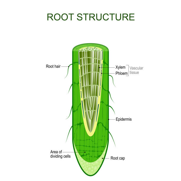
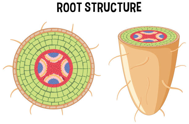
Root structure. Plant anatomy. The cross-section of the root with area of dividing cells, Xylem, Phloem, cap, epidermis, and hairs. Vector illustration for biological, science, and educational use
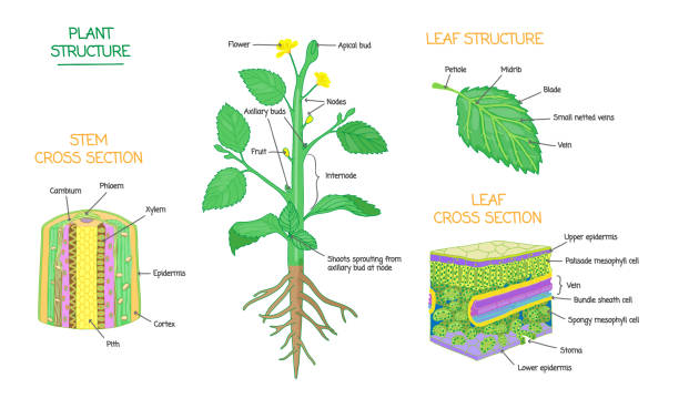
Plant structure and cross section diagrams, botanical microbiology vector illustration schemes collection. Stem and leaves labeled closeup drawings with layers and cells. Educational biology poster.
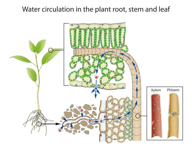
Water enters the root hairs by osmosis, moving from high water concentration to low water concentration.Minerals dissolve easily in water and move into the root by diffusion - either by passive transport or by active transport.Water moves across the ground tissue and into xylem tissue. Water is then transported up the plant.

Section of the stem plant. Xylem, phloem
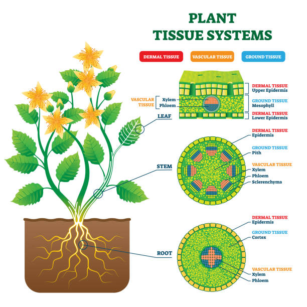
Plant Tissue Systems vector illustration. Labeled biological structure scheme. Anatomical diagram with leaf, stem and root microscopic graphic. Plant inner vascular, dermal and ground cross section.

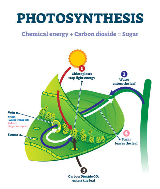
Plants contain a vast network of conduits, which consists of xylem and phloem tissues. The xylem and phloem tissues extend throughout the plant. These conducting tissues start in the roots and transect up through the trunks of trees, branching off into the branches and then branching even further into every leaf

Metabolism and transport in plants. Transpiration

Plant diagram xylem and phloem

Cross section cut of plant stem. Xylem, phloem
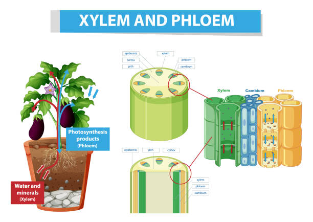
Diagram showing xylem and phloem in plant illustration

Plant vascular tissue Phloem. Cross section showing vascular bundles. Translocation in vascular plants

The primary components of vascular tissue are the xylem and phloem. These two tissues transport fluid and nutrients internally. There are also meristem associated with vascular tissue: the vascular cambium and the cork cambium

Xylem and phloem. biological structure scheme of inner vascular in Plant. cross section. function of xylem is to transport water and nutrients from roots to stems and leaves. phloem for transport organic compounds made during photosynthesis

Parts of a green leaf. Epidermis, mesophyll, veins
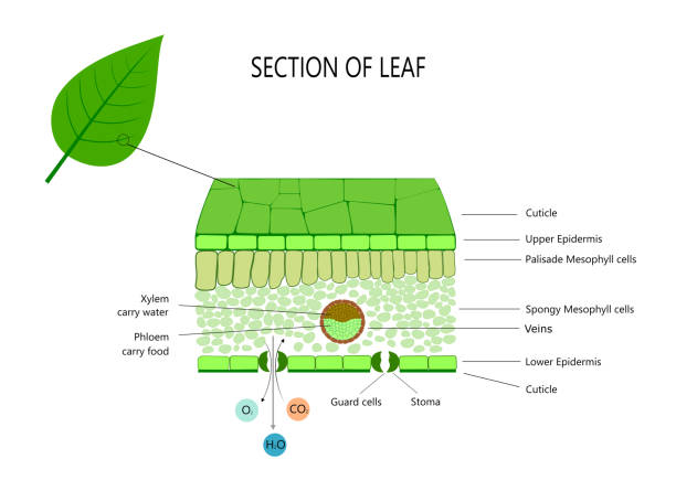
Leaf structure for school and education. Leaf tissue, cells and trasport in and out leaf

Plant vascular tissue Xylem. Cross section showing vascular bundles. Translocation in vascular plants

Diagram showing xylem and phloem of plant illustration

Diagram showing xylem and phloem in plant illustration

Cross section of leaf, with your structures, cells and tissue

Root structure. monocot and dicot stems. cross sections of plants roots. Vector diagram for educational, biological, and scientific use

Internal structure of stem diagram illustration

Diagram showing xylem and phloem in plant illustration
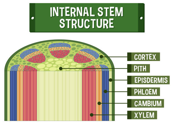
Internal structure of stem diagram illustration
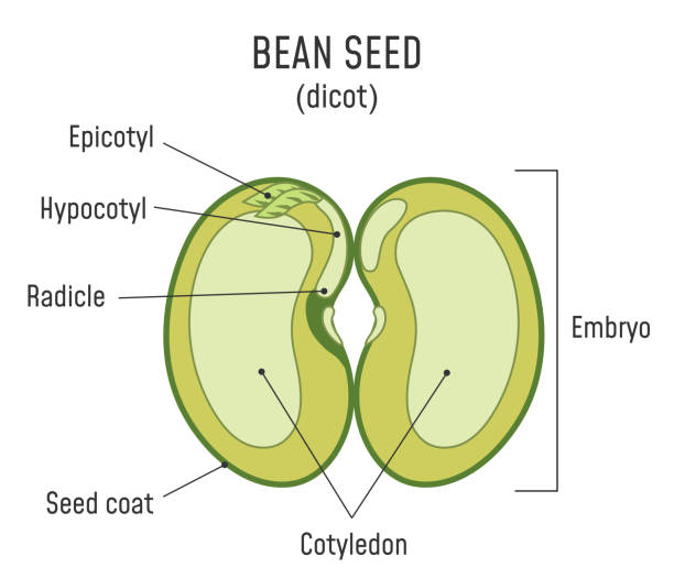
Bean Seed Structure. Anatomy of grain. Dicot seed diagram.
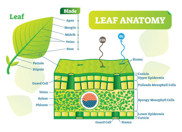
Leaf anatomy vector illustration diagram. Biological macro scheme poster with leaf inner layers, veins and breathing oxygen exchange.

Animal cells are generally smaller than plant cells. Animal cells range from 10 to 30 micrometers in length, while plant cells range from 10 and 100 micrometers in length. Animal cells come in various sizes and tend to have round or irregular shapes. Plant cells are more similar in size and are typically rectangular or cube shaped.

Plant tissue systems diagram shows leaf, stem, and root cross-sections, highlighting xylem, phloem, and epidermis. Outline diagram

Plant vascular tissue. Xylem and phloem. Cross section showing vascular bundles. Translocation in vascular plants

Internal structure of stem diagram illustration
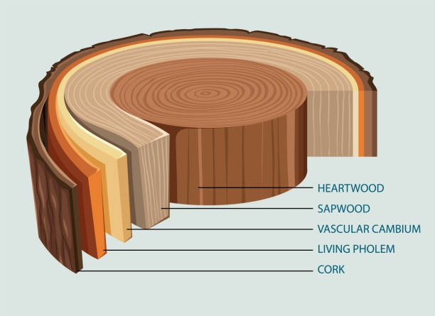
Anatomy of tree trunk illustration
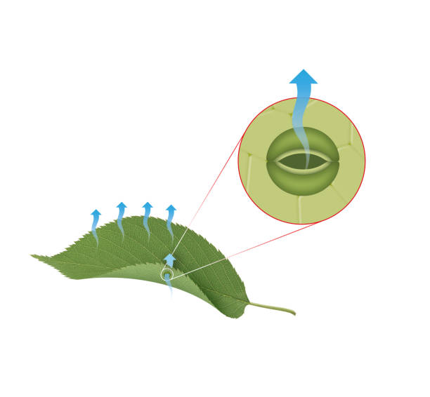
Transpiration is the loss of water from a plant in the form of water vapor
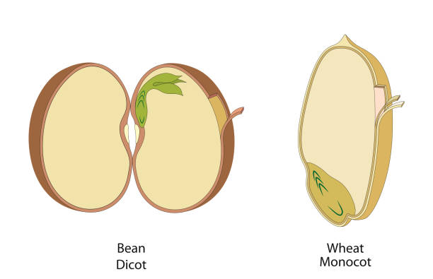
The structure of the bean seed in dicots and monocots

Types of plant tissues. Anatomy of a Plant Body. Cross section of Root, stem and leaf of a green plant. Vector illustration. Poster for education and biology
This figure illustrates in a schematic way what goes on in a leaf through the processes of photosynthesis and respiration

Structure of the slice of the tree layers in cross section. Tree trunk different layers scheme. Cross section of woody stems infographics. Education biology, dendrochronology poster, illustration. Stock vector

A leaf is made of many layers that are sandwiched between two layers of tough skin cells (called the epidermis)

Diagram showing xylem and phloem in plant illustration
Internal structure of root diagram illustration
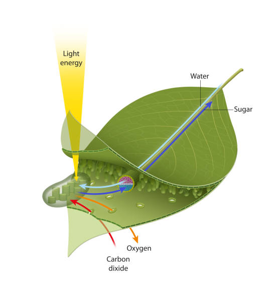
Photosynthesis leaf vector illustration. Labeled educational scheme where light energy converts to chemical sugars. Natural botanic process visualization with stages explanation. Closeup plant system.

Chloroplasts are critical organelles that perceive and convey metabolic and stress signals to different cellular components, while remaining the seat of photosynthesis and a metabolic factory

Illustration of a leaf anatomy on a white background
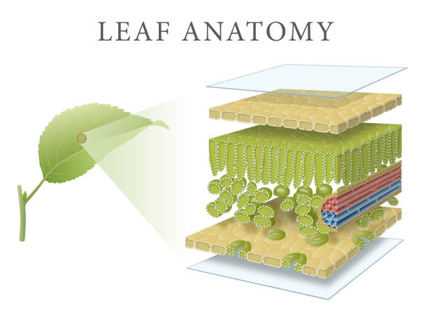
The fine scale structure of a leaf featuring the major tissues; the upper and lower epithelia (and associated cuticles), the palisade and spongy mesophyll and the guard cells of the stoma. Vascular tissue (veins) is not shown. Key plant cell organelles (the cell wall, nucleus, chloroplasts, vacuole and cytoplasm) are also shown.

Anatomy of tree trunk illustration
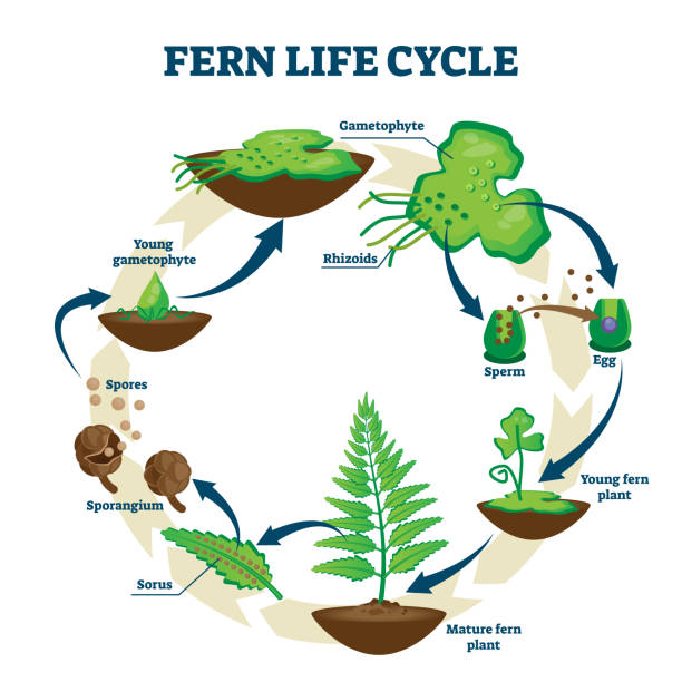
Fern life cycle vector illustration. Labeled educational development process scheme. Different plant stages examples with gametophyte, rhizoids, sorus and spores. Self reproduction explanation scheme.

Root hair cells are elongated structures that extend from the surface of plant roots. They are tubular in shape and have thin walls, which maximize their surface area for absorption. This elongated shape allows them to penetrate between soil particles and increase contact with water and mineral ions.

Anatomy of tree trunk illustration

The only tissue present in ground tissue of monocot leaf is mesophyll. Mesophyll is present between the upper and lower epidermis and is not differentiated into palisade and spongy parenchyma. They are made up of chlorenchyma (chlorophyll containing parenchyma) cells and form the photosynthetic tissue of the leaf.

Corn Seed Structure. Anatomy of grain. Monocot seed diagram.

Anatomy of tree trunk illustration

Cross section of some phloem cells. Phloem Tissue in Plants

Internal structure of plant diagram illustration

Root hair cells are specialized cells found in the epidermis of plant roots. They are crucial for the absorption of water and nutrients from the soil.

Multicellular organisms require transport systems to supply their cells and remove waste products. Plants transport substances through xylem and phloem

Flowering plants are divided into monocots (or monocotyledons) and dicots (or dicotyledons). This comparison examines the morphological differences in the leaves, stems, flowers and fruits of monocots and dicots

graphical diagram of Photosynthesis

