
Images
Browse 320+ retinal hemorrhage stock photos and images available, or start a new search to explore more stock photos and images.


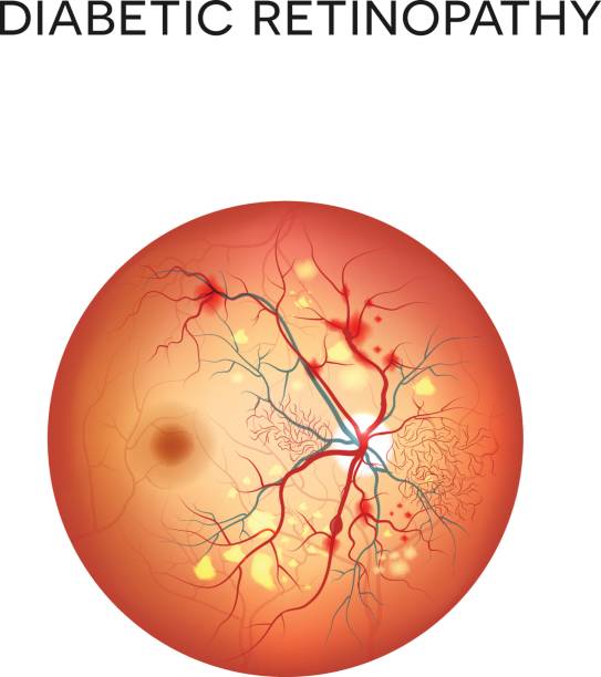
Diabetic retinopathy image, medical infographic

Our creative library is free of AI-generated contentChoose your visuals with confidence knowing our creative library is free from AI-generated content, so your searches only return safe, high-quality visuals you can trust.

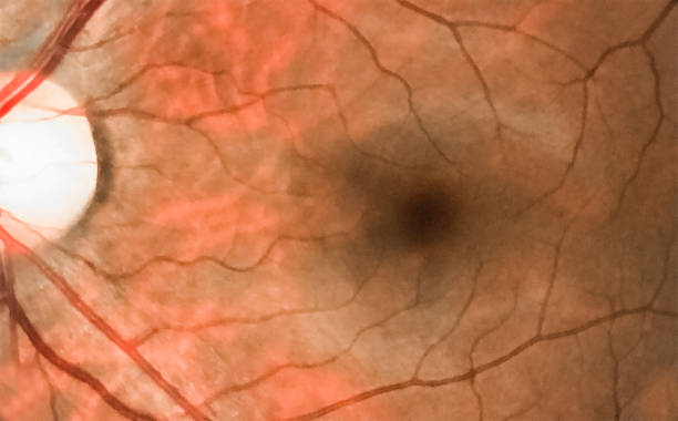
Macular degeneration Drusen, Atrophy, Subretinal hemorrhage. Normal retina

Person with symptoms of conjunctivitis due to allergies

Diabetic retinopathy anatomical poster. Human eye disease, vision loss or blindness. Damaged blood vessels in the retina. Proliferative or nonproliferative eye condition medical vector illustration

Diabetic retinopathy. Retinal damage. Cross section of human eye. Diabetes. Close-up of a macula, optic disc, choroid, retina, sclera, and fovea. Medical condition. Microaneurysm of the small blood vessels. Vector poster

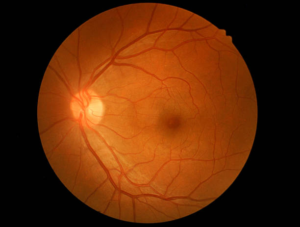
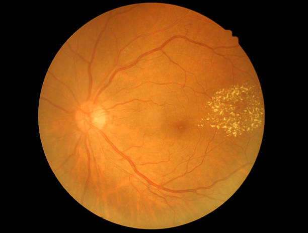
Wide angle retinal ultra high resolution wide field fundus imaging closeup. It is intended for diagnostics of retinal diseases.


Hemorrhage Eyeball or Subconjunctival Hemorrhage is a condition where a tiny blood vessel breaks just underneath the clear surface of your eye. It can be caused by violent coughing, powerful sneezing, straining, vomiting, or even rubbing your eye too hard.

Woman feels pain in eyes covering reddened pupil with hand and needs help of ophthalmologist. Girl is experiencing vision problems due to pain or inflammation of eyes after getting infection

Hemorrhage Eyeball or Subconjunctival Hemorrhage is a condition where a tiny blood vessel breaks just underneath the clear surface of your eye. It can be caused by violent coughing, powerful sneezing, straining, vomiting, or even rubbing your eye too hard.


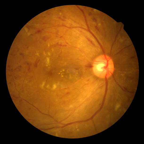
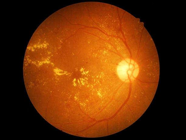
Fundus photography Madical Retina Abnormal isolated on white background.Retina of diabetes check up medical healthcare concept.

Subconjunctival bleeding, also known as subconjunctival hemorrhage, is bleeding from a small blood vessel over the whites of the eye. It results in a red spot in the white of the eye. There is generally little to no pain and vision is not affected. Generally only one eye is affected


Diabetic retinopathy anatomy. Healthy eyeball and damaged organ. Human eye disease, vision loss or blindness. Proliferative or nonproliferative eye condition. Retina medical flat vector illustration
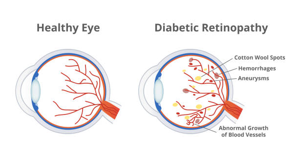
Vector illustration of diabetic retinopathy, a complication of diabetes caused by high blood sugar and normal healthy eye isolated on a white background. Cotton wool spots, hemorrhages, aneurysms, and abnormal growth of blood vessels.


Diabetic retinopathy. The eye condition that affect people with diabetes. Illustration of the retina of the eye

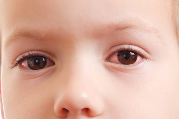
Child conjunctivitis red eye with infection, baby face, health.

Diabetic retinopathy (diabetic eye) . Spots floating in the vision, blurred vision, blindness. A complication of diabetes, caused by high blood sugar levels damaging the back of the eye (retina).

Retinopathy is damage to the retina of the eyes, which cause vision impairment. Anatomy of the human eye. Vertical section of the eye and eyelids. Schematic diagram. detailed illustration.

Subconjunctival bleeding, also known as subconjunctival hemorrhage, is bleeding from a small blood vessel over the whites of the eye. It results in a red spot in the white of the eye. There is generally little to no pain and vision is not affected. Generally only one eye is affected


Close-up of woman's eye with red inflamed and dilated capillaries magnified with a magnifying glass. Disease of retina of the eye. Conjunctivitis, keratitis, dry eye syndrome

Close-up of woman's eye with red inflamed and dilated capillaries. Hemorrhage under the conjunctiva. Disease of retina of the eye. Conjunctivitis, keratitis, dry eye syndrome, trauma, uveitis

Blood in the eye from a subconjunctival hemorrhage usually disappears within a week or two.Human eye and blood close up.

attractive young brunette ponytail female asian doctor wear white coat and stethoscope pen pointing explaining brain model to gray hair old sick at clinic - an xray film display on computer


Close up shot of a very red and bloodshot eye, Taken with a macro lens, the image shows an abstract pattern of blood swirls within the eye.


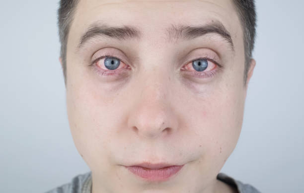
Copy space for advertisement. Tired eyes after working at the computer. Close-up videos and macro. Close up of two annoyed red blood eyes of male affected by conjunctivitis or after flu, cold, allergy.

Medical worker in protective , mask and hat looking in surgical microscope and looking through it on patient


Wet and dry macular degeneration anatomical poster. Human eye anatomy, retina, macula and fovea structure. Central vision disease diagram. Age related eye problems AMD medical flat vector illustration

Blood in the eye from a subconjunctival hemorrhage usually disappears within a week or two.Human eye and blood close up.

Vector medical poster normal eye and eye with diabetic retinopathy.

Macular degeneration poster. Human vision concept, eye disease. Deficiency of retina, macula and fovea. Normal vision and central vision defect. Age related eye problems flat vector illustration


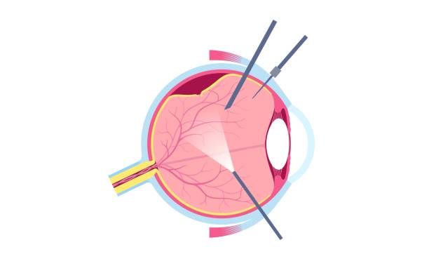
Vitrectomy surgical procedure. Ophthalmology clinic, remove the vitreous gel from the eye, treating retinal detachment, macular holes, or vitreous hemorrhage, improving vision flat vector illustration

Close-up of woman dripping her eyes with medicinal drops natural tear. Disease of eye retina. Conjunctivitis, keratitis, dry eye syndrome, trauma. Treatment of red inflamed and dilated capillaries


View inside human eye disorders - showing retina, optic nerve and macula.Eye treatment concept.

Diabetic retinopathy anatomical poster. Human eye disease, vision loss or blindness. Damaged blood vessels in the retina. Proliferative or nonproliferative eye condition medical vector illustration


Cropped shot of woman dripping her eye with medicinal drops natural tear. Disease of retina of eye.Conjunctivitis, keratitis, dry eye syndrome, trauma. Treatment of red inflamed and dilated capillaries
of6Next