
Isolated on white background. Isometric vector illustration
Browse 200+ blood on microscope slide stock illustrations and vector graphics available royalty-free, or start a new search to explore more great stock images and vector art.
Isolated on white background. Isometric vector illustration
A set of hematology icons that include editable strokes or outlines using the EPS vector file. The icons include a lab worker in a hazmat suit doing scientific research on a vial of blood, hand holding a test tube of blood, blood being applied to a microscope slide for examination, microscope with blood on a slide being examined, hematologist holding up a test tube of blood, person donating blood, finger being pricked to test blood, syringe drawing blood, blood donee recipient, hematologist with patient chart, nurse administering blood transfusion to patient, blood glucose monitor, syringe drawing blood from a patients arm, unit of blood in a bag, medical research on blood, hand checking pulse of patient, researcher at microscope examining blood, blood on test strip, human heart, person with a blood clot in his leg, blood clot in veins, recipient receiving blood donation from donor, blood donation, wound on foot with blood and a hematologist examining blood platelets with a magnifying glass.

Infographic template. Blood donation concept with 4 steps. Vector illustration

Blood sample under a microscope showing red blood cells with other substances (oxygen, carbon dioxide, hemoglobin). Microscope slide with blood sample.
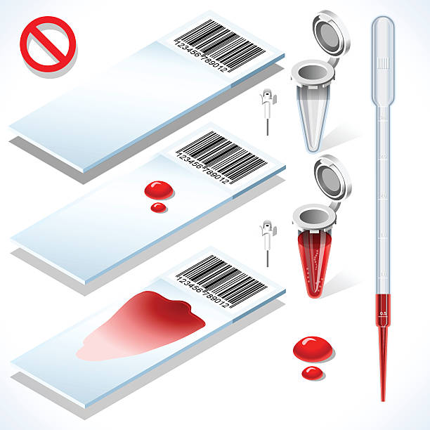
Hematology Test Kit 3D Isometric Set. Laboratory Equipment for Medical Analysis or Scientific Translational Research. White Slides Blood Sample Filled Tube Empty Eppendorf and Pipette

EPS, High and Low Resolution JPGs. Stage your own crime scene investigation with these highly-detailed CSI elements. Included are: Shoeprint, magnifying glass (with wooden handle), detailed fingerprint, chalk outline (with body silhouette), "POLICE LINE DO NOT CROSS" yellow cordon, eyedropper, tweezers, blood spatter, microscope, test tube, blood droplets, hypodermic needle, microscope slides, and blood specimen bottles.

Hepatology concept vector for medical website, landing page. Concept of hepatitis A, B, C, D, cirrhosis, world hepatitis day. Tiny doctors treat the liver.

Simple diagram showing basic structures of the connective tissue
A set of icons. File is built in the CMYK color space for optimal printing. Color swatches are global so it’s easy to edit and change the colors.

Blood sample under a microscope showing red blood cells with cartoon germs. Microscope slide with blood sample.

Blood test with blood sampling and medical equipment. Color vector icons set
A set of icons. File is built in the CMYK color space for optimal printing. Color swatches are global so it’s easy to edit and change the colors.
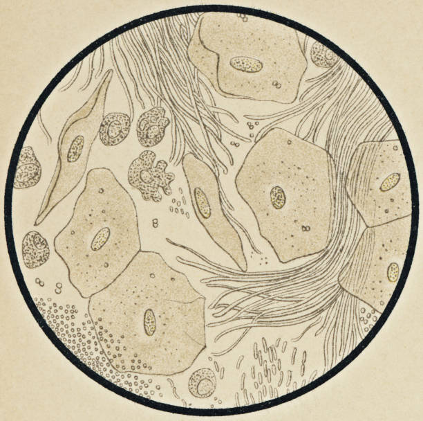
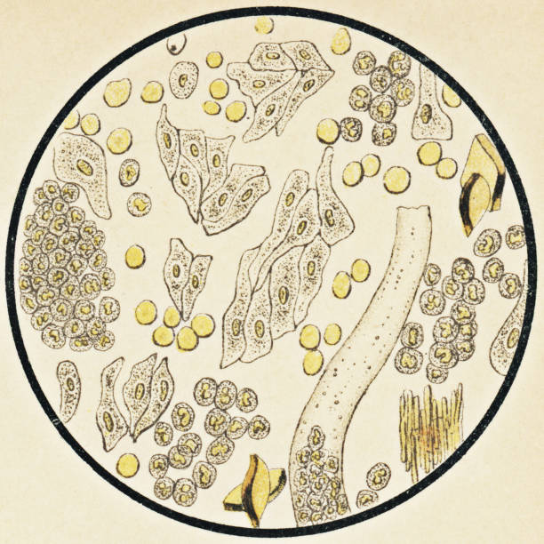
Microscopic view of human cells and oral microbiome found in the gingival tissue of the gums. Vintage etching circa mid 19th century.

Microscopic view of human blood cells with corynebacterium diphtheriae bacteria found in the blood from a patient with Diphtheria. Vintage etching circa mid 19th century.

Hematology concept with red blood cell tube laboratory analysis flat examine. Hematology blood red cell liquid
A set of icons. File is built in the CMYK color space for optimal printing. Color swatches are global so it’s easy to edit and change the colors.

A set of icons. File is built in the CMYK color space for optimal printing. Color swatches are global so it’s easy to edit and change the colors.
A set of icons. File is built in the CMYK color space for optimal printing. Color swatches are global so it’s easy to edit and change the colors.

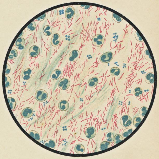
Microscopic view of human white blood cells and streptococcus pyogenes bacteria from a patient that developed a group A streptococcal infection with pneumonia. Vintage etching circa mid 19th century.
Blood cell count or biochemical blood test and medical equipment silhouettes. Black and white vector icons set isolated on white background
Microscopic view of human blood cells and orthomyxoviridae bacteria (influenza) found in the sputum mucus from a patient with acute bronchitis, stained with acid fuchsin. Vintage etching circa mid 19th century.

Detailed illustration of a Hematology Test Complete Set This illustration is saved in EPS10 with color space in RGB.
Microscopic view of human blood cells and mycobacterium tuberculosis bacteria found in the sputum mucus from a patient with Tuberculosis, Ziehl-Neelsen staining method used. Vintage etching circa mid 19th century.

Microscopic view of human white blood cells and burkholderia mallei bacteria from a patient with glanders. Vintage etching circa mid 19th century.
Microscopic view of human urinary sediment from a patient with pyelonephritis. Vintage etching circa mid 19th century.

Hand drawn cute illustration drop of blood on a glass slide. Flat vector laboratory microscope slide in simple colored doodle style. Blood test, medicine sticker, icon. Isolated on white background.

Microscopic view of human urinary sediment from a patient with albuminuria. Vintage etching circa mid 19th century.

Microscopic view of diminished multinucleates in human blood cells found in a patient with typhoid fever. Vintage etching circa mid 19th century.
A set of icons. File is built in the CMYK color space for optimal printing. Color swatches are global so it’s easy to edit and change the colors.
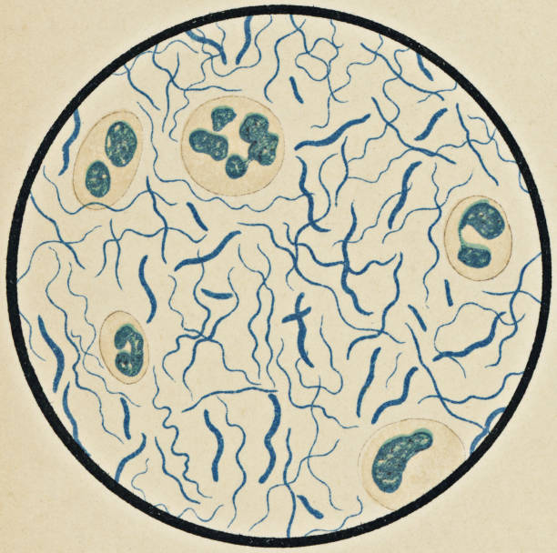
Microscopic view of borrelia spiral bacteria found in a patient with Norma disease, stained with gentian violet. Vintage etching circa mid 19th century.
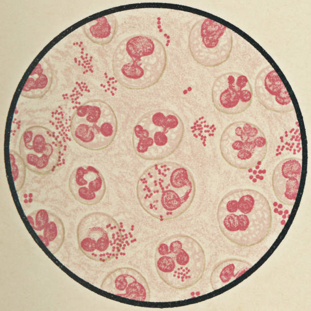
Microscopic view of eosinophil cells and micrococci bacteria found in sputum mucus from a patient with asthma, stained with methylene blue and eosin. Vintage etching circa mid 19th century.

Hematology concept with red blood cell tube laboratory analysis flat examine. Hematology blood red cell liquid

Microscopic view of human blood cells and bacteria found in sputum mucus. Vintage etching circa mid 19th century.

Set of blood donation items. Medical and health care objects.

Microscopic view of human blood cells and mycobacterium tuberculosis bacteria found in the sputum mucus from a patient with Tuberculosis, Ziehl-Neelsen staining method used. Vintage etching circa mid 19th century.

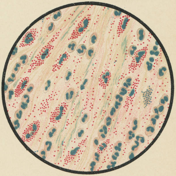
Microscopic view of myelocyte human white blood cells in mitosis (cell division) found in the blood from a patient with chronic myelogenous leukemia. Vintage etching circa mid 19th century.

Microscopic view of human urinary sediment from a patient with a bladder infection (cystitis). Vintage etching circa mid 19th century.

Microscopic view of frog blood cells. Vintage etching circa mid 19th century.

Microscopic view of human blood cells found in the blood from a patient with chronic myelogenous leukemia, stained with Ehrlich triacid stain. Vintage etching circa mid 19th century.

Microscopic view of mouse blood cells with pneumococcus bacteria, stained using the Gram’s Method. Vintage etching circa mid 19th century.

Microscopic view of human blood cells and staphylococcus aureus bacteria from a patient with a ruptured lung abscess, Gram staining method used. Vintage etching circa mid 19th century.

Microscopic view of human urinary sediment from a patient with amyloidosis of the kidney. Vintage etching circa mid 19th century.

Microscopic view of human white blood cells and mycobacterium tuberculosis bacteria found in the pus from a patient with urogenital tuberculosis. Vintage etching circa mid 19th century.

Microscopic view of human white blood cells and salmonella typhi bacteria from a patient with typhoid fever. Vintage etching circa mid 19th century.


