Cytoplasm stock illustrations
Browse 1,900+ cytoplasm stock illustrations and vector graphics available royalty-free, or search for cytoplasm translation or cell cytoplasm to find more great stock images and vector art.
Most popular
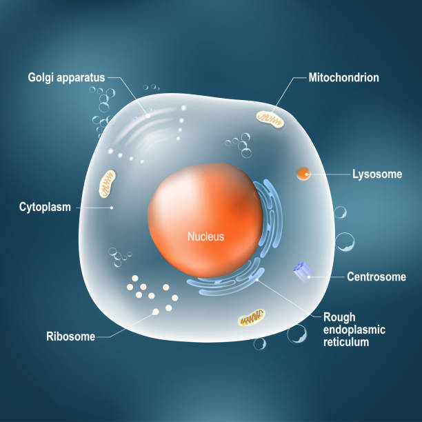
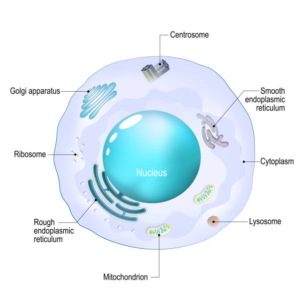
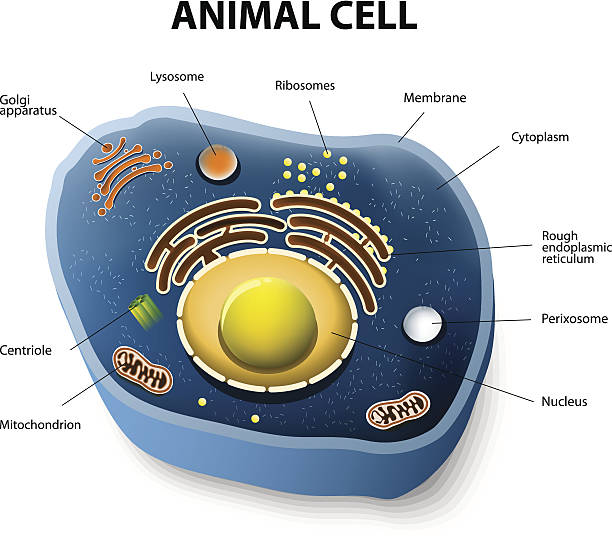
Anatomy of cell. All organelles: Nucleus, Ribosome, Rough endoplasmic reticulum, Golgi apparatus, mitochondrion, cytoplasm, lysosome, Centrosome. Animal cell on the dark background. Illustration easy editable for Your color
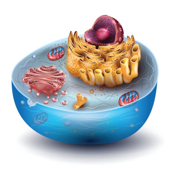
Eukaryotic cell diagram, vector illustration, text on own layer
Gradient and transparent effect used.
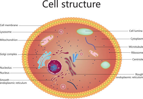
Cell structure, cross section of the cell detailed colorful anatomy with description
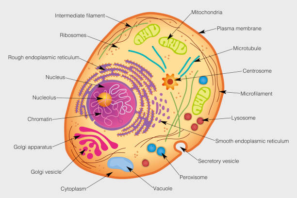
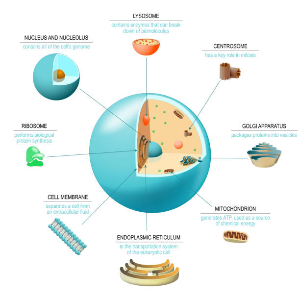
The graphic shows the elements of a human cell with their names on a gray background. Vector image

translation (biological protein synthesis). Number 1: syntesis of mRNA from DNA in the nucleus. 2 The mRNA decoding ribosome by binding of complementary tRNA anticodon sequences to mRNA codons. 3-5 ribosomes synthesize proteins in the cytoplasm .

Extracellular matrix labeled infographic vector illustration scheme. Biological diagram with collagen fiber, fibronectin, phospholipid bilayer and cytoskeleton filaments.
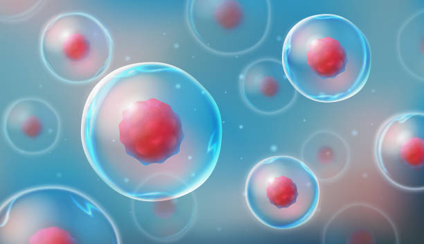
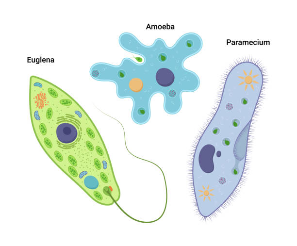
Cells under a microscope. Research of stem cells. Cellular Therapy. Cell division. vector illustration on a light background.


Animal cell anatomy. Structure and organelles of Eukaryotic cell. Vector poster for education. illustration
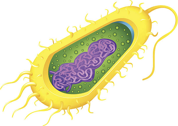
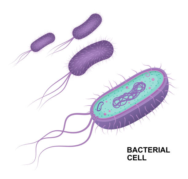
Illustration of a bacteria cell
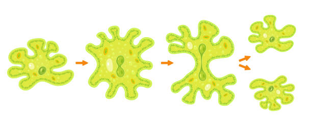
Amoeba binary fission infographic. Vector illustration of reproduction of simplest bacteria. Formation of unicellular organisms.
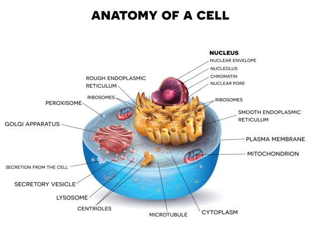
Cell organelles. Structure and anatomy of a animal cell. realistic cell on a white background. Vector illustration. Poster for education
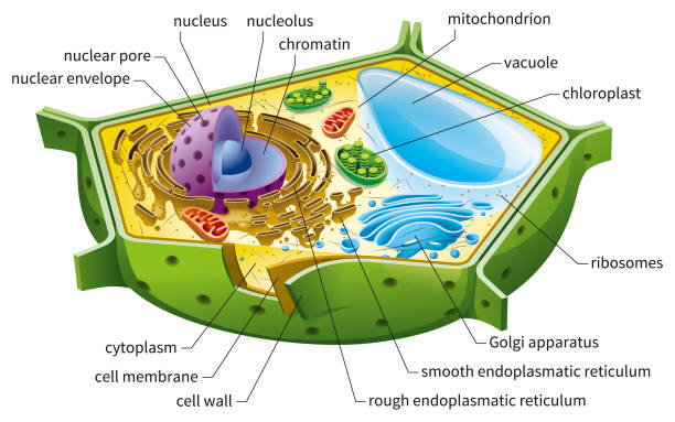
Eukaryotic cell diagram, vector illustration, text on own layer


Active and passive transport as molecules ATP movement in outline diagram. Labeled educational scheme with closeup cellular model vector illustration. Facilitated diffusion compared process example.

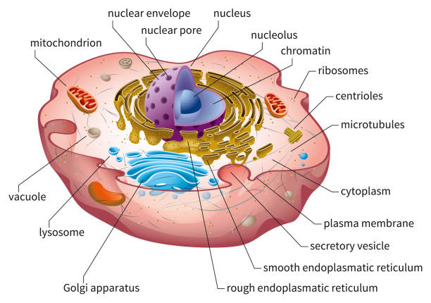
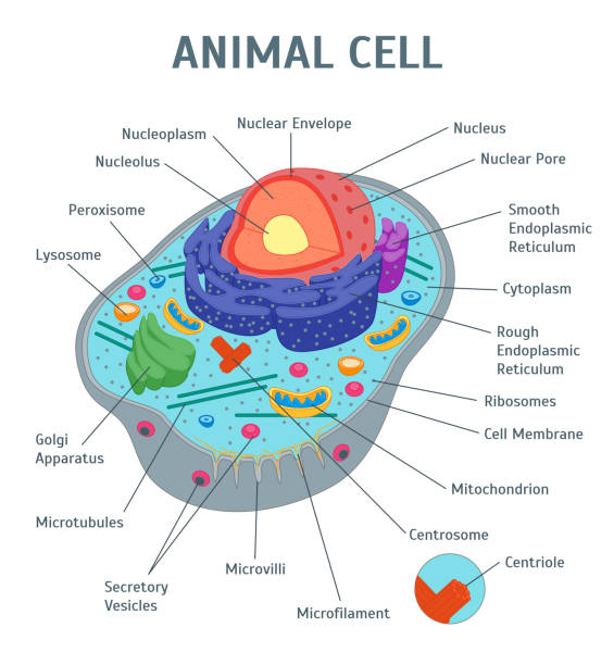
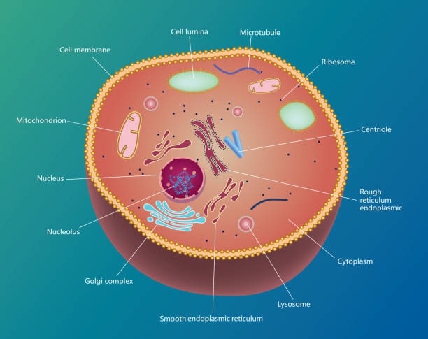
Cell anatomy. Structure and organelles of human's cell. Cross sections of animal cell: nucleus, nucleolus, mitochondria, centresome, golgi apparatus, endoplasmic reticulum, ribosome and membrane. poster biological diagram. vector illustration


Internal anatomy of the prokaryotic cell. Different types of bacteria. Comparison with the eukaryotic cell.
Different human cell types icon set. Medicine and biology illustrative symbol. Health, anatomy and science. Biology vector isolated on white background.

Cancer and normal cells. comparison and difference between healthy tissue and tumor. details about chromatin, nucleus and cytoplasm. Vector illustration
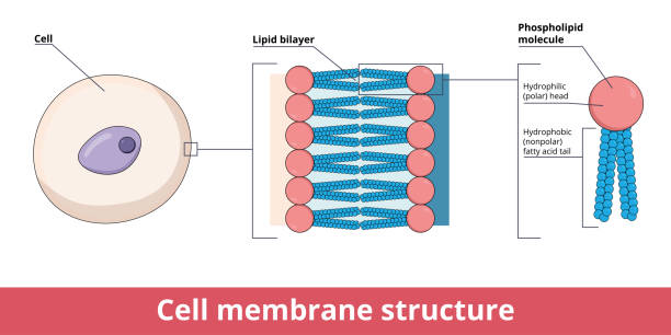
Cell membrane structure, that is represented by lipid bilayer and its phosphatidylcholine (a phospholipid), that is composed of polar hydrophilic “head” and nonpolar hydrophobic “tail.”

The process of karyokinesis (or mitosis) is divided into five stages: prophase, prometaphase, metaphase, anaphase, and telophase.
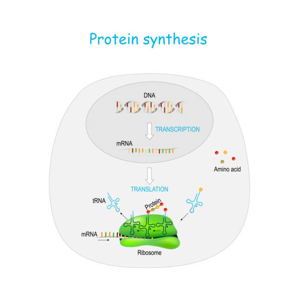
Protein synthesis in ribosome. transcription and translation. synthesis of mRNA from DNA in the nucleus. The mRNA decoding ribosomes. steps diagram with cycle explanation
Cell structure, cross section of the cell detailed colorful anatomy
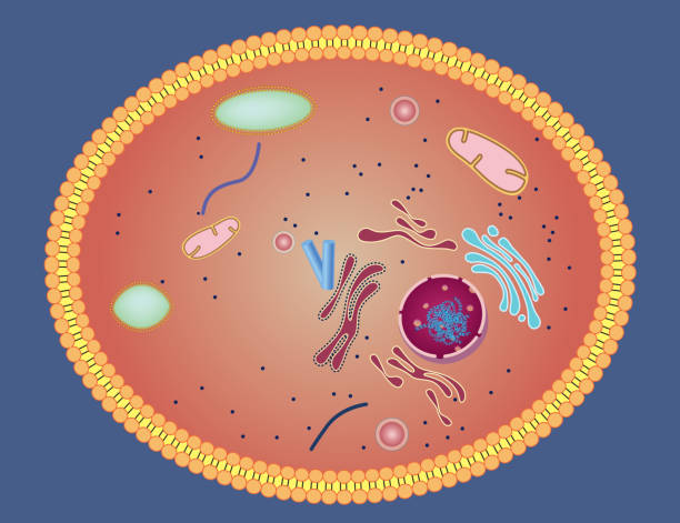
human or animal cell. cross section. structure of a Eukaryotic cell. Vector diagram
Cartoon Animal Cell Anatomy Banner Card Poster Scientific or Education Concept Flat Design Style. Vector illustration of Structure
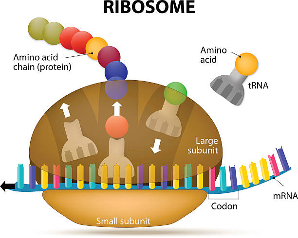
Ribosome during protein synthesis. The Interaction of a Ribosome with mRNA. Process of initiation of translation
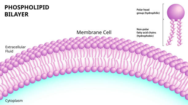
Structure of the Phospholipid Bilayer in the Cell Membrane - Medical Vector Illustration

bacteria and fungal (yeast). comparison of cell structure. Similarities and differences. cross section and anatomy of cell. Biology Chart. Vector illustration on a white background. detailed diagram for use in education
Cosmetic properties line icons. Vector illustration include icon - cream, body lotion, lifting, moisture, anti wrinkle outline pictogram for skincare product. 64x64 Pixel Perfect, Editable Stroke.
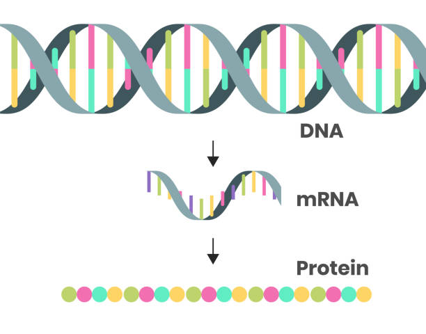
Protein syntesis schematic illustration. Vector illustration of the DNA, mRNA and polypeptide chain

Differentiation of monocytes. Osteoclast, Macrophage and Dendric cell
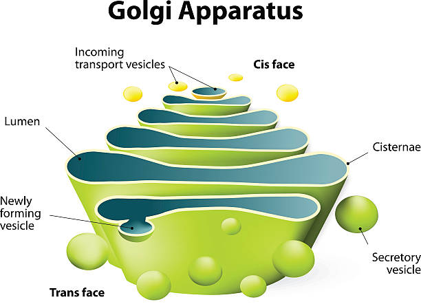
Golgi apparatus. Golgi Complex plays an important role in the modification and transport of proteins within the cell
Gradient and transparent effect used.
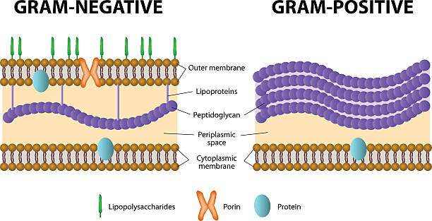
Bacteria. Difference of Gram-positive from Gram-negative bacterial.
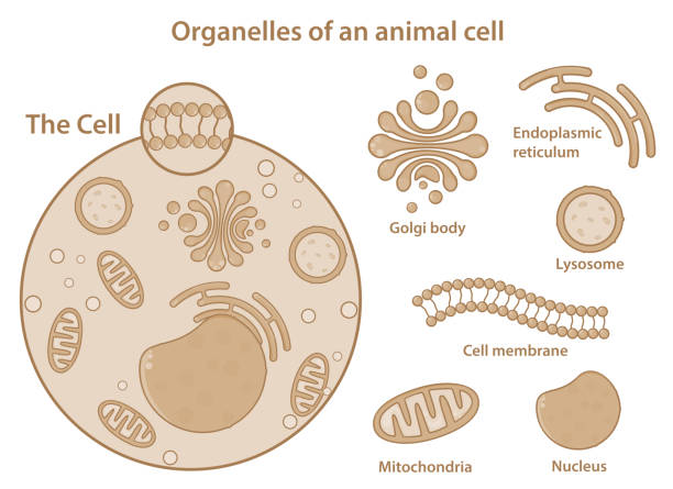
Organelles of an animal cell showing different components present in a eukaryotic cell. Simple and clear medical illustration. Major parts of a cell only. Aesthetic graphics.
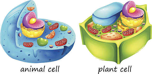
lllustration of the animal and plant cells on a white background

Cell membrane. GABA receptor and various sita for ligands bind. Top view of ion channel which illustrates the five combined subunits that form Cl ion channel pore.
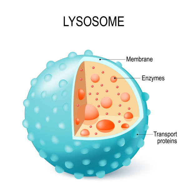
Anatomy of the Lysosome: Hydrolytic enzymes, Membrane and transport proteins. This organelle use the enzymes to break down and digest food particles, engulfed viruses or bacteria in the cell. Vector diagram for medical use
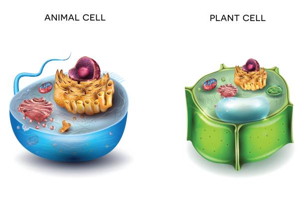
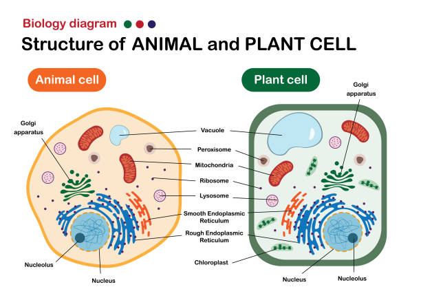
Animal Cell and Plant Cell structure, cross section detailed colorful anatomy.

Mitochondria under microscope on dark blue backgound in futuristic glowing low polygonal style. Medical research concept, Science, biology research banner. Modern abstract design vector illustration
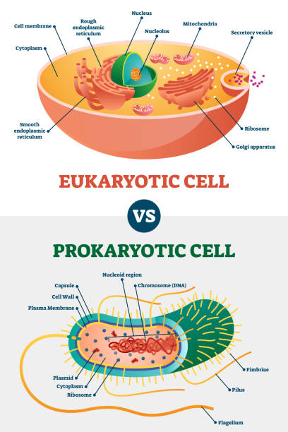
Eukaryotic vs Prokaryotic cells, educational biology vector illustration diagram. Microbiology scheme with cell type examples. Cell membranes, cytoplasm, chromosomes, ribosomes and various organelles.
Muscular tissue seamless pattern. Stock vector illustration of muscle cells in rows. Medicine and biology collection
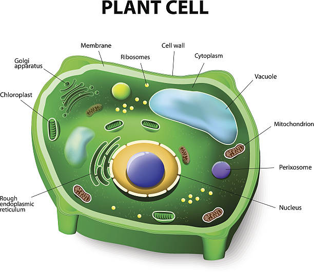
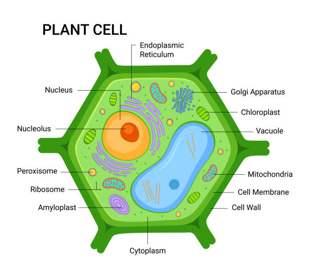
Plant cell structure. Vector diagram
Gradient and transparent effect used.
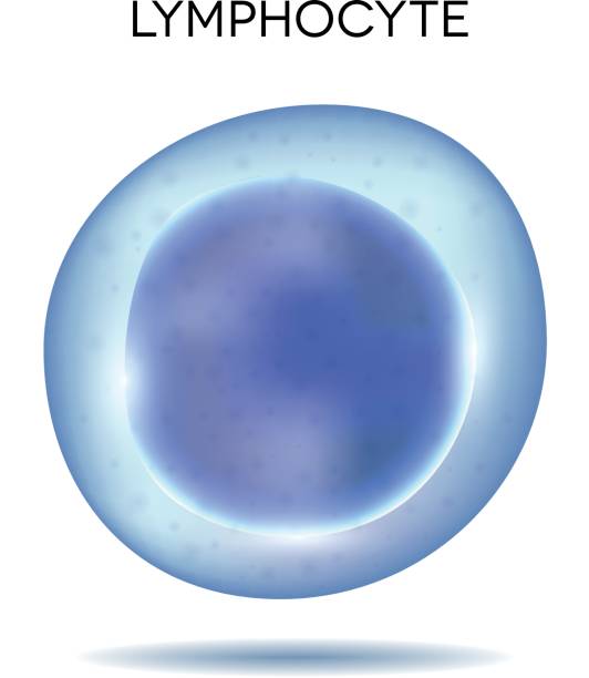
Human blood cell Lymphocyte
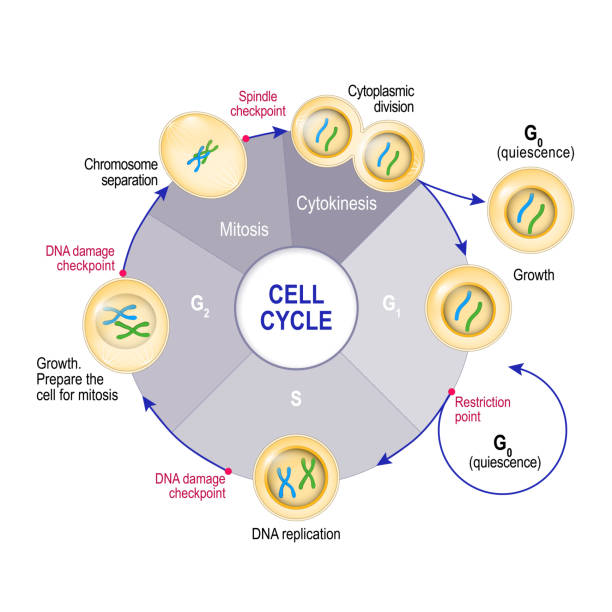
Cell Cycle (Cell division): from quiescence, Growth and DNA replication to Mitosis and Cytokinesis. Cell cycle checkpoints: DNA damage, Spindle checkpoint, Restriction point. Vector illustration for educational, medical and science use
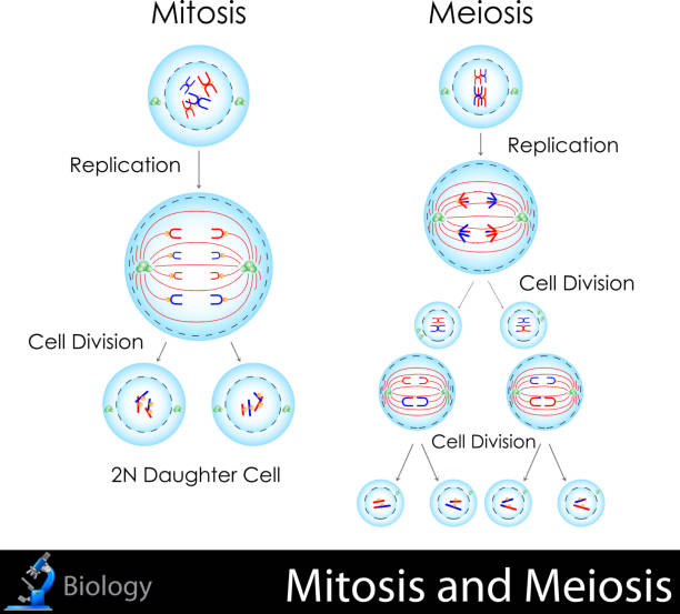
easy to edit vector illustration of mitosis and meiosis

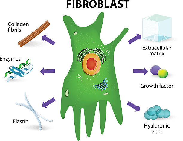
Fibroblast. Structure and function. Human skin cell
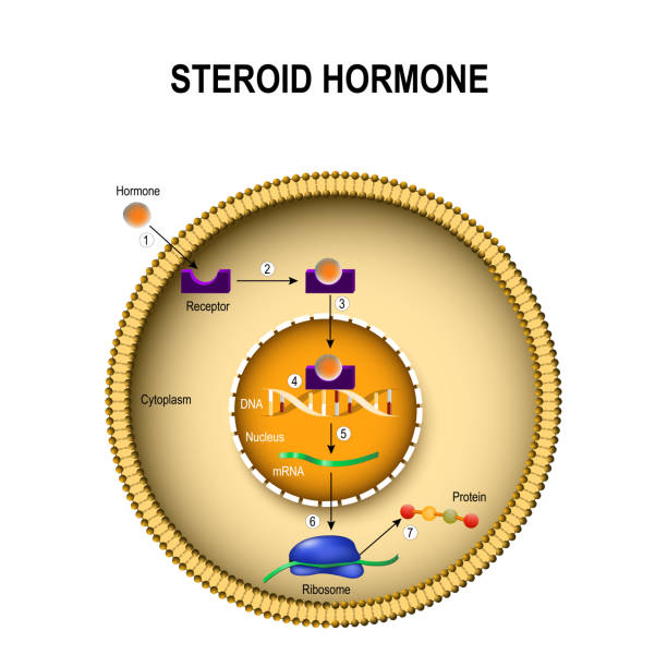
How steroid hormones work. interaction of the hormone with the intracellular receptor. Human endocrine signaling system
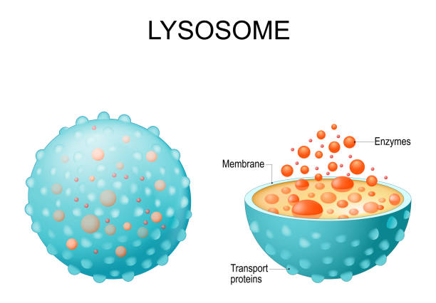
Lysosome. appearance, exterior and interior view. Cross section and Anatomy of the Lysosome: Hydrolytic enzymes, Membrane and transport proteins. Vector illustration
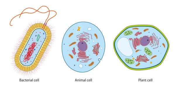
Difference between bacteria, animal and plant cells
Next