Knee Joint Diagram stock illustrations
Browse 810+ knee joint diagram stock illustrations and vector graphics available royalty-free, or start a new search to explore more great stock images and vector art.
Most popular
Knee anatomy. Structure of leg joint. Major parts. Vector poster with text label for medical education
knee anatomy. side and front view. Cross section of the joint showing the main parts: femur, fibula, articular capsule, menisci, muscles and ligaments. vector illustration
Knee Cartilage Anatomy - Illustration as EPS 10 File
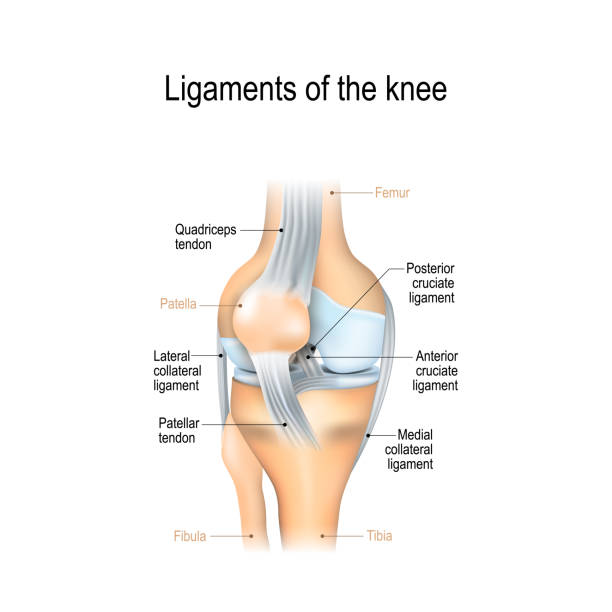
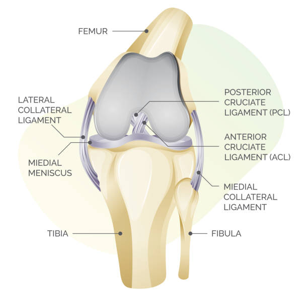
Ligaments of the knee. Anterior and Posterior cruciate ligaments, Patellar and Quadriceps, tendons, Medial and Lateral collateral ligaments. joint anatomy. Vector illustration for biological, medical, science and educational use
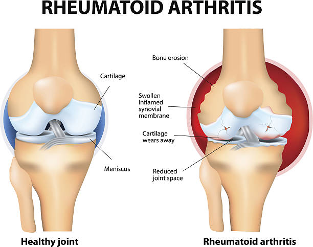
Rheumatoid Arthritis or RA is an inflammatory type of arthritis that usually affects knees. Rheumatoid arthritis of the knee the auto immune disease. The body's immune system mistakenly attacks healthy tissue.

The knee joint joins the thigh with the leg and consists of two articulations: one between the femur and tibia and one between the femur and patella. It is the largest joint in the human body. Illustration anatomy body.

Shin splints vector illustration. Leg muscle sport trauma and bone pain labeled diagram. Isolated femur, patella, fibula, tibia and foot bones with shown injury location.

Knee bone anatomy concept. Descriptions of the human leg bones and joints. Meniscus, ligaments, tendons, patella anatomy. Medical anatomical poster for clinic. X ray isolated flat vector illustration.

Medical infographic with orthopedic anatomy charts. Human silhouette in motion with marked spine, pelvis, knee, foot, shoulder, elbow, hand bones and joints. Orthopedics medical group design
leg back muscles 3d medical vector illustration on white background eps 10
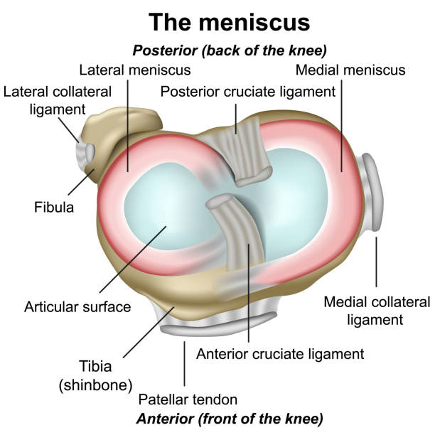
Meniscus knee anatomy medical vector illustration isolated on white background infographic eps 10 ligament

Types of joints. Knee joint, Articulations of foot, Elbow joint. Set icons. black and white. Flat vector illustration.

coxa saltans syndrome 3d medical vector illustration on white background eps 10

Knee Joint - Pain - Stock Illustration as EPS 10 File

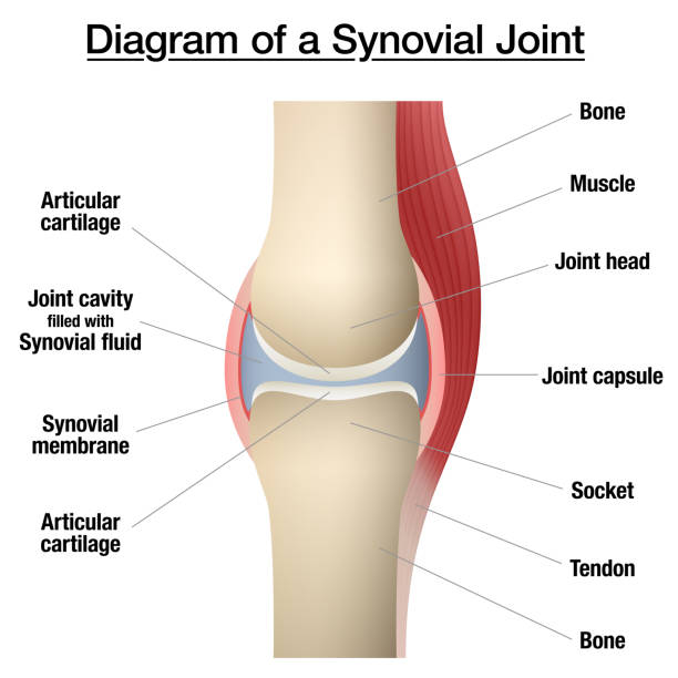
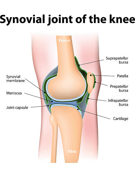
Synovial joint anatomy. joint capsule with synovial fluid and membrane. Vector illustration
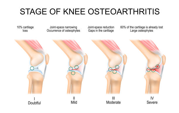
knee Osteoarthritis. Stages of OA. Kellgren and Lawrence criteria for assessment stage of osteoarthritis. The classifications are based on osteophyte formation and joint space narrowing. side view. Vector

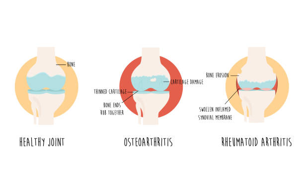
Vector diagram with healthy joint and joint with osteoarthritis, rheumatoid arthritis. Medical poster, design

Stages of knee osteoarthritis. From Minimum disruption to lost a cartilage and joint-space reduction. Anatomy of knee joint. Vector illustration
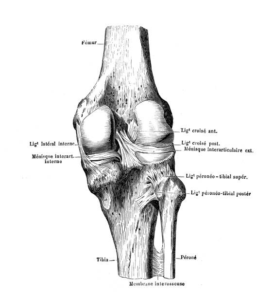
illustration of a knee
Knee bones vector. Human bone and joint icon set. Rheumatology and traumatology, vector design and illustration. Vector illustration

Arthrosis medical illustration diagram with damaged knee structure and healthy knee comparison. Bone exposure, osteophytes, cartilage fragments, erosion of cartilage and inflamed joint capsule.
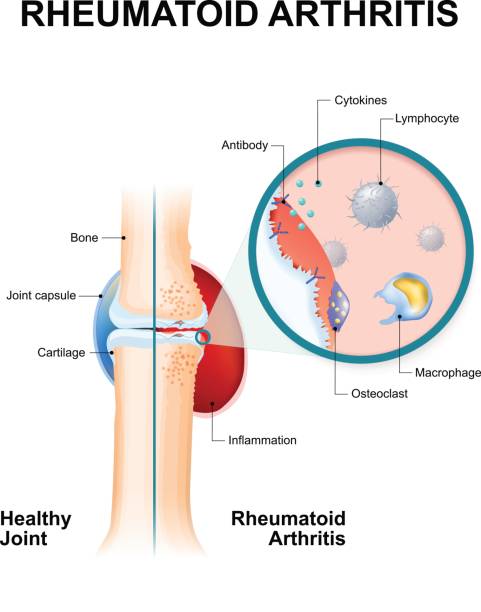
normal joint and one with rheumatoid arthritis. Rheumatoid Arthritis (RA) is an inflammatory type of arthritis that usually affects knees. the auto immune disease. The body's immune system mistakenly attacks healthy tissue.


Arthrosis medical vector illustration diagram with damaged knee structure and healthy knee comparison. Bone exposure, osteophytes, cartilage fragments, erosion of cartilage and inflamed joint capsule.

Front and side anatomical view of an human knee. Digital illustration.


Anatomy of human knee vector sketch of leg bones and joint, medicine design. Side and front view of knee bones, hand drawn femur, patella, tibia and fibula, tibial plateau and lateral condyle

Baker's cyst. fluid collection behind the knee. cross section of Human joint. Flat vector diagram like x-ray illustration.

Stem cell therapy ( Injections) for repair of knee cartilage. The cell mixture is combined with a fibrin glue component and implanted.

Lateral collateral tear 3d medical vector infographic isolated on white background eps 10
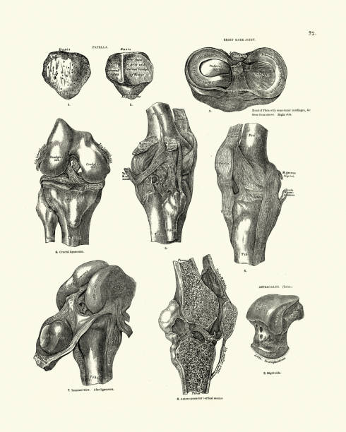
Vintage engraving of human bones, Knee joint, Patella, Talus, Victorian anatomical drawing, 19th Century. Descriptive Atlas of Anatomy, 1880
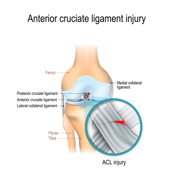
Anterior cruciate ligament injury. joint anatomy. Vector illustration for biological, medical, science and educational use

Body movement types with medical physical motion explanation outline diagram. Labeled educational anatomy scheme with healthy leg, arm or shoulders rotation, extension or inversion vector illustration


tear of a meniscus is a rupturing of one or more of the fibrocartilage strips in the knee. Human Joint, and Traumatic force in sports or physical exertion. Torn meniscus.
Synovial bursa of the human knee. Synovial bursa is a sac filled with lubricating fluid, located between tissues such as bone, muscle, tendons, and skin, that decreases rubbing.
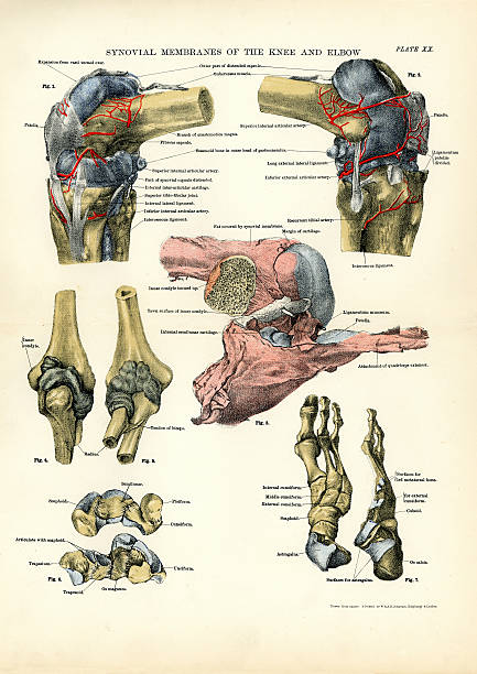
Victorian engraving of the human Synovial membranes of the Knee and Elbow

Injury of knee bone and leg while human running. Illustration about medical and sport.
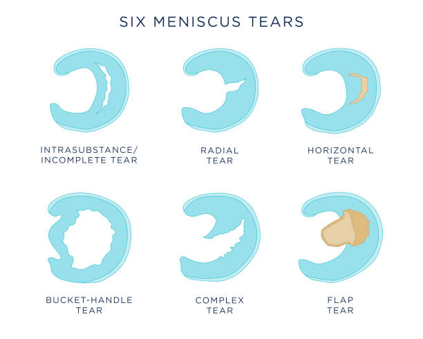
Flat vector illustration showing six different types of (knee) meniscus tears, including intrasubstance tear, radial tear, horizontal tear, bucket-handle tear, complex tear, and flap tear.
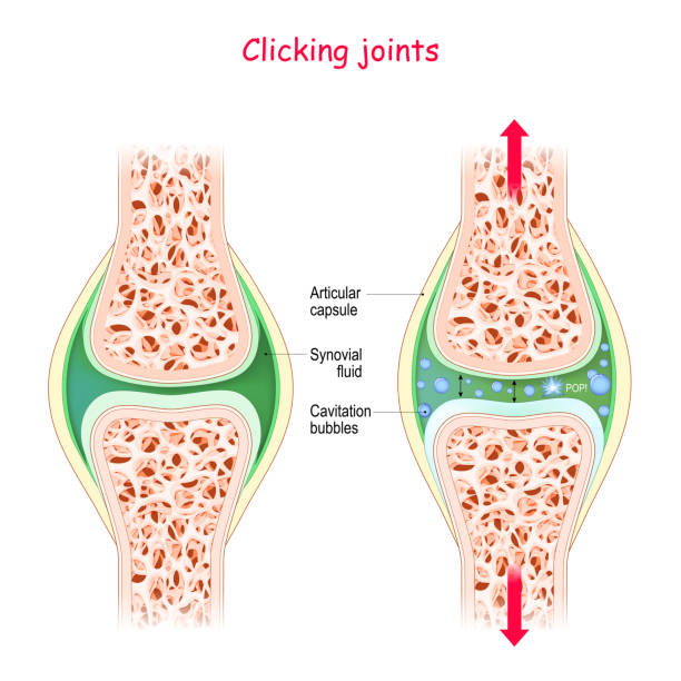
Joints and popping sound. Physiological Mechanism of cavitation. manipulate the joint we will get sound this is rapid movement of gas bubbles within the synovial solution. Cracking joint

Infographic Treatment Method Rheumatoid Arthritis. 3d Banner Vector Illustration Human Knee Joint Diseases. Information Joint Treatment Medicine Injection for Pain Relief. Packaging Painkiller Pils

Woman doing Exercise with speed jumping rope in 3 step. Illustration about workout with lightweight equipment.
Leg vector in front view. Illustration about human legs composition.
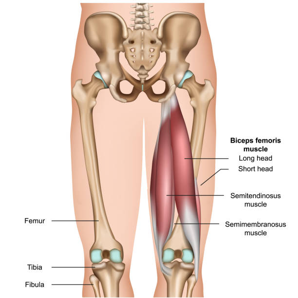
hamstring muscle anatomy 3d medical vector illustration on white background eps 10
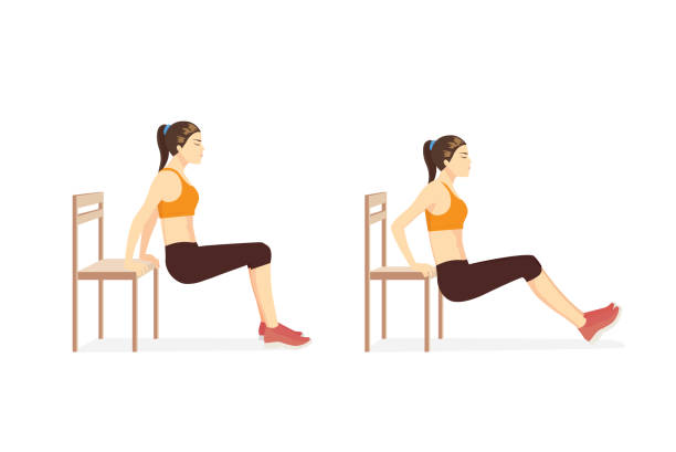
Woman doing Triceps Dips with bench in 2 step for exercise guide. Illustration about workout for build strength triceps brach ii muscle.

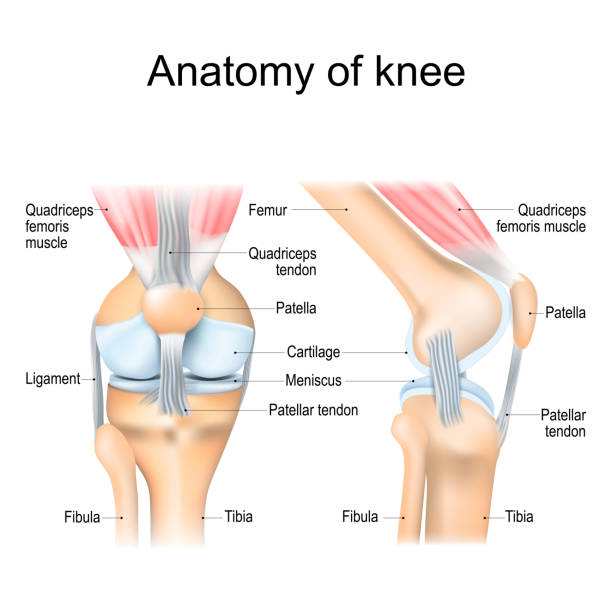
Knee anatomy including ligaments, cartilage and meniscus. Detailed Anatomy of the Knee Joint. Muscles and Tendons

Human Knee Rheumatoid Arthritis vector diagram illustration
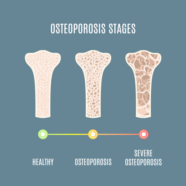
Osteoporosis process infographic of bone tissue close-up with different density. Skeletal system disease stages. Senior osteopathy medical concept. Vector illustration.

Human renal system vector illustration isolated on white background
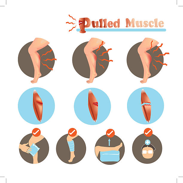
Muscle Strain degree and Treatment isolated on white background.Vector illustrations

Incorrect sleeping positions can affect your whole body. Illustration about healthy lifestyle.

Woman doing full plank passe twist guide in 2 step for exercise diagram. Illustration about workout posture for fat reducetion.
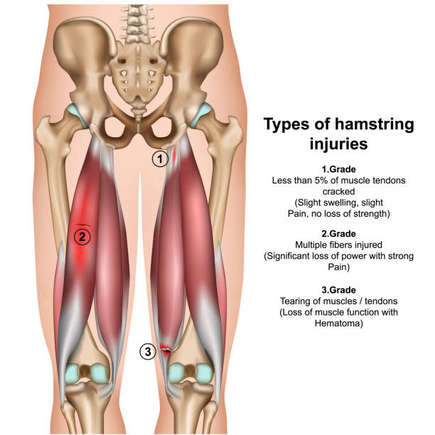
types of hamstring injurys 3d medical vector illustration on white background eps 10

Woman doing Hip Dip Fitness with bench in 2 steps for exercise guide. Illustration about workout for fit triceps brach ii muscle.

Autoimmune disorders. For example Gonorrhea (sexually transmitted infection) and Arthritis. Antigens of bacterium Neisseria gonorrhoeae are similar to self-molecules of healthy joint cells. normal immune response can result in the production of antibodies that bind to healthy cells of joint, and caused of inflammation

The human knee vector diagram illustration
Next