Lumbar Vertebra stock illustrations
Browse 370+ lumbar vertebra stock illustrations and vector graphics available royalty-free, or search for spine or lumbar fusion to find more great stock images and vector art.
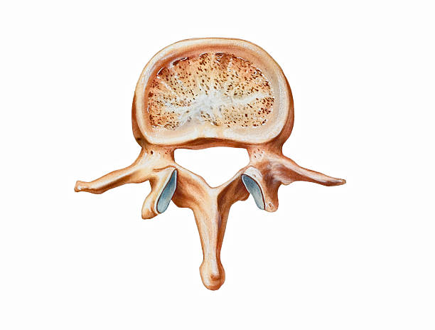
The second lumbar vertebra (L2, top view). Shown are the vertebral body, pedicle, superior articular process, vertebral foramen, spinous process, lamina, mamillary process, and transverse process.
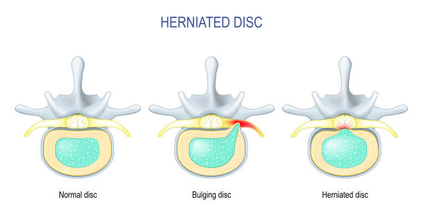
Spinal disc herniation. Difference Between Bulging disc and Herniated Disc. Vector illustration

Lumbar region of the human spine

Vertebral column: cervical, thoracic and lumbar spine, sacrum and coccyx. Numbering order of the vertebrae of the human spinal column. Vector diagram for medical use

Illustration of anterior and lateral views of "lumbar vertebrae, sacrum, coccyx"

Diagram of a human spine with names of all sections of the vertebrae.


Engraving from "Die descriptive und Topographische Anatomie des Menschen"; by Dr. C. Heintzmann. Published by Wilhem Braumüller, Vienna (1884). Photographed and edited by J. C. Rosemann.

Deformation of the intervertebral disc due flexion and extension motion

Illustration of anterior and lateral views of "lumbar vertebrae, sacrum, coccyx"
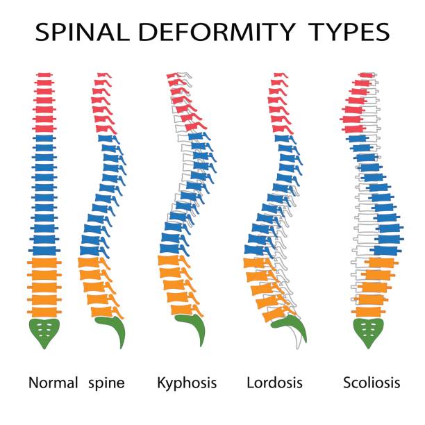
Illustration of spinal deformity types. Kyphosis, lordosis and scoliosis.
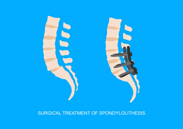
Vector illustration of slipped lumbosacral vertebra or spondylolithesis and surgical fixation of spine.

http://thebrainstormlab.com/banners/ami_banner.jpgPhotographer's Description:


t is an illustration that understands the structure of the pelvic muscles

Concept for a spine clinic, chiropractic practice or medical device company or product.


Engraving from "Die descriptive und topographische Anatomie des Menschen"; by Dr. C. Heintzmann. Published by Wilhem Braumüller, Vienna (1884). Photographed and edited by J. C. Rosemann.


The intervertebral disc is a fibrocartilaginous structure that joint the vertebral bodies of the spinal column and provides it resistance and protection to the effect of axial loads


Herniated spinal disc - Degenerative, Protrusion, Extrusion, Sequestration - vector patient-friendly diagram, hand drawn. Infographic of stages hernia of intervertebral disk

Engraving from "Die descriptive und Topographische Anatomie des Menschen"; by Dr. C. Heintzmann. Published by Wilhem Braumüller, Vienna (1884). Photographed and edited by J. C. Rosemann.

anatomy of human spine, spinal cord, thoracic, cervical, lumbar, pelvic bone, internal organs body part orthopedic health care, vector illustration cartoon flat character design clip art isolated

Spine one line on white background, simple sketch of part of skeleton. Human bones, spine, rehabilitation. Medical drawing, anatomy, osteopathy. Vector stock illustration.


Medical illustration of human lumbar vertebrae bone. Vintage etching circa 19th century.

Illustration of types of scoliosis of spine on the white background.

Vintage engraving of Hyoid bone, Atlas, Seventh cervical vertebra, Dorsal vertebra, peculiar dorsal vertebra, Lumbar vertebra, Victorian anatomical drawing, 19th Century. Descriptive Atlas of Anatomy, 1880

Corset for correction of posture on the green background. Vector illustration


man having chest pain in the upper and middle rib cage, cartilage ,costochondritis, medical internal organs nervous system anatomy health care symtomps, flat vector illustration cartoon design
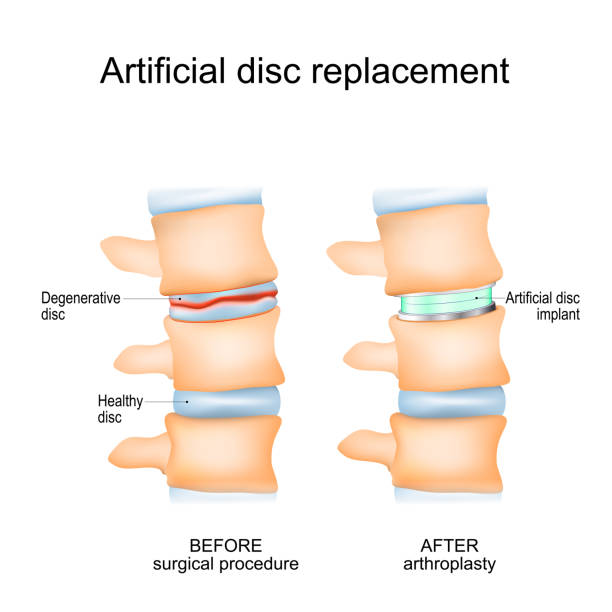
Intervertebral disc arthroplasty. Close-up of a human spinal column before Surgical procedure and after Arthroplasty. Degenerative Disc and Artificial disc replacement. Vector poster

Illustration of a Scheme for understanding homologies in vertebrae in all regions of the spine

A rear skeletal - Neck, Shoulder, Rid and Spine detail. 2 red circular glow highlights indicate potential injury /pain areas..

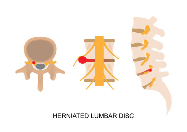
Vector illustration of back pain caused by herniated lumbar vertebral disc. Intervertebral disc protusion causing nerve root compression.


Illustration of the entire spine including cervical, thoracic, lumbar, sacral, and coccyx
Backache - Spinal Arthritis- Stock Icon as EPS 10 File

Engraving from "Die descriptive und Topographische Anatomie des Menschen"; by Dr. C. Heintzmann. Published by Wilhem Braumüller, Vienna (1884). Photographed and edited by J. C. Rosemann.


The human spine column. Vector illustration in natural color isolated on a dark grey background. Front view. Medicine, anatomy and biology concept.
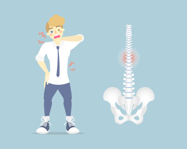
man having back pain, backache, neck, upper, lower, waist pain, anatomy of human spine, health care symptoms orthopedic concept, internal organs body part, flat vector illustration character design
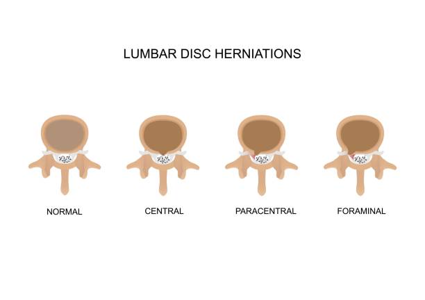
Vector illustration types of lumbar intervertebral disc herniation. Central, paracentral and foraminal disc herniation.

Vertebral column: cervical, thoracic and lumbar spine, sacrum and coccyx. Kyphosis, and Lordosis. Numbering order of the vertebrae of the human spinal column. Vector diagram for medical use

Vector illustration of lumbar fusion surgery with screws

A color illustration that shows the structure of the pelvis

The lumbar region is sometimes referred to as the lower spine, or as an area of the back in its proximity. Vector graphic.

A Text book of Naked Eye Anatomy

spinal disease. Lumbar section through a magnifying glass. human spinal column. Illustration highlighting lumbar spine. Vector diagram for medical use


Illustration of anterior and lateral views of "lumbar vertebrae, sacrum, coccyx"


Thoracic region of the human spine

The human spine column. Vector illustration in natural color isolated on a white background. Front view. Medicine, anatomy and biology concept.


Woman with pain in the cervical and lumbar vertebrae. Back pain, muscle pain, osteoarthritis, rheumatoid arthritis. Medicine. Illustration, vector
Next