Cardiac Muscle Tissue Pictures, Images and Stock Photos
Browse 5,800+ cardiac muscle tissue stock photos and images available, or search for smooth muscle tissue or skeletal muscle to find more great stock photos and pictures.
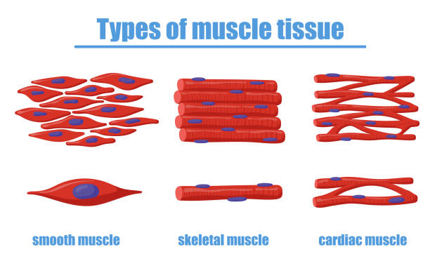
Different types of muscle tissue vector illustration. Smooth, skeletal and cardiac muscles of human body. Can be used for anatomy, biology, education, science concept
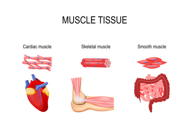
Types of muscle tissue. Skeletal muscle (elbow joint), smooth (gastrointestinal tract) and cardiac muscle (heart). Human internal organs and Muscle cells. vector illustration for medical, educational and science use

Types of muscle tissue. Skeletal muscle, smooth muscle, cardiac muscle. Vector scheme.
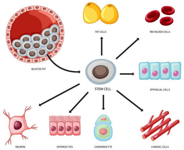
Types of stem cells on white background illustration


Types of Muscle Tissue illustration
Vector icons set and pattern depicting stem cell possibilities.
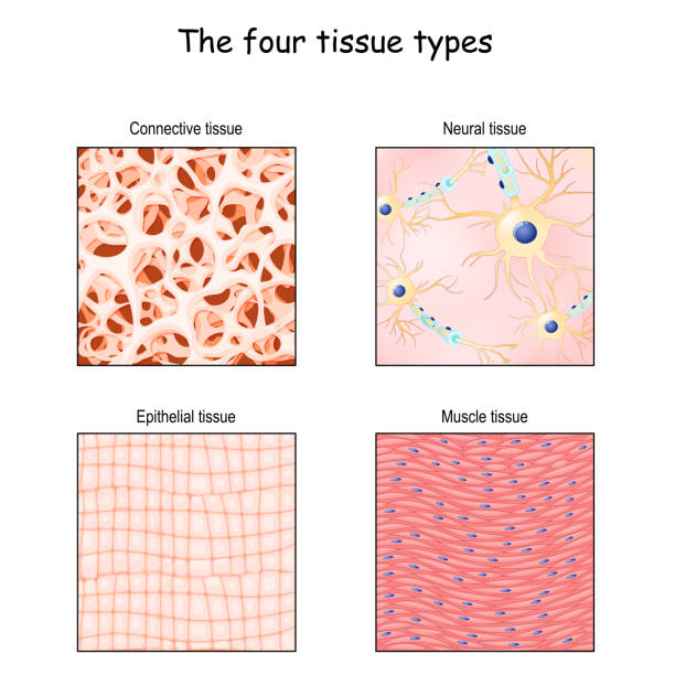
Tissue types. connective, muscle, nervous, and epithelial. Close-up of cells in different tissue. anatomical fiber parts: Epithelium, bone matrix, neurons, and smooth muscle. vector illustration. structural diagram for Science and educational use

Wall heart structure. Human organ anatomy diagram. Medical cardiology with internal membrane and visceral or parietal pericardium. Flat vector infographics with veins, arteries and myocardium

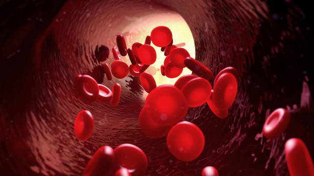
3d illustration blood in blood vessel
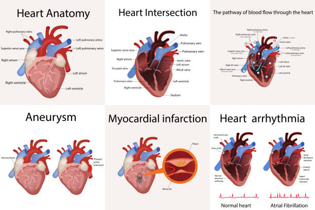
heart anatomy and types of heart disease vector illustration isolated on white
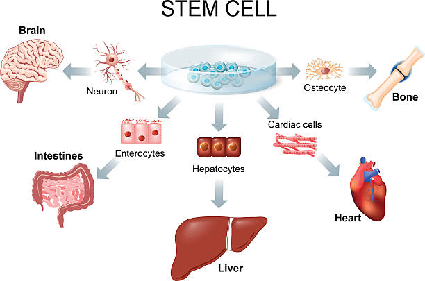
stem cell application. Using stem cells to treat disease
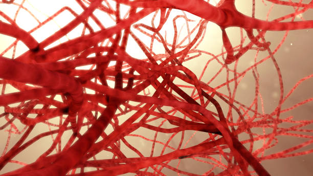
3d illustration blood vessel

Muscular Health Concept. Tiny Doctors Characters at Huge Board with Infographics Presenting Skeletal, Cardiac and Smooth Musculature. Medicine, Muscles Anatomy. Cartoon People Vector Illustration
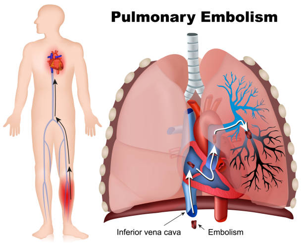
Pulmonary embolism medical vector illustration with description on white background eps 10
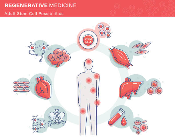
Vector illustration showing adult or somatic stem cell possibilities.
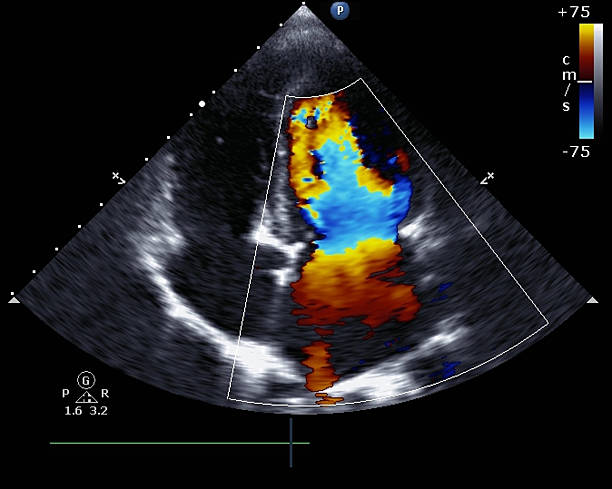
Transthoracic two-dimensional color Doppler echocardiography
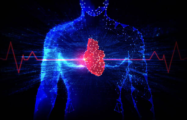
Future Technologies in Cardiology and Healthcare - Emerging Technologies to Treat Heart Diseases - Electrophysiology - Innovation in the Medical Fields - Conceptual Illustration


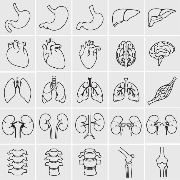
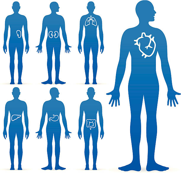
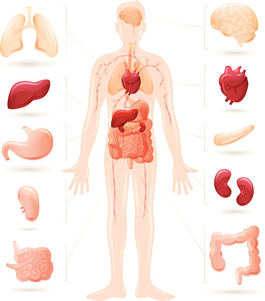
Internal human organs. Anatomy set. Vector illustration of outline medical icons for infographic.

Cardiac sarcoidosis is a rare condition which affects a small number of people who suffer from a condition called sarcoidosis – an inflammatory condition that can affect multiple organs. The disease tends to affect younger people, generally between 25 and 45 years old. Sarcoidosis is characterized by the presence of granulomas. These are ball-like collections of inflammatory cells that cluster around and react to a foreign substance. The inflammation associated with granulomas can damage virtually every part of the heart, including the electrical system, muscle, valves, arteries and surrounding tissue called the pericardium.
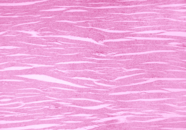
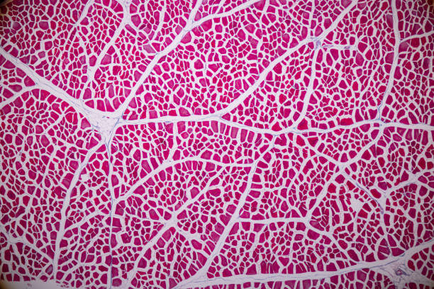
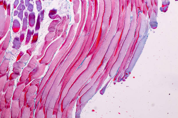
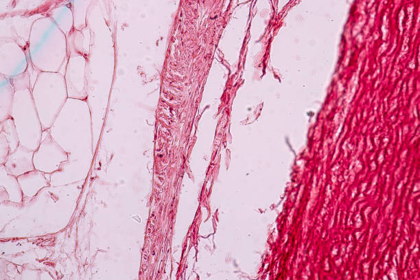
Cardiac muscle section under light microscope. Heart muscle, also myocardium, a vertebrate muscle. Striated muscle that constitutes the main tissue of the walls of the heart. Micrograph. Macro photo.
Human Internal Organs. Vector icons Set. Editable Stroke. 36 icons set. Colon, heart, female breast, mammary glands, trachea, follicle, brain, kidneys, eye, testicles, nerve, synapse, muscle, stomach, female reproductive system, male reproductive system, liver, gallbladder, lungs, spine, thyroid gland, pancreas. You can find more unique icon sets at the link: https://www.istockphoto.com/collaboration/boards/qUfvBxVnEU64XaERvnM_Fw
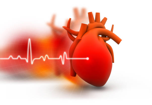
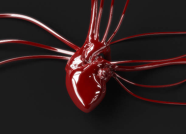
Human heart on abstract dark background


Air Transport of Organ Donation for Transplantation September Green - September 27 - National Organ Donor Day - Commercial Aviation in Brazil and worldwide, has great importance in the transport of organs for transplants in receiving patients. Studies show that commercial air carriers carry more than 9,000 organs a year on board airplanes with passengers, helping in the health system of the Brazilian government (SUS). And these airplanes take priority over the landing and take-off operation due to the time of urgency of withdrawal from the donor organ to the recipient. As is the case of the lung and heart that must be reimplanted in the maximum 4 hours in the patient.
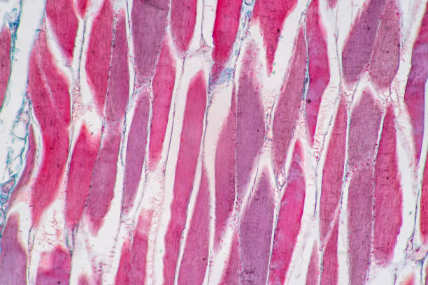
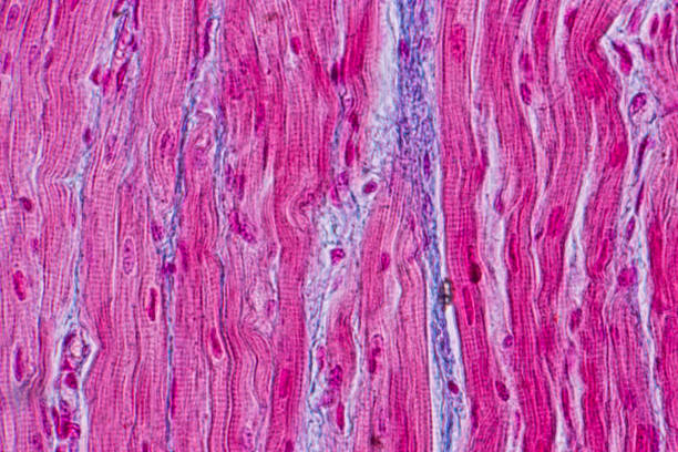
Characteristics of anatomy and Histological sample Striated (Skeletal) muscle of mammal Tissue under the microscope.

musculus cardiacus cells section under microscopy
Human heart on abstract dark background. 3d illustration
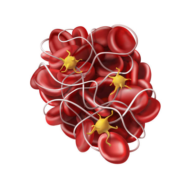

Image of surgeon hand holding aortic valve implant in the operating room
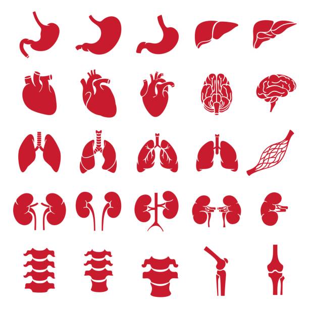
Internal human organs. Anatomy set illustration. Vector of outline medical icons for infographic.
Human Organs. Vector Icons Set. Human Internal Organ.

Striated muscle human under the microscope for education.

Microscopic photograph of a professionally prepared slide of ventricular choroid plexus. The choroid plexus is a plexus of cells that produces the cerebrospinal fluid in the ventricles of the brain. The choroid plexus consists of modified ependymal cells.

Red blood pressure it have dense oxygen from the heart to tissue, cells or body parts. Unconscious it caused by blood oxygen level minimum or lack of oxygen. Illustration human body.
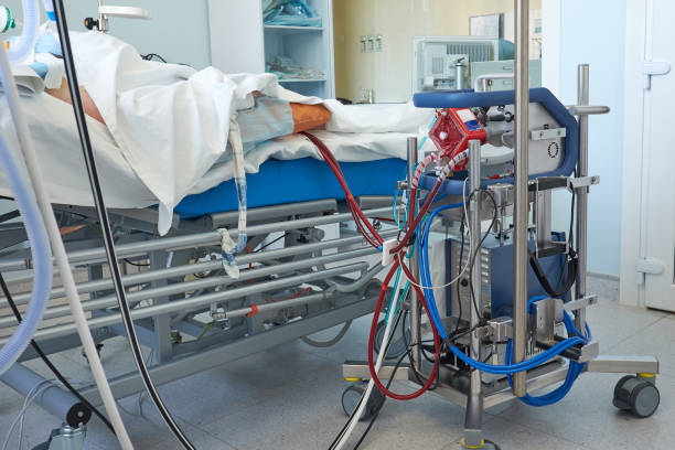
Extracorporeal membrane oxygenation. Working ecmo machine in intensive care department in patient with RDS caused by virus pneumonia
Human body diagrams. EPS 10 file. Transparency used on shadows.
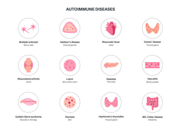
Autoimmune disorders diseases, set with icons of symptoms. Illness when the immune system attacks joints, blood, skin, internal organs. Medical poster for clinic or education flat vector illustration

3d illustration blood cells in blood vessel

Vector icon set of human internal organs like heart spleen lungs stomach thyroid intestine bladder gallbladder pancreas kidneys and liver in flat style
Human heart with vessels and cut rib cage. On white background.

A mammography x-ray image of 2 breasts in shape of heart

Female body on a white background

Engraved Illustrations of Human Body Nervous and Blood flow System Diagram Engraving from Iconographic Encyclopedia of Science, Literature and Art, Published in 1851. Copyright has expired on this artwork. Digitally restored.


3d printing human body. 3d printed body parts with laptop and 3d printer on table, indoors.
Detailed human body diagram. Each element grouped and labeled for easy use and editing.
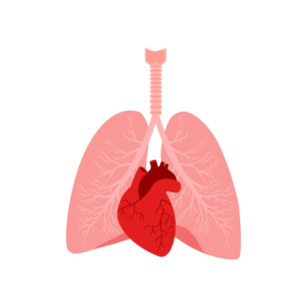
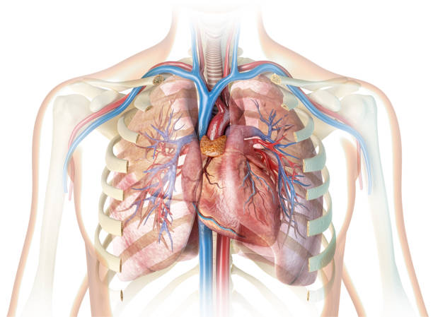
Heart and lungs. Internal organs in a male human body. Anatomy of people.Part of the human heart. Anatomy. Diastole and systole.Filling and pumping of Human Heart structure anatomy anatomical diagram

Cardiac hemochromatosis or primary iron-overload cardiomyopathy is an important and potentially preventable cause of heart failure. This is initially characterized by diastolic dysfunction and arrhythmias and in later stages by dilated cardiomyopathy.

Types of heart diseases: hypertrophic cardiomyopathy and dilated cardiomyopathy. healthy heart and heart with Pathology. vector illustration for medical use
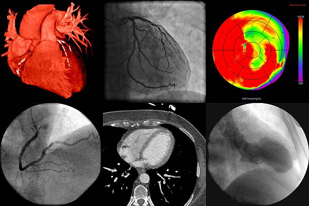
A series of cardiac imaging with different techniques
Next