
Human Foot X Ray Image Ankle The Human Body Pictures, Images and Stock Photos
Browse 650+ human foot x ray image ankle the human body stock photos and images available, or start a new search to explore more stock photos and images.
Most popular
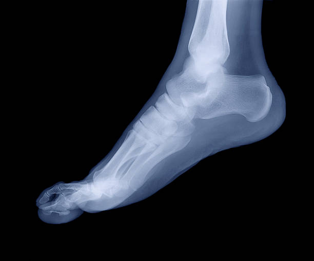
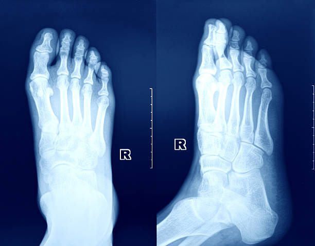
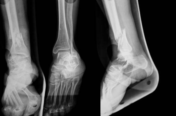
X-ray of a human ankle and foot.
Common types of bursitis. Inflamed bursa in human body. Elbow, shoulder, knee pain. Ankle hip and foot inflammation. Treatment and injection concept. Anatomical poster flat medical vector illustration

Xray of a foot on a ball with grass.
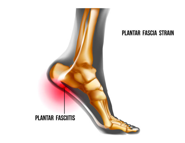
Plantar fasciitis inflammation and ruptures strain. Bones ot Foot pain realistic illustration. Medial view. Anatomy of joints, human leg black and yellow transparente skeleton. For medical orthopedic advertising. Vector illustration stock vector.

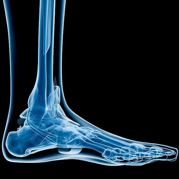
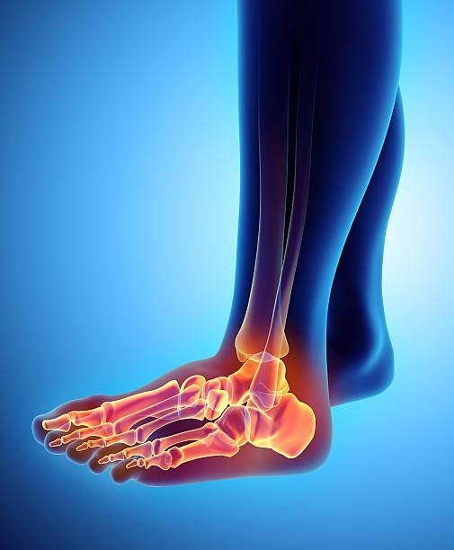
Digital medical illustration: Lateral (side) x-ray view (orthogonal) of human foot and ankle. Featuring:
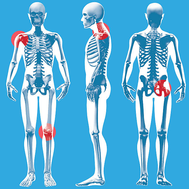
A stylized x-ray view of a Figure standing (dot pattern grid version) from the Front, Side and Rear. The 4 red circular highlights indicate potential injury /pain areas.
3D illustration of Foot Skeleton - Part of Human Skeleton.
xray background
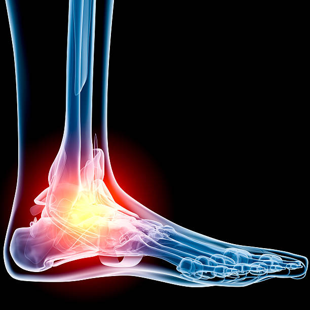
Digital medical illustration: Lateral (side) x-ray view (orthogonal) of human foot and ankle. With pain zone in ankle. Featuring:


football player with Sprained Ankle. character design. injury during workout - vector illustration

3D Isometric Flat Vector Concept of Podiatrist, Podiatric Physician Doctor, Treatment of Disorders of the Foot, Ankle, and Lower Extremity.

Lateral view of heel of human foot in X-ray (blue on black background), with pains on hell and Calcaneus bone.
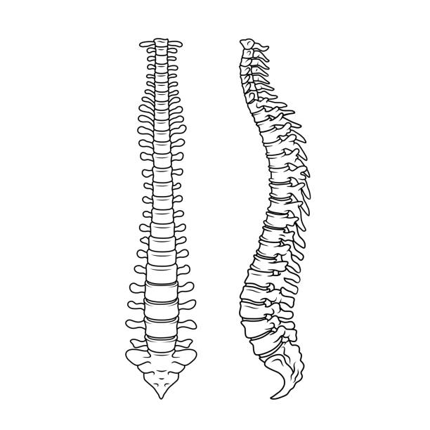

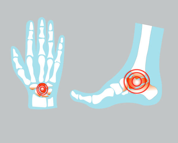
Set of human joints, knee joint, elbow joint, ankle joint, wrist, skeletal spinal bone structure of Human Spine, emblem or sign of medical diagnostic center or clinic, flat vector illustration.
xray background

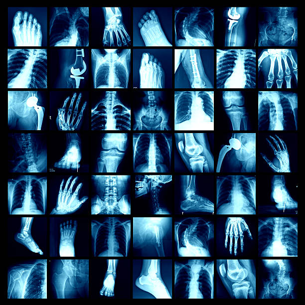
X-ray multiple part of human with multiple disease (stroke, arthritis, gout, rheumatoid, brain tumor, osteoarthritis, etc)


Right and wrong support of the foot.
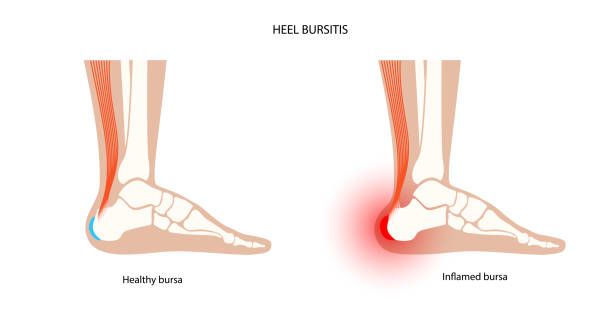
Heel bursitis inflammation. Inflamed bursa in human ankle. Achilles tendon and foot disease, pain and deformity. Diagnosis and treatment. Anatomical musculoskeletal poster, medical vector illustration


Doctor orthopedist on a computer shows a heel spur on an x-ray of the foot

X-Ray image of the Foot.


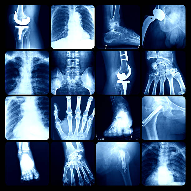
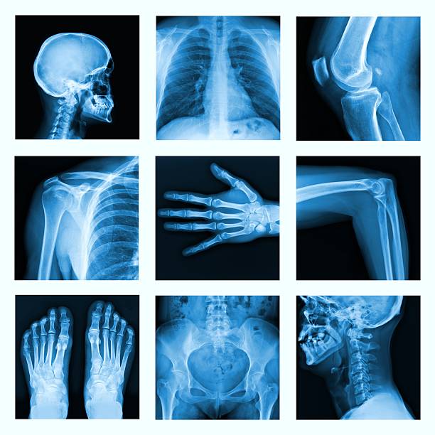
Collection X-ray "Multiple part of human,orthopedic surgery and multiple disease

Collection X-ray multiple human's organ & orthopedic surgery & Multiple disease (Pulmonary tuberculosis , Gout , Rheumatoid arthritis ,Spondylosis , Fracture bone , Stroke , Brain tumor , etc)

Collection X-ray multiple part of human & Orthopedic surgery & Multiple disease (Osteoarthritis knee,spondylosis,Stroke,Fracture bone,Pulmonary tuberculosis, etc)
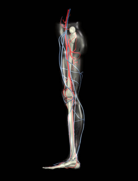
Side view of leg bones, arteries and veins. Anatomically accurate and highly detailed.

Inspection of the toes with the hands to detect the presence of calluses, warts or athlete's foot

x-ray image of human hand and foot.

bone of the hand and foot with arm and leg, anatomy, internal organs body part orthopedic health care, vector illustration cartoon flat character design clip art


x-ray 57 year old man's right foot - top view
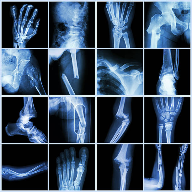
Collection X-ray multiple bone fracture (finger,spine,wrist,hip,leg,clavicle,ankle,elbow,arm,foot)


Set of xray of human skeletal, human joints, knee joint, elbow joint, ankle joint, wrist, skeletal spinal bone structure of Human Spine, emblem or sign of medical diagnostic center or clinic, flat vector illustration.


Digital medical illustration: Anterior (front) perspective 45 degree rotation (Anterior oblique 45) x-ray view of human foot and ankle. Featuring:


xray background

Supinated foot, arch deformation, bottom and back view. Foot weight distribution. For medical orthopedic advertising. Vector illustration stock vector.

ultrasound of child's feet -sole- diagnosis,close up

specialist watching images of foot at x-ray film viewer

Set of xray of human,human joints,knee joint,elbow joint, ankle joint, wrist, skeletal spinal bone structure of Human Spine, emblem or sign of medical diagnostic center ,flat vector illustration.


3D illustration of human leg with arteries, veins and bones. Knee is hilighted. Highly detailed.

Digital Tablet with X-ray and Stethoscope


3D illustration of Foot Skeleton - Part of Human Skeleton.
Next