
Pics For Ulna Bone Pictures, Images and Stock Photos
Browse 8,500+ pics for ulna bone stock photos and images available, or start a new search to explore more stock photos and images.
Most popular
Orthopedic surgeon studying a x-ray of a broken radius bone in theater after correctional surgery.
X-ray image of forearm, AP and lateral view, show fracture of ulna and radius

In vertebrates, thoracic vertebrae compose the middle segment of the vertebral column, between the cervical vertebrae and the lumbar vertebrae.
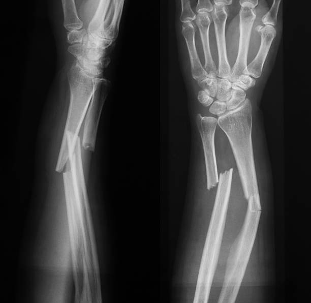
An x-ray image of an boken arm with double fracture: radius and ulna.
Tennis elbow injury medical vector illustration on white background eps 10

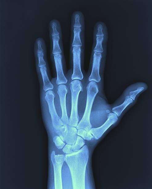
Hand x-ray showing bones and hand silhouette. EPS 10 file. Transparency effects used on highlight elements.
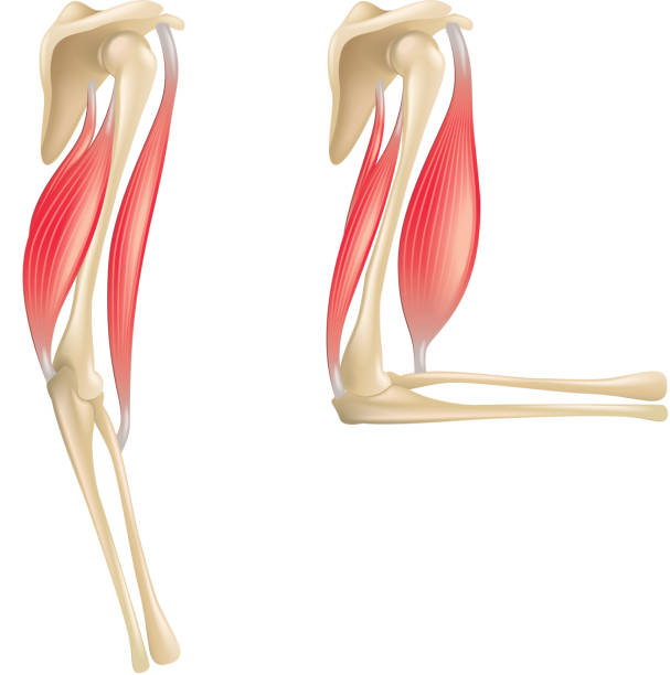
Anatomy of the elbow muscles medical vector illustration eps 10
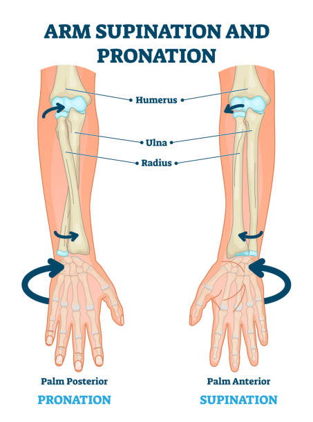
Arm supination and pronation vector illustration. Labeled anatomical scheme. Medical diagram with inner bones and joints. Compared palm posterior and anterior. Hand rotation movement biological terms.
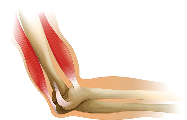
Diagram of a human elbow

Scaphoid bone fracture

Olecranon bursitis. student elbow. medical condition. inflammation of the bursa located under the elbow Olecranon. trauma or repetitive smaller traumas. Anatomy of a elbow joint. parts of the arm, bones Muscle and Distal tendon of triceps. Vector illustration for medical use
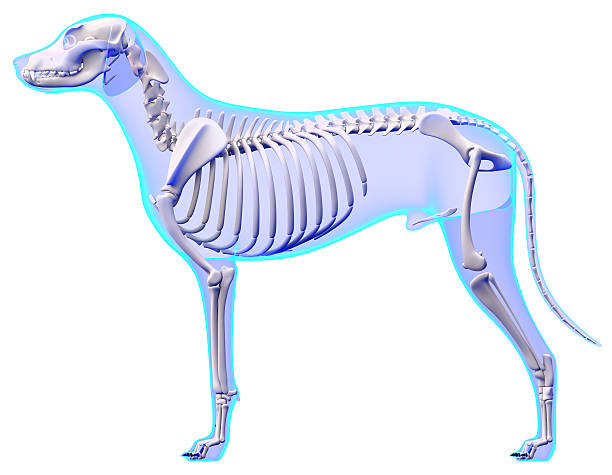
Dog Skeleton Anatomy - Anatomy of a Male Dog Skeleton

X-Ray image of the Human elbow bone. Front view.
Elbow joint anatomy isolated on white photo-realistic vector illustration

3D Illustration of Human Skeleton System Rib Cage Anatomy

Human body, 3d illustration. Full figure male muscular and skeletal systems, front view on white background."n
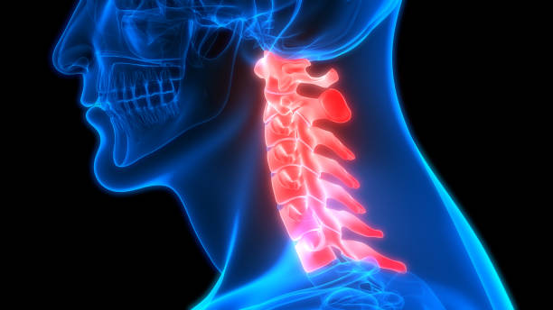
3D Illustration of Human Skeleton System Vertebral Column Cervical Vertebrae Anatomy
Elbow joint vector illustrated diagram, medical scheme. Educational injury information.
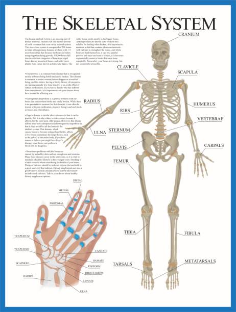
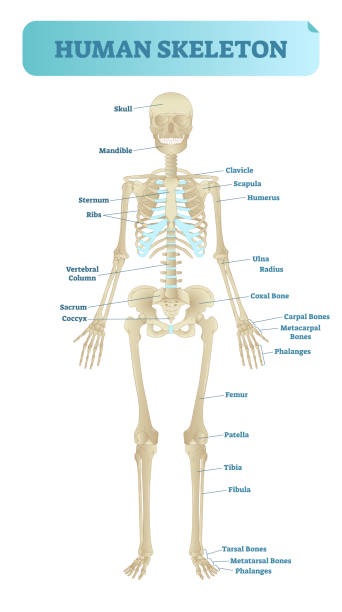
Human skeletal system poster containing detailed information about the skeletal structure. The poster contains a detailed illustration of the human hand.

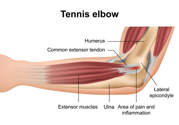
Tennis elbow diagram showing a detail of the damaged tendon tissue. Digital illustration.


3D Illustration Concept of Human Skeleton System Femur Bone Joints Anatomy

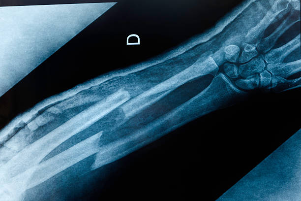
Film X ray fracture radius

Two fracture in human arm

3D Illustration of Human Skeleton System Appendicular and Axial Skeleton Anatomy


3D Illustration Concept of Human Skeleton System Skull Bone Parts Mandible Bone Anatomy
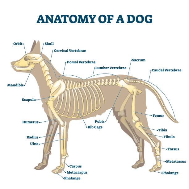
Anatomy of dog skeleton with labeled inner bone scheme vector illustration. Zoological structural examination with inside location description for study handouts. Educational skull, vertebrae location

3D Illustration of Human Skeleton System Appendicular and Axial Skeleton Anatomy

3D Illustration Concept of Human Skeleton System Skull Bone Parts Mandible Bone Anatomy

cubital tunnel syndrome, inflamed ulnar nerve medical vector illustration on white background eps 10 background

Anatomy of dog paws with forelimb and hindlimb bones vector illustration. Educational labeled skeleton comparison with zoological inside structure scheme. Animal legs inner closeup examination model.
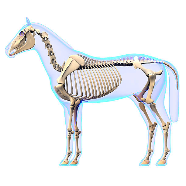
Horse Skeleton Side View - Horse Equus Anatomy - isolated on white
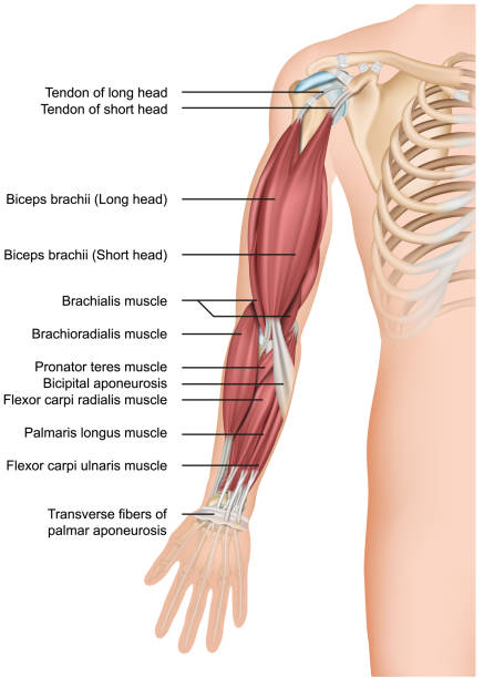
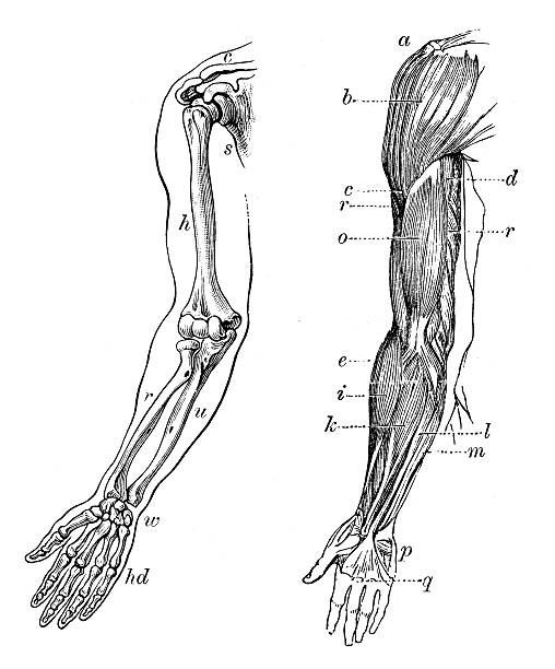
Arm muscle anatomy 3d medical vector illustration forearm eps 10

Pain in human arm. Medial epicondylitis golfer elbow. Trauma or inflammation in hand. Muscular system and skeletal anatomical poster. Structure of muscle groups and bones isolated vector illustration.

Magnifier on hand magnifying inner arm for looking wrist fracture. Illustration about medical concept.

3D Illustration of Spinal cord (Cervical Vertebrae) a Part of Human Skeleton Anatomy

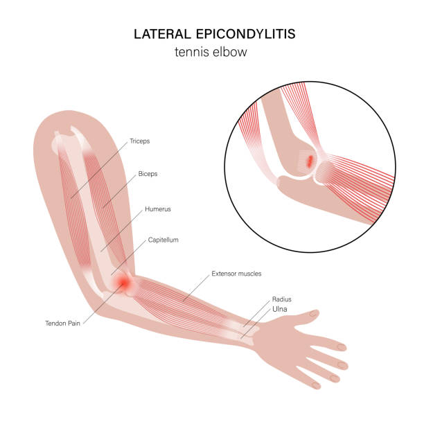
Tennis elbow or lateral epicondylitis. Vector illustration for medical use

Dog X Ray Showing Radius and Ulna Fracture. Lateral View.
Skeleton hand with bones icon. Vector illustration.

3D Illustration of Human Vertebral Column Anatomy
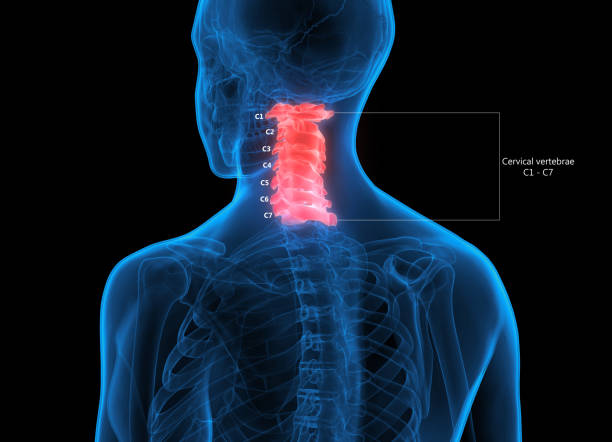
3D Illustration of Human Skeleton System Vertebral Column Cervical Vertebrae Anatomy
Pain in human arm. Lateral epicondylitis tennis elbow. Trauma or inflammation in hand. Muscular system and skeletal anatomical poster. Structure of muscle groups and bones isolated vector illustration
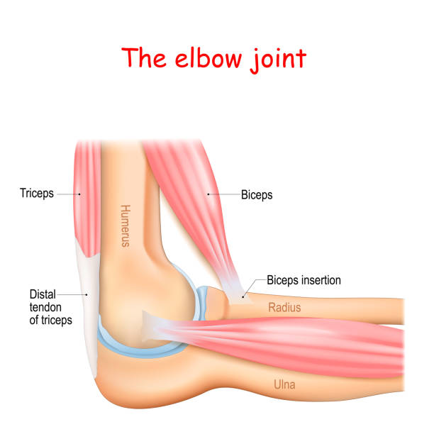
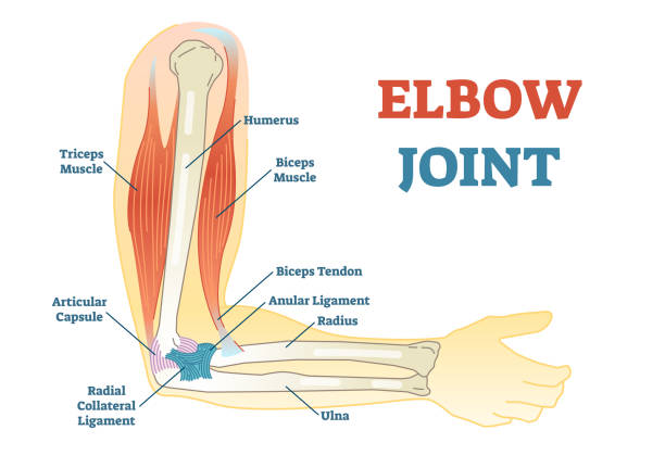
Anatomy of a elbow joint. parts of the arm. bones (humerus, radius, ulna) Muscle (Triceps, Biceps) and Distal tendon of triceps.
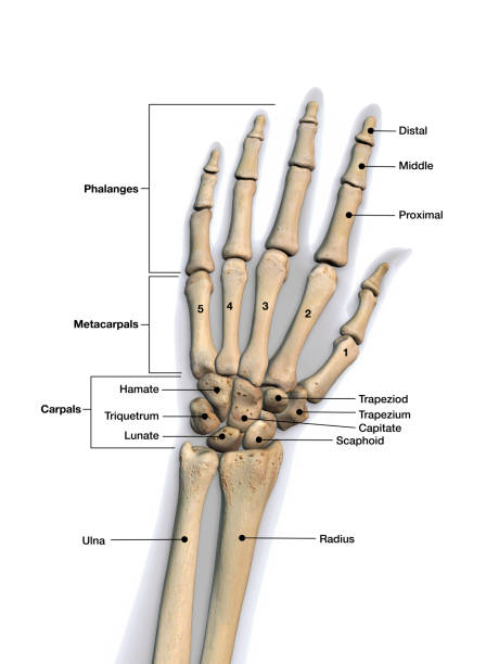
Skeletal bones of wrist and hand with labeling. Dorsal (back) view.

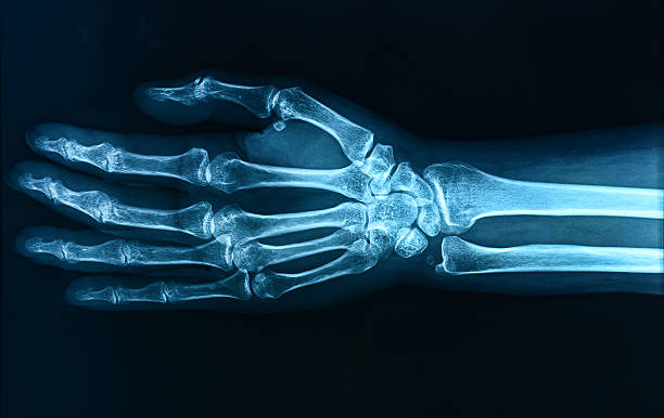
Xray of hand XXXLarge

3D Illustration Concept of Human Skeleton System with Nervous System Anatomy
3D Illustration Concept of Human Skeleton System With Nervous System Anatomy

X-Ray image of the Human elbow bone. Front view.
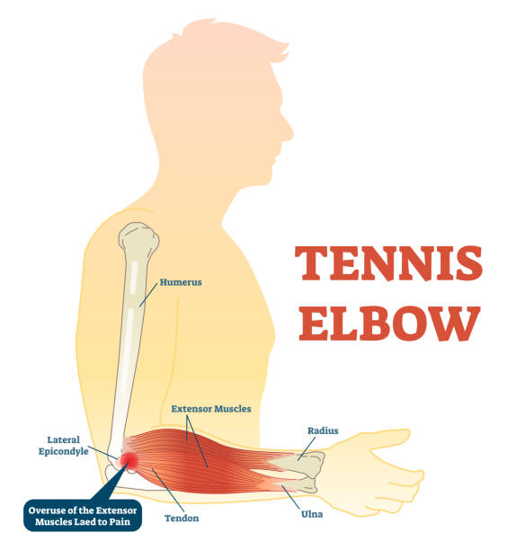
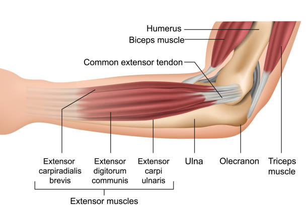
Tennis elbow medical fitness anatomy vector illustration diagram with arm bones, joint and muscles. Overuse of extensor muscles leading to pain.
Full human skeleton anatomical model. Medical vector illustration poster, educational information. Head, ribcage, arms, hips, legs and other main bone structure.
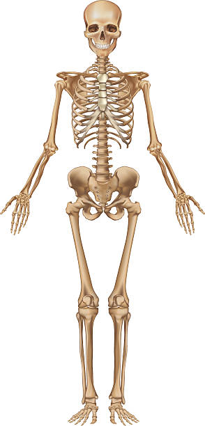
"Human skeleton, male, front view. Every single bone is a single object that can be modified individually."


Hand and wrist bones vector sketch of human anatomy and medicine design. Hand drawn arm of skeleton with radius, ulna, finger phalanges and palm metacarpals, trapezoid, scaphoid and carpal bones
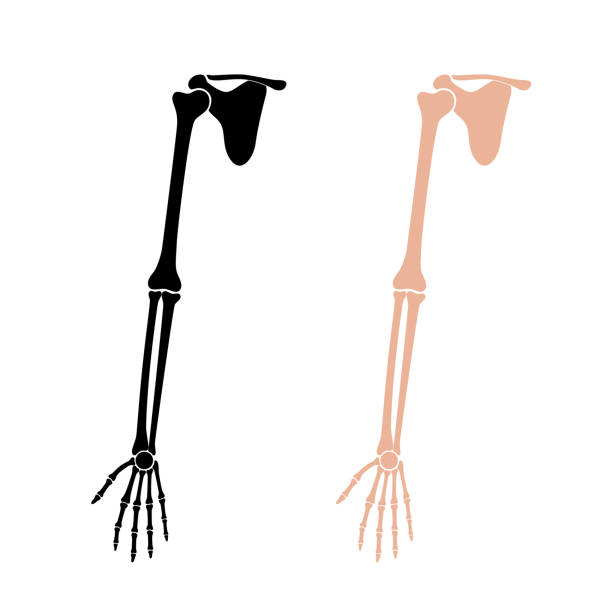
Human arm anatomy. Wrist and Hand Bones. Vector illustration isolated on white background. skeletal system silhouette. Medical, educational and science banner
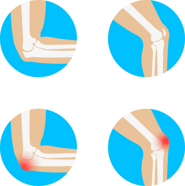
Knee and Elbow vector illustration. Elbow pain. Knee pain. Anatomy of knee and elbow.

Next