Renal Medulla Photos Pictures, Images and Stock Photos
Browse 70+ renal medulla photos stock photos and images available, or start a new search to explore more stock photos and images.
Most popular
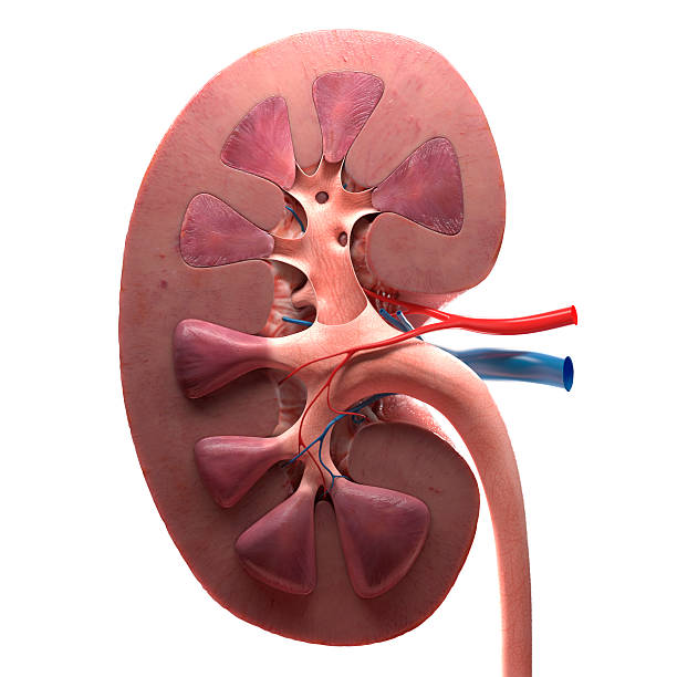
3d kidney on white background


Anatomical Kidney model showing blood vessels and anatomical structure

Hemodialysis machines with tubing and installations. Health care, blood purification, kidney failure, transplantation, medical equipment concept. Close up
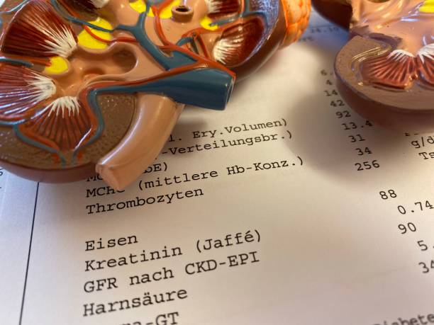


Kidney model on lab results showing kreatinin and GFR

Hemodialysis machines with tubing and installations. Health care, blood purification, kidney failure, transplantation, medical equipment concept. Close up


Kidney model on lab results showing kreatinin and GFR

Anatomical Kidney model with blurred laboratory equipment in background

Anatomical Kidney model with blurred laboratory equipment in background

Hemodialysis machines with tubing and installations. Health care, blood purification, kidney failure, transplantation, medical equipment concept. Close up

Anatomical Kidney model with blurred laboratory equipment in background

Anatomical Kidney model with blurred laboratory equipment in background

Renal medulla. 0.5 µm thick section of material embedded in plastic, stained with toluidine blue. The image shows two types of cross sectioned tubules. The thinner tubules, lined by a very flattened simple squamous epithelium, in which the nuclei make prominence towards the lumen are thin portions of a loop of Henle. The thickest tubules, with collapsed lumen, lacking a brush border, and showing basal infoldings, are the thick ascending portion of the loops of Henle.

Anatomical Kidney model showing blood vessels and anatomical structure

Anatomical Kidney model showing blood vessels and anatomical structure


Hemodialysis machines with tubing and installations. Health care, blood purification, kidney failure, transplantation, medical equipment concept. Close up


Low magnification micrograph showing the cortex of a human kidney stained with hematoxylin-eosin. Bundles of parallel tubules are vertically crossing the renal parenchyma, coming from the renal medulla. They are the medullary rays, which serve to define the renal lobules. A renal lobule comprises the portion of the renal cortex whose nephrons drain their urine into the collecting tubules of a medullary ray. Large rounded formations (renal corpuscles) and abundant tubules with a very tortuous trajectory (convoluted tubules) are observed in the renal cortex, generally in cross or oblique sections.

Hemodialysis machines with tubing and installations. Health care, blood purification, kidney failure, transplantation, medical equipment concept. Close up


Hemodialysis machines with tubing and installations. Health care, blood purification, kidney failure, transplantation, medical equipment concept. Close up


Human kidney model in laboratory. Test tubes in background

Human kidney model in laboratory. Test tubes in background

Human kidney model in laboratory. Test tubes in background


Proximal convoluted tubes in a kidney of an experimental animal injected with Trypan blue, a vital stain. Trypan blue, filtered through the glomerulus, is reabsorbed as it passes through the proximal convoluted tubule and incorporated into endocytic vacuoles to which the lysosomes subsequently fuse. These vacuoles appear on the image as blue spots in the cytoplasm of the cells in the convoluted tubule. A distal convoluted tubule remains in the lower left corner of the image, lacking an endocytic apparatus and therefore not marked by Trypan blue. A Feulgen method, which stains the DNA magenta, has been used to mark the nuclei.
Model of the human kidney - view from inside

Low magnification micrograph of the renal cortex. The presence of renal corpuscles stands out, in which, even at this magnification, the uriniferous or Bowman space located around the glomerular vascular tuft can be clearly distinguished. The space between the glomeruli is occupied by sections of convoluted tubes. Crossing the image obliquely, two groups of tubules sectioned longitudinally are observed. They are the medullary rays, also called columns or pyramids of Ferrein. They contain collecting ducts and straight portions of the proximal and distal convoluted tubules. The vessels in the lower right corner are an arcuate artery and vein.

Anatomical Kidney model with blurred laboratory equipment in background
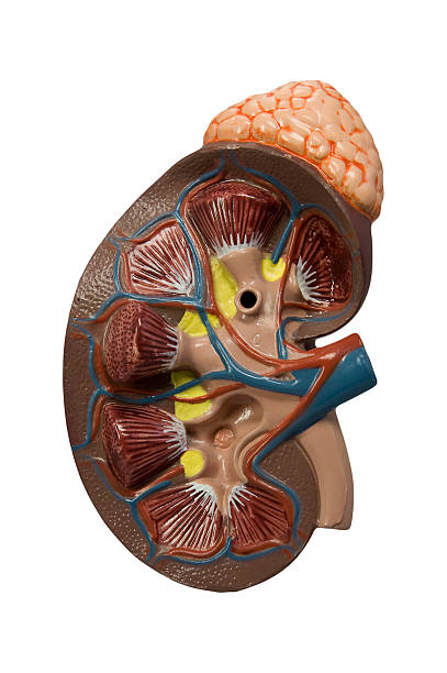
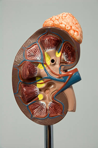
Model of the human kidney and suprerenal gland isolated on white

Kidney medulla (near renal papilla) showing cross-sectioned Bellini ducts lined by a simple columnar epithelium with dark nuclei. Among them, there are capillaries (some of them with blood) and many sections of Henle's loops.

Inner area of the renal medulla. 0.5 µm thick section of material embedded in plastic, stained with toluidine blue. The image shows three types of tubes cut longitudinally. One, of which two examples are seen in the upper area of the image, has its light occupied by blood, in which the red blood cells stand out. They are capillaries whose thin endothelium is difficult to identify even at this magnification. The second type, which crosses the center of the image horizontally, is also lined by a very flattened simple squamous epithelium, in which the nuclei make great prominence towards the light. Unlike the previous capillaries, the lumen of this tubules does not have blood-cells. It is the thin portion of a loop of Henle. Below are two collecting ducts with simple cuboidal epithelium of cells with a domed apical pole.

MRI of the upper abdomen coronal view on tablet is a non-invasive imaging technique providing detailed visuals of organs like the liver, pancreas, and kidneys.


Light micrograph cross section of rat kidney stained with hematoxylin and eosin displaying the peripheral renal cortex (with many glomeruli) and, in the central medulla, a Malpighian pyramid leading to a renal calyx.

Inner area of the renal medulla in cross section. 0.5 µm thick section of material embedded in plastic, stained with toluidine blue. There are two types of tubes that are difficult to distinguish between them. The first, the most abundant in the image, shows a relatively wide lumen and an extremely thin lining (endothelium). They are capillaries and venules that, in the image, are empty of blood except for one located in the center. The second type is smaller in diameter and generally shows two or three cells per cross section. They are flattened cells, although with a thickness greater than that of capillaries. They are Henle's loops of which only two are seen in the image. Among the tubes, some interstitial cells endowed with blue grains are identified."n"n


Inner area of the renal medulla. 0.5 µm thick section of material embedded in plastic, stained with a silver method. Two longitudinally sectioned Bellini ducts are seen at the left and right edges of the image. They are lined by a tall, simple prismatic epithelium that, at this point, is made up of only clear main cells. The space among the ducts is occupied by very thin tubes, in which two types are distinguished. One, the most abundant, is lined by a very thin endothelium (it appears as an almost imperceptible line); They are blood vessels, both capillaries and venules. The second type is smaller in diameter and has a higher epithelium, in which the cytoplasm is visible: they are loops of Henle. Finally, in the space among these tubes, cells endowed with dark grains are observed. They are interstitial cells, secreting prostaglandins and medulipin.


Hemodialysis machines with tubing and installations. Health care, blood purification, kidney failure, transplantation, medical equipment concept. Close up


Hemodialysis machines with tubing and installations. Health care, blood purification, kidney failure, transplantation, medical equipment concept. Close up

Very low magnification micrograph of the cortex of a human kidney stained with hematoxylin-eosin. The renal parenchyma contains large rounded formations (renal corpuscles) and abundant tubules with a very tortuous path (convoluted tubules) so that generally only short sections of them are observed. The image also shows two groups of tubules longitudinally sectioned, with a vertical, slightly oblique path. They are the medullary rays, columns or pyramids of Ferrein, formed by collecting ducts and straight portions of the proximal and distal convoluted tubules.


Inner area of the renal medulla in cross section. 0.5 µm thick section of material embedded in plastic, stained with toluidine blue. There are two types of tubes that are difficult to distinguish between them. The first, the most abundant in the image, shows a relatively wide lumen and an extremely thin lining (endothelium). They are capillaries and venules that, in the image, are empty of blood except for one located in the center. The second type is smaller in diameter and generally shows two or three cells per cross section. They are flattened cells, although with a thickness greater than that of capillaries. They are Henle's loops of which only two are seen in the image. Among the tubes, some interstitial cells endowed with blue grains are identified.

Next