
Skin Biopsy Pictures, Images and Stock Photos
Browse 1,600+ skin biopsy stock photos and images available, or search for skin biopsy icon or collagen skin biopsy to find more great stock photos and pictures.
Most popular
An injection of a painkiller to the site of a mole removal surgery.

Skin Biopsy. Punch biopsy take skin sample. Skin before and after medical procedure. Close up of Single use dermal biopsy punch. Cancer Care.

Nurse hand patching mole removal surgery incision as epidermal birthmark concept

Checking benign moles. Close up detail of the bare skin on a man back with scattered moles and freckles. Sun effect on skin. Birthmarks on skin

Close-up surgeon burns a mole on the back of the patient. Mole Removal Surgery Procedure.

Male doctor look at magnifying glass on patient skin against hospital office background. Lesions danger problem therapy concept

Close-up photo of dermatologist looking at birthmark on patient's skin

Close-up surgeon burns a mole on the back of the patient. Mole Removal Surgery Procedure.

Pre-cancerous tissue removed from a man's back
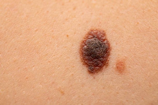
Close up picture of dangerous brown nevus on human skin - melanoma
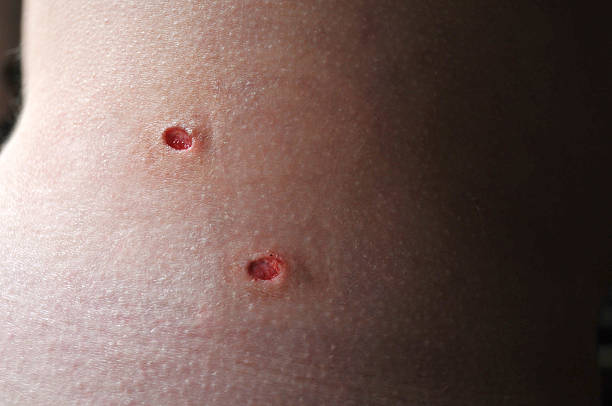
Horizontal image of two punch biopsy wounds. The wounds have been cleaned and the blood has begun to clot. The skin samples will be tested for melanoma. The photograph was taken with low light, which creates a shallow depth of field, but the wounds themselves are in sharp focus.
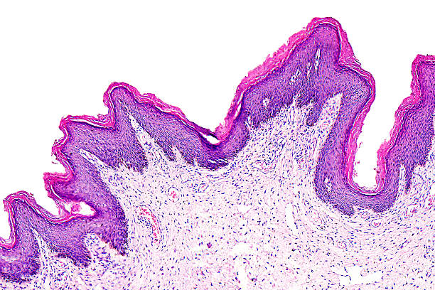
Skin papilloma of a human, highly detailed segment of panorama. Photomicrograph as seen under the microscope, 10x zoom.
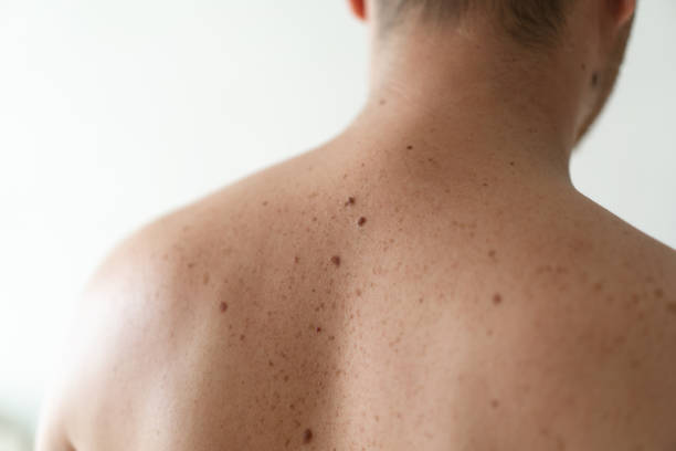
Checking benign moles. Close up detail of the bare skin on a man back with scattered moles and freckles. Pigmentation. Birthmarks on skin

Beautiful young blonde woman receiving mole or birthmark removal treatment at beauty clinic. Medical surgery for body and face corrections concept. View from above.

Skin cancer diagnosis. Doctor detecting asymmetric evolving spot on skin. Melanoma, dermatology, mole screening, birthmark. For topics like prevention, medicine, physical exam


Cropped photo of a nurse handing stainless steel surgical instruments on a tray to a doctor

Request for biopsy - Lipoma

Human stratified squamous epithelium under microscope, light micrograph

Pre-cancerous tissue removed from a man's back; iodine and fresh stitches visible.

A medical professional removes pre-cancerous tissue from a man's back; iodine and fresh stitches visible.

Close-up surgeon burns a mole on the back of the patient. Mole Removal Surgery Procedure.

Squamous epithelial cells under microscope view for education histology. Human tissue.

taken with a macro lens, 5 surgical stitches inserted after biopsy and the removal of a mole

The doctor uses a syringe to take a skin biopsy from the patient. The concept of malignant subcutaneous lesions
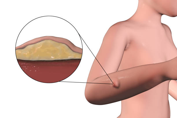
Lipoma, a growth of fat tissue under human skin, 3D illustration showing lipoma on the female arm and its cross-section

Close up of the sutures and bruise from a skin biopsy

skin biopsy with punch biopsy and suture repair

Adipose tissue human, Soft palate human, Bone human and Striated (skeletal) muscle human under the microscope in Lab.
Cosmetology. Skin care. Vector illustration

Spot on skin has traces of nevi and birthmarks. Dermatology and medical concept. Woman with traces of melanoma on skin. Skin with white patches blemishes dermatology concept

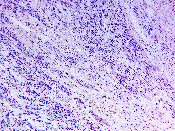
Cancer, malignant tumor, cancerous cell growing and spreading into surrounding and underlined tissues, 3D illustration

Cancer cell, malignant tumor cell, 3D illustration


Macro black birthmark on skin.

Male doctor look at magnifying glass on patient skin against hospital office background. Lesions danger problem therapy concept

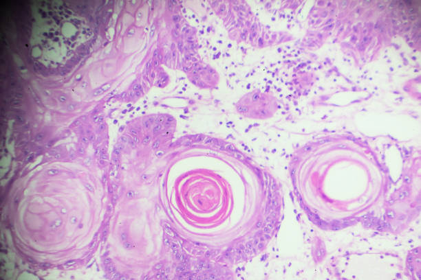
Melanoma of a human, photomicrograph panorama as seen under the microscope, 200x zoom.
Types of biomaterial a set of line icons in vector, illustration of human sperm and hair, blood and nails, cerebrospinal fluid and bone marrow

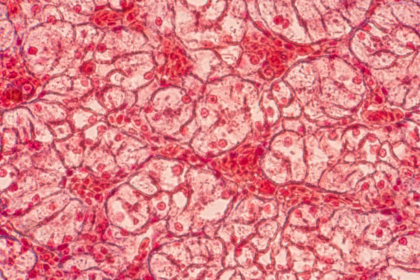
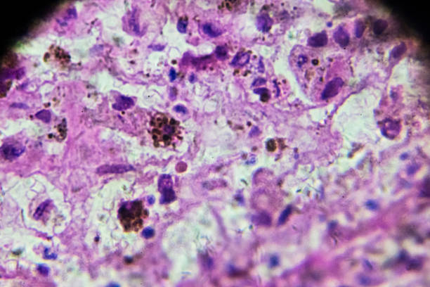
Squamous Cell carcinoma under microscopy,different area and
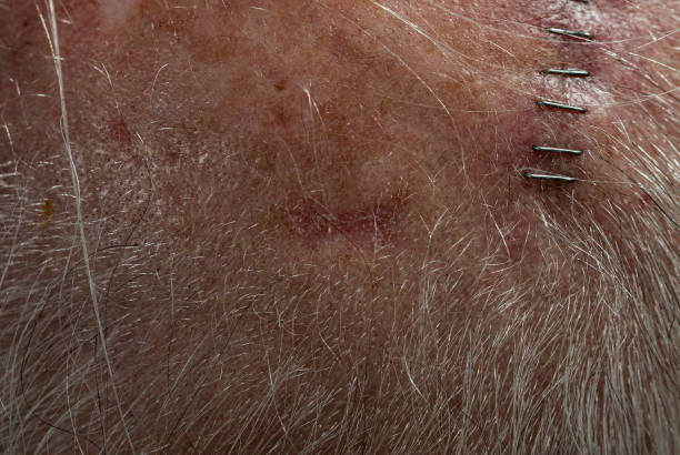
View of 65 year old male's scalp with squamous cell carcinoma site (horizontal) at center along hariline. Site is approx 3/4" across, 1/4" high. Subsequent Mohs surgery at site was successful in clearing the affected area. Vertical row of staples to right of center are closing incision from previous successful Mohs surgery on another squamous cell site.
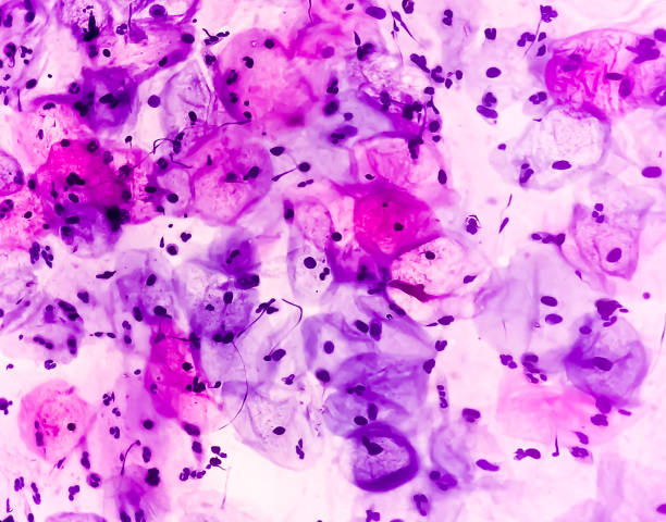
Melanoma biopsy under microscopy zoom in different regions

Close up detail of the bare skin on a man back with scattered moles and freckles. Checking benign moles. Sun effect on skin. Birthmarks on skin


Adipose tissue human, Soft palate human, Bone human and Striated (skeletal) muscle human under the microscope in Lab.


The nurse's hands are patching the postoperative seam with a plaster. Standard hospital dressing.
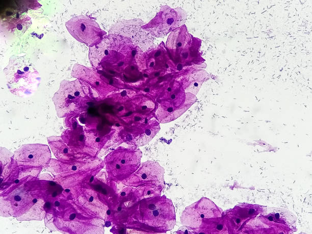
Squamous epithelial cells. Micrograph close up image at medical laboratory.


Scar from surgery with thread. Long surgical suture, skin wound is sewn with threads, a week after surgery


Next