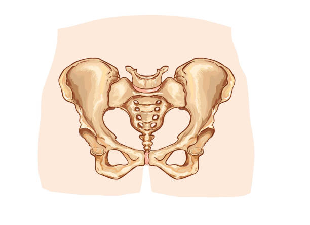
human pelvis anatomy 3d medical vector illustration on white background eps 10
human pelvis anatomy 3d medical vector illustration on white background eps 10
Human man skeleton pain, fracture or inflammation, parts of male body on x ray view. Vector isolated flat illustration of skull and bones on reontgen. Medical, educational or science banner.
Osteoarthritis - Bone Death - Hip Joint - Stock Illustration as EPS 10 file
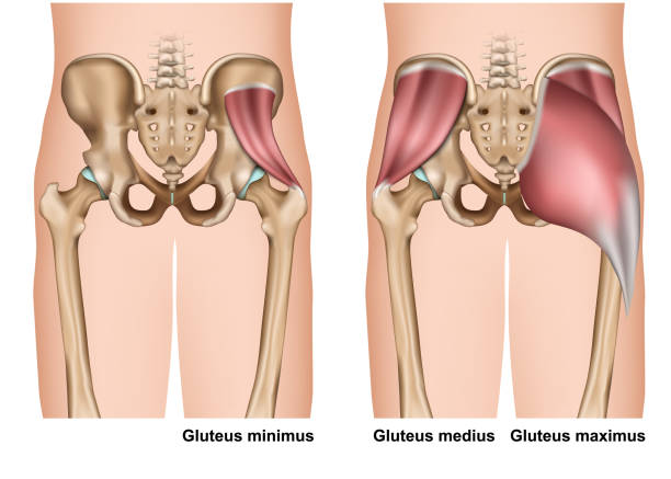
gluteus muscle anatomy 3d medical vector illustration on white background eps 10
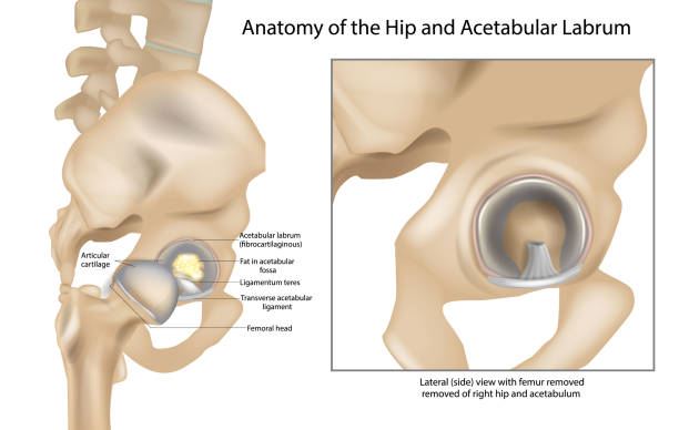
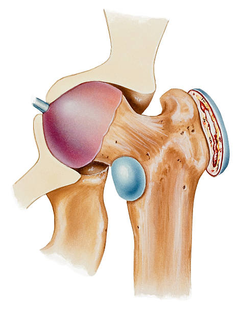
Anatomy of the Hip and Acetabular Labrum. Ligamentum teres and Articular cartilage. Lateral view with femur removed of right hip.
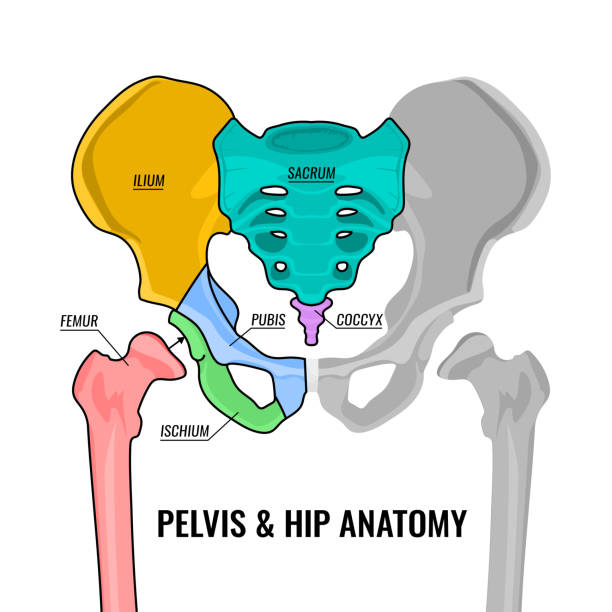
Human male anatomy scheme. Main pelvis bones - sacrum, ilium, coccyx, pubis, ischium and femur. Vector illustration isolated on a white background.
Set line icons of orthopedics isolated on white. Vector illustration

Types of joint pain medical medical icons set
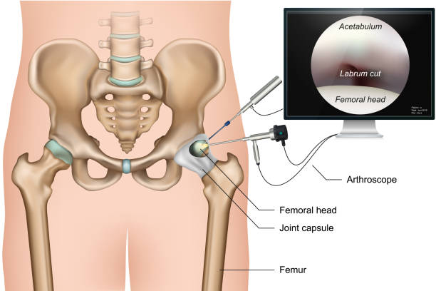
Hip arthroscopy 3d medical vector illustration on white background eps 10
The hip is the body’s second largest weight-bearing joint (after the knee). It is a ball and socket joint at the juncture of the leg and pelvis.
Cartoon Color Osteoporosis Bones Ad Poster Card Skeletal Health Concept Flat Design. Vector illustration of Spongy Texture Bone
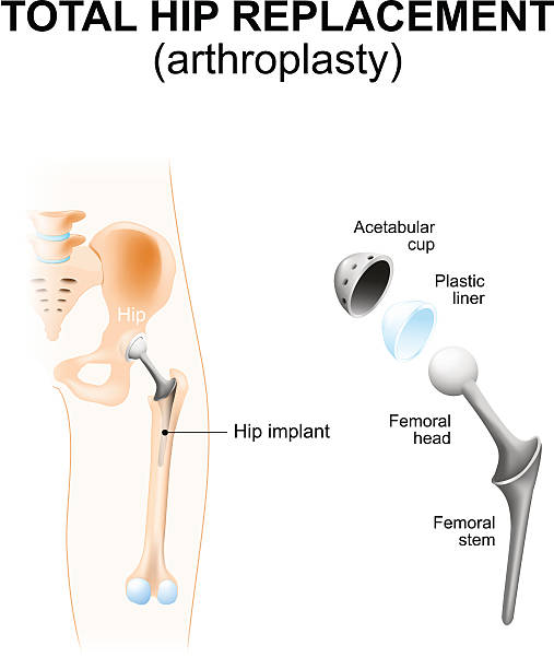
Total hip replacement or arthroplasty and hip Implant

Hip osteoarthritis. Synovial joints degenerative disease. Editable vector illustration in realistic style isolated on a grey background. Medical, healthcare, physiology concept. Scientific infographic

Hip joint with osteoarthritis with femoral head and the acetabulum of the pelvis. A comparison between a healthy hip joint and one with osteoarthritis, highlighting the key differences in structure. This can help patients understand diagnosis. Vector illustration with labeling and annotations for easily identifiable.
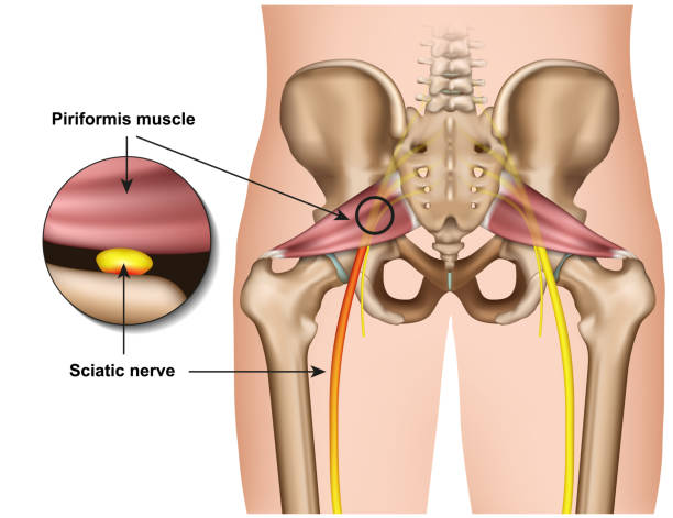
piriformis syndrome 3d medical vector illustration on white background eps 10

Arthritis of the hip joint, healthy joint and unhealthy joint with damaged cartilage and osteophytes.

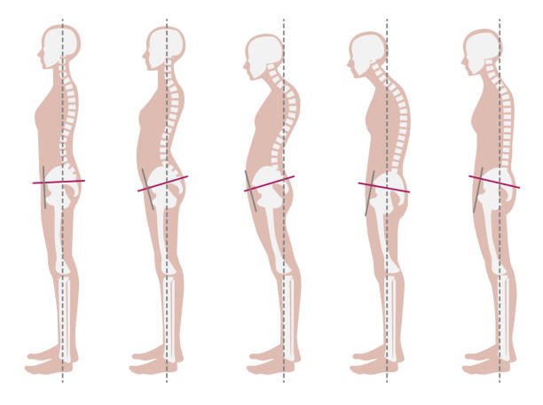
Various shapes of human's back
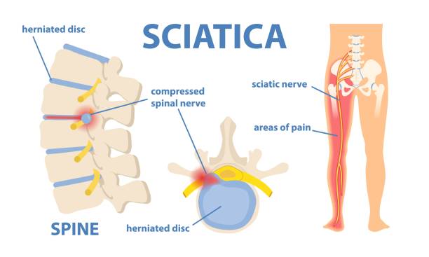
Sciatic nerve pain in the lower back through hip, thigh, knee to leg. Educational or informational poster. Flat vector medical illustration isolated on white background

Hip Trochanteric bursitis is inflammation of the bursa. Illustration of the Healthy and inflamed trochanteric bursa. Greater trochanteric pain syndrome.
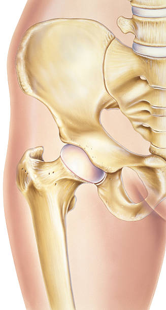
"Shown is a normal human hip with the bones and joints visible, specifically the pelvis, acetabular (socket), femoral head (ball), femur, cartilage and base of the spine."
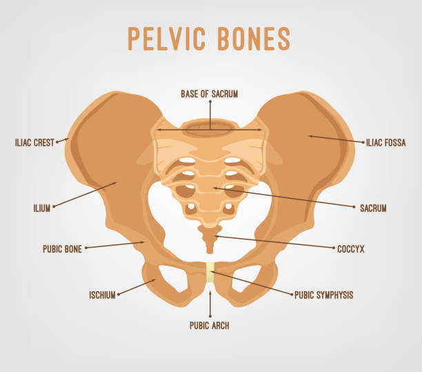
Human male anatomy scheme. Main pelvic bones - sacrum, ilium, coccyx, pubis, ischium. Vector illustration isolated on a white background.
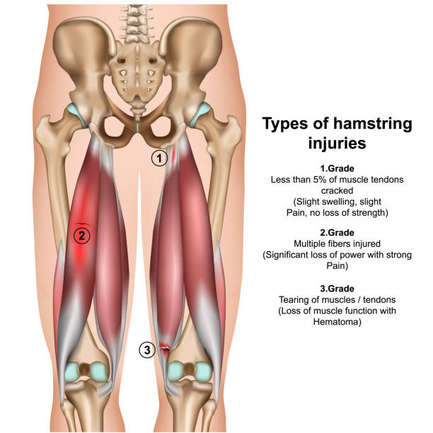
types of hamstring injurys 3d medical vector illustration on white background eps 10

piriformis muscle 3d medical vector illustration on white background eps 10
Bursitis of the hip bursa(e), which can cause severe pain. A bursa is a fluid filled sac that facilitates the smooth motion of joints. Shown is bursitis of the trochanteric bursa between the bony prominence of the outside of the hip and the tendon that passes over it (right, blue) and iliopsoas bursitis (center, blue)..
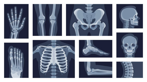
Collection different human body parts roentgen pictures vector flat illustration. Set x rays shot of head, hands, legs, torso, chest, hip, elbow, knee, spine skeleton character isolated on white
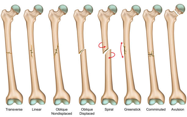
Bone fracture types medical vector illustration eps 10

Hip Osteoarthritis Infographic. Realistic bones scheme. Lower back and joint pain. Editable vector illustration isolated on a light background. Medical, healthcare, elderly diseases graphic concept.

Groin strain trauma and pulled or torn muscle injury anatomy outline diagram. Labeled educational medical sport problem explanation with body hips and leg overstretching condition vector illustration.
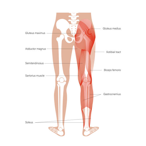
Human muscular system of legs in back view. Gluteus medius, gluteus maximus gastrocnemius and other muscles. Pelvis, leg and hip bones skeleton poster. Bodybuilding and strong body vector illustration

t is an illustration that understands the structure of the pelvic muscles

A skeleton poster design similar to posters found in medial offices on a transparent background. File includes EPS Vector file and high-resolution jpeg.

Sciatica vs piriformis medical muscle conditions comparison outline diagram. Labeled educational scheme with hip anatomy and compressed nerve or herniated disc caused acute pain vector illustration.
Vector Line icons set. One icon consists of a single object. Files included: Vector EPS 8, HD JPEG 3000 x 3000 px

Hip Labral Tears. Labrum torn from socket and Repaired labrum. Surgery for Repairing a Torn Hip Labral. Vector

Hip joint anatomy. Ball-and-socket joint. Vector poster

A skeleton poster design similar to posters found in medial offices. File includes EPS Vector file and high-resolution jpeg.
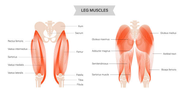
Gluteus medius, gluteus maximus and quadriceps muscles. Human muscular system. Pelvis and hip bones skeleton anatomical poster. Bodybuilding, workout, strong body concept. Isolated vector illustration
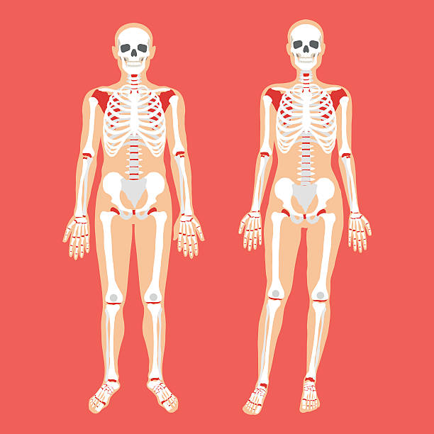
Human anatomy and skeletal system. Female and male bodies and skeletons. Modern concepts for web banners, infographics, websites, printed materials. Flat style design vector illustration

Male hip bone anatomy. Anterior view with primary bones names. Vector illustration with human skeleton scheme isolated on a white background.
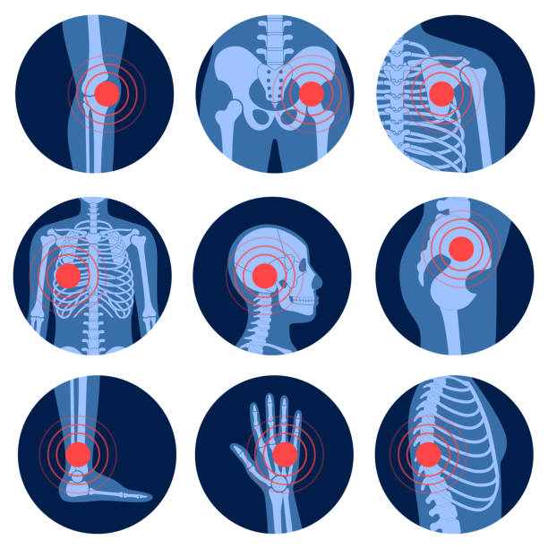
Human skeleton pain points icon set vector flat illustration. Chronic anatomy body injury in circle shape isolated on white. Healthcare medical studying educational acupuncture scheme. Health medicine
A set of human body parts and organ icons that include editable strokes or outlines using the EPS vector file. The icons include a human nose, ear, mouth, tongue, skull, eye, throat, hand, head, knee, leg, lungs, tooth, arm, muscle, foot, hip, brain, heart, knee, wrist, spine, liver, pancreas, gall bladder, stomach and kidney.

Pelvis Fracture. pelvic bones: sacrum, ilium, coccyx, pubis, ischium and femur. Vector illustration isolated on a white background

coxa saltans syndrome 3d medical vector illustration on white background eps 10
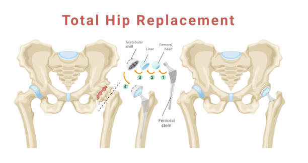
Total hip replacement surgery with anatomical acetabular prosthesis medical poster isometric vector illustration. Medicine procedure with new artificial bone structure skeleton joint education scheme
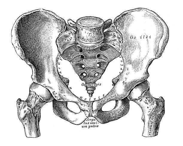
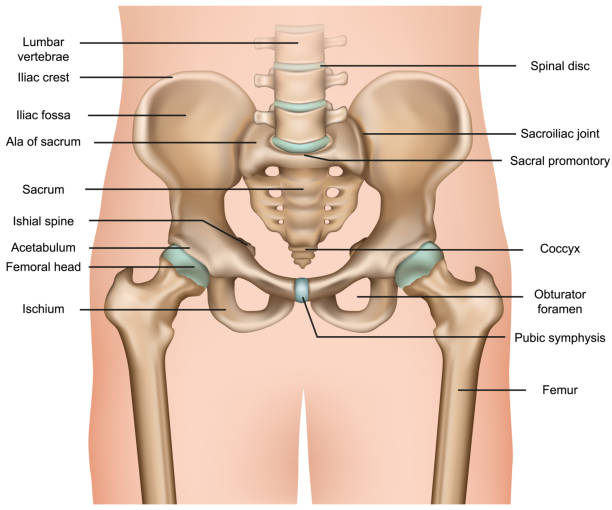
Human anatomy scientific illustrations with latin/italian labels: Pelvis (male)
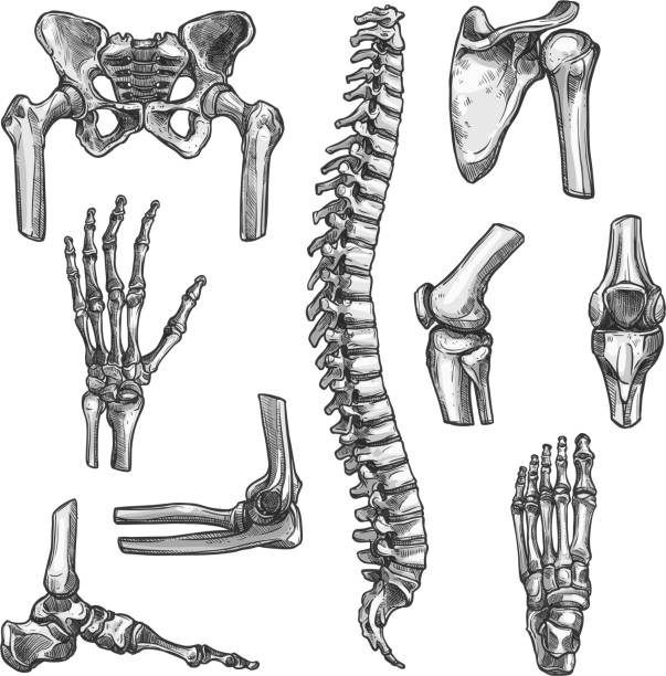
Bone and joints sketches set. Human skeleton hand, knee and shoulder, hip, foot, spine, leg and arm, finger, elbow, pelvis, thorax, ankle, wrist icon for orthopedics and rheumatology medicine design
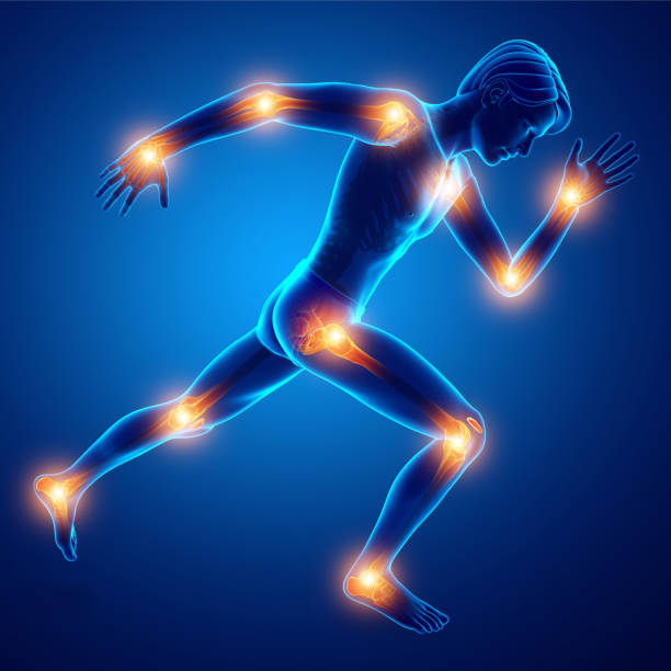
3d Illustration of Male Joint Pain

Arthritis, osteoarthritis medical infographic design. Joint replacement, implantant. Vector illustration

Femoral neck fracture. Types of hip fractures. Subtrochanteric, Intertrochanteric, Transcervical and Subcapital neck fracture, Fracture of the greater and lesser trochanter. Anatomy

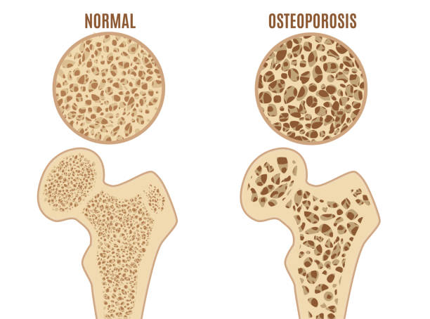
Osteoporosis disease poster. Systemic skeletal disorder of spine, wrist, and femur, loss of bone mineral density. Increased risk of hip fracture. Deterioration of bone tissue flat vector illustration

"Osteoarthritis is a noninflammatory degenerative joint disease characterised by the breakdown of the joint's cartilage. Cartilage that cushions the bones of the hip starts to erode, eventually allowing the bones to grind or rub together and causing hip pain and stiffness.The exact cause of osteoarthritis is unknown. Shown are the pelvis, acetabular (socket), femoral head (ball), femur, cartilage and base of the spine."
© 2025 iStockphoto LP. The iStock design is a trademark of iStockphoto LP. Browse millions of high-quality stock photos, illustrations, and videos.
Do Not Sell or Share