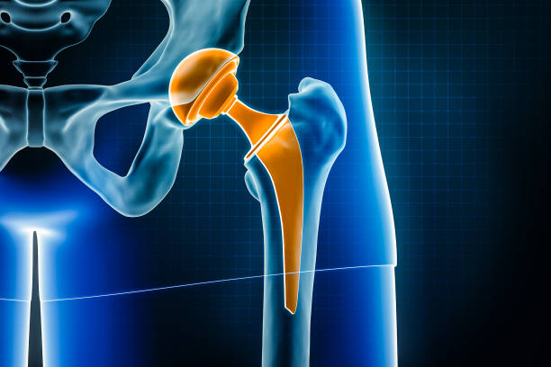
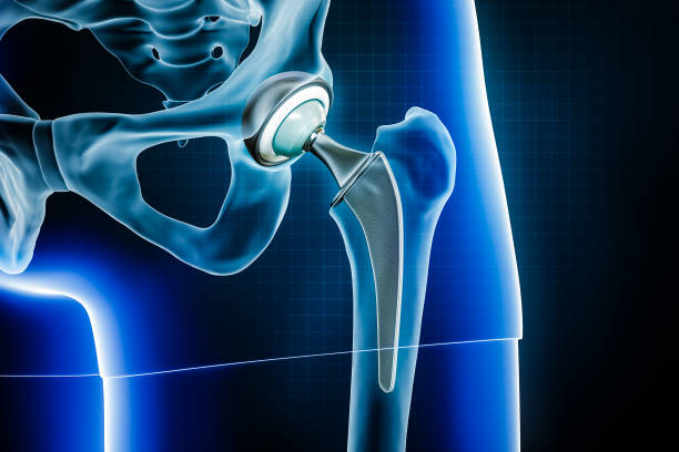
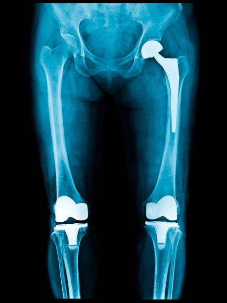
Plain X ray of both knee joints. view fracture and displacement of the patella bone or knee cap. X-ray image of the knee joint, close up
Browse 13,700+ x ray arthritis stock photos and images available, or start a new search to explore more stock photos and images.
Plain X ray of both knee joints. view fracture and displacement of the patella bone or knee cap. X-ray image of the knee joint, close up
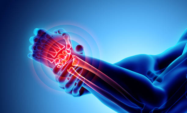
Wrist painful - skeleton x-ray, 3D Illustration medical concept.

Wrist painful - skeleton x-ray, 3D Illustration medical concept.
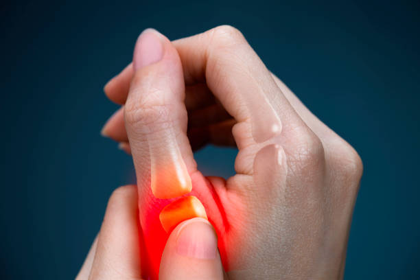
Pain, Finger, Thumb, Human Hand, Medical X-ray

X-ray of the human pelvis with both hip joints AP view. No bony injury, erosion, porosis, and sclerosis were seen. Both hip and S.I. joints have a normal appearance. No soft tissue lesion was seen.

Radiology and radio surgery X-ray web banners for medical technology. Vector design for scientific medicine center in orthopedics and traumatology therapy and diagnostics with X-ray bones and joints

X-ray of the human pelvis with both hip joint. No bony injury, erosion, porosis and sclerosis seen. Normal finding.
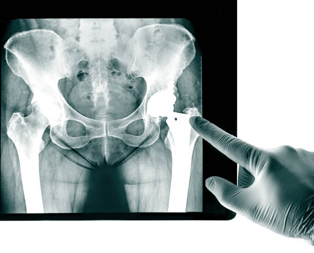
X-ray of the pelvis Endoprosthesis, the socket of the hip joint used for Hip Joint rheumatic diseases and Rheumatoid Arthritis

concept of orthopedic technology or bones and joints medical treatment, graphic of running man with x-ray interface
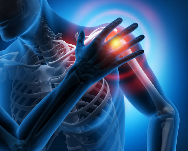
Pain in the Shoulder Joint - Medical Illustration - 3D Rendering

medical consultation - closeup side view asian male doctor with x ray image on computer screen is explaining about hallux valgus surgery before and after comparison to elderly man patient

Human skeleton chest x ray image torso roentgen shot medical analyzing vector flat illustration. Anatomical bone medicine laboratory radial scan picture for disease diagnostic health care checkup

3d render human spine anatomy with pain symptoms - back view - Grayscale Image

Human joint pain concept as skeleton and muscle anatomy of the body with a group of sore joints as a painful injury or arthritis illness symbol for health care and medical symptoms with 3D illustration elements.

"Spondylosis" (Left image) , Patient was operated and internal fixed. (Right image)

Doctors point to x-ray medical imaging with hand bone disease vertical concept

Toe bone has Hallux valgus deformity or Bunion disease

Heel spur, injury to the plantar ligament of the leg. Polygonal design of interconnected lines and points. Blue background.
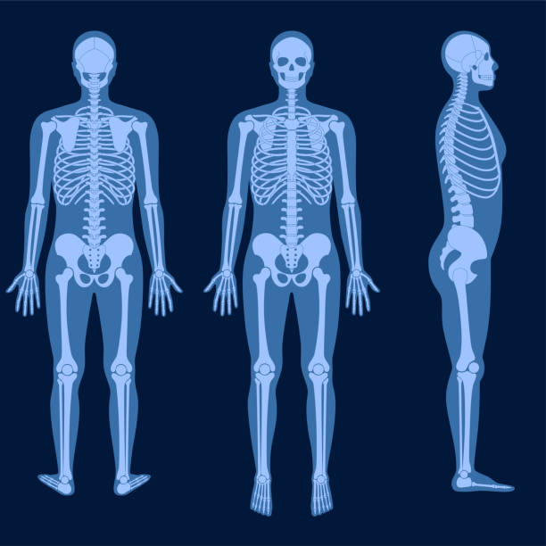
Human man skeleton anatomy in front, profile and back on x ray view. Vector isolated flat illustration of skull and bones in male body. Medical, educational or science banner

x-ray image show fracture of the radius bone

X-ray of whole leg, lower extremity. Scanogram of lower extremity.

knee joint pain cause by knee truma,gout,rheumatoid,osteoarthritis of knee
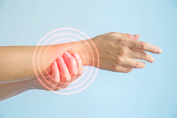
Wrist, Pain, Arm, Medical, Tomography

Dark navy blue vector realistic medial and top x ray of skeleton of foot. Human leg bones. Anatomy of joints. For advertising or medical publications. Illustration stock vector.
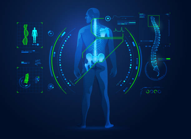
concept of chiropractic technology or spine medical treatment, graphic of human back bone with x-ray interface
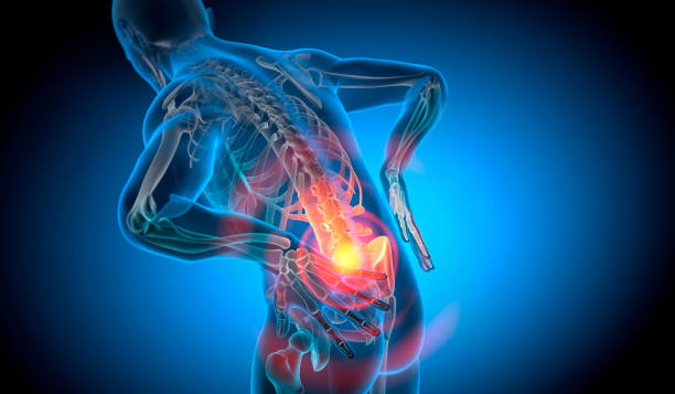
X Ray 3D Rendering - Man with severe pain in the spine and intervertebral discs - Herniated disc

Osteoarthritis Knee ( OA Knee ) ( Film x-ray both knee with arthritis of knee joint : narrow knee joint space ) ( Medical and Science background )

3D illustration, shoulder painful skeleton x-ray, medical concept.
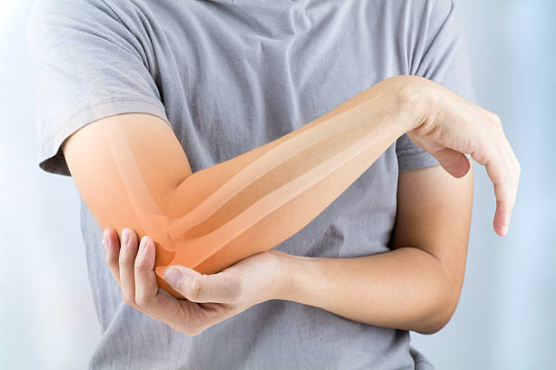
elbow bones injury white background
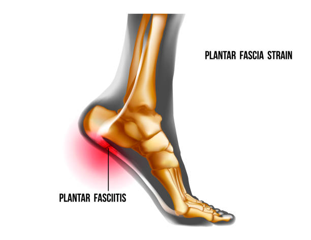
Plantar fasciitis inflammation and ruptures strain. Bones ot Foot pain realistic illustration. Medial view. Anatomy of joints, human leg black and yellow transparente skeleton. For medical orthopedic advertising. Vector illustration stock vector.
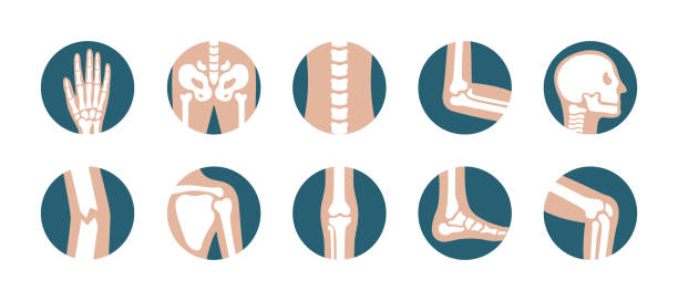
Vector illustration flat design of knee, leg, pelvis, scapula, skull, elbow, foot and hand icons
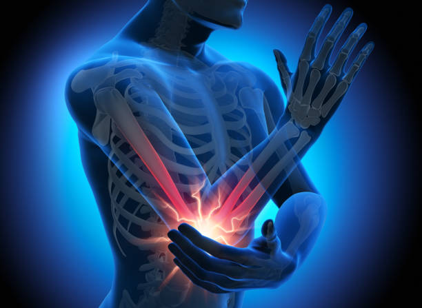
Pain in the Elbow Joint - Medical Illustration - 3D Rendering

Rheumatoid arthritis, osteoarthritis, rheumatism of wrist and fingers diagnosis vector illustration. Cartoon tiny people research xray of patients hands with autoimmune disease, joint inflammation

3D illustration of Humerus - Part of Human Skeleton.

Human spine x ray image anatomical cartoon bone science medical healthcare vector flat illustration. Spinal xray roentgen radiography shot side view orthopedic surgery diagnostic physiology research

Explore the intricacies of the human body's joints with this captivating image. Titled "Joints in the Human Body," it provides a detailed look at the anatomy of joints, bones, and skeletal structures. This medical concept delves into issues like arthritis, therapy, and health, employing advanced 3D technology to create a futuristic representation. Ideal for medical and scientific presentations, this image offers a comprehensive understanding of the human body's vital structural components.

Sports nutrition. Dietary supplement to maintain joint health and prevent injuries of the articular-ligamentous apparatus, mixed media.
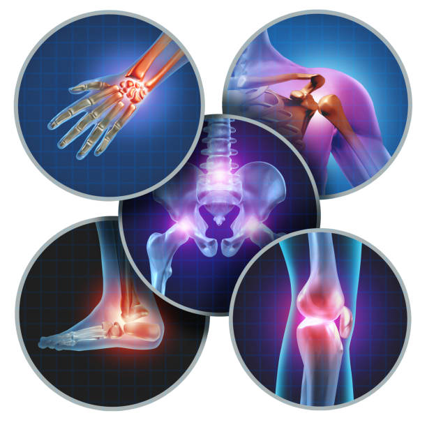
Human painful joints concept with the skeleton anatomy of the body with a group of sores with glowing joint pain and injury or arthritis illness symbol for health care and medical symptoms.