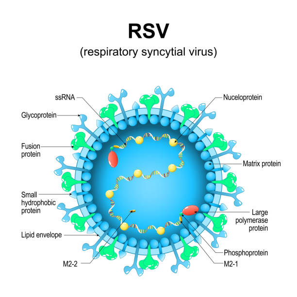
Respiratory syncytial virus. RSV structure. Close-up of a orthopneumovirus. Virion anatomy. Magnified of virus that causes infections of a human respiratory tract. Vector diagram
Browse 340+ glycoprotein stock illustrations and vector graphics available royalty-free, or search for spike glycoprotein or coronavirus glycoprotein to find more great stock images and vector art.
Respiratory syncytial virus. RSV structure. Close-up of a orthopneumovirus. Virion anatomy. Magnified of virus that causes infections of a human respiratory tract. Vector diagram
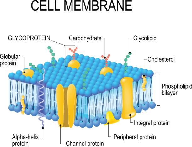
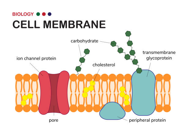
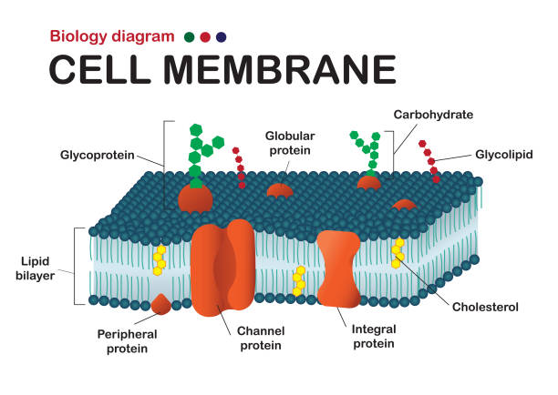
Cell membrane. A detailed diagram models of membrane Structure

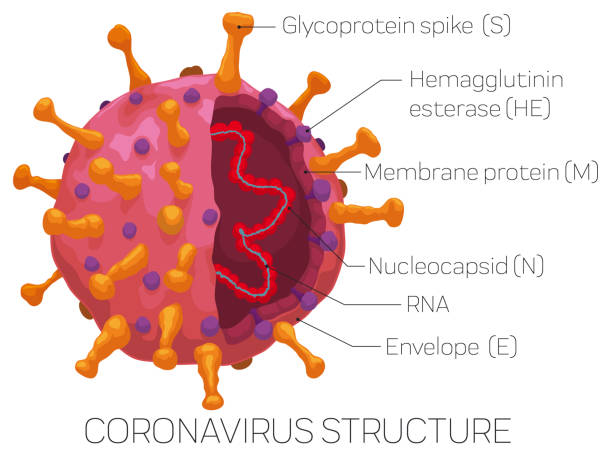
Coronavirus model sliced, one with external part and the other with internal view to show its parts: glycoprotein spike, hemagglutinin esterase, membrane protein, enveloped, nucleocapsid and RNA.
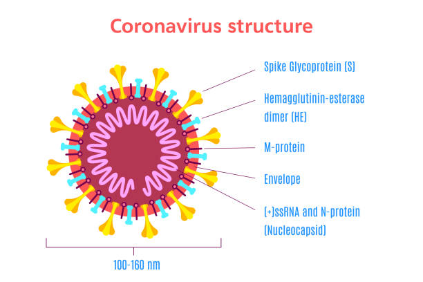
Infographic with Coronavirus sliced showing its parts, detailed for a easy recognition of this virus: glycoprotein spike, hemagglutinin esterase, membrane protein, envelope, nucleoprotein and RNA.

Structure of the AIDS virus. Vector
Coronavirus virion structure diagram isolated on white background. Infographic template. Stock vector illustration in flat style
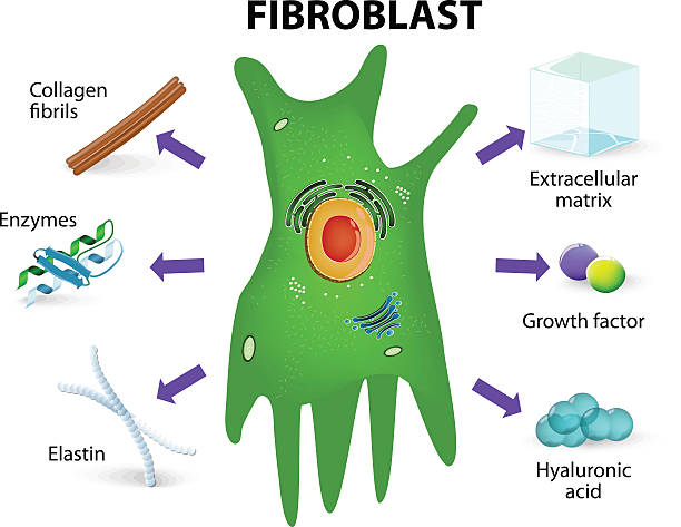
Fibroblast. Structure and function. Human skin cell
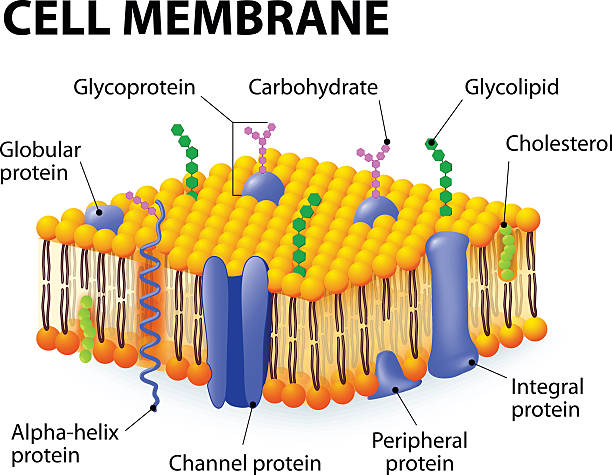
Cell membrane. A detailed diagram models of membrane Structure
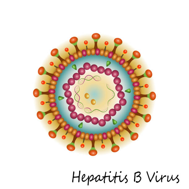
Colorful Hepatitis B Virus particle structure isolated on white background. Vector illustration
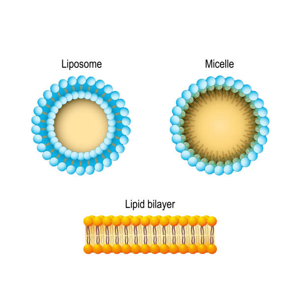
Cell membrane (Lipid bilayer), Micelle, Liposome. Phospholipids aqueous solution structures. A detailed diagram models of membrane Structure. Vector illustration for biology, scientific, and medical use.

Cytomegalovirus. structure of the virion. herpesvirus type 5. view outside and cross-section
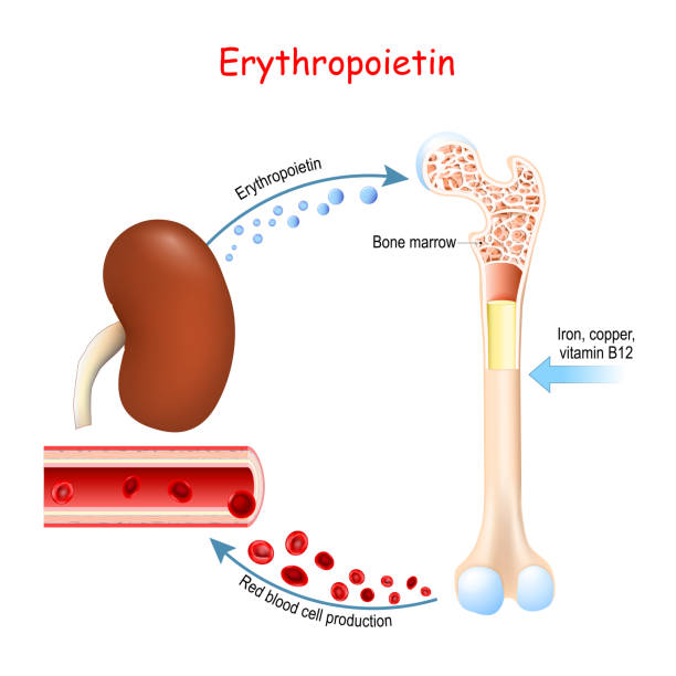
Erythropoietin. Glycoprotein cytokine secreted by the kidney in response to cellular hypoxia that stimulates red blood cell production (erythropoiesis) in the bone marrow. Vector illustration
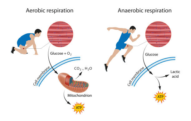
There are two types of cellular respiration: aerobic and anaerobic. One occurs in the presence of oxygen (aerobic), and one occurs in the absence of oxygen (anaerobic)
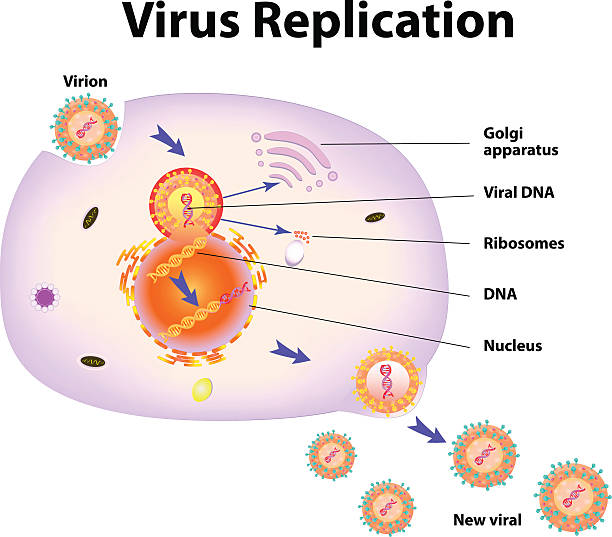
Scheme of virus replication cycle. Vector illustration
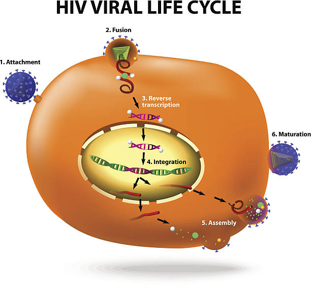
HIV viral life cycle. Active infection of t cell by HIV. Vector diagram

Structure of cell membrane. close-up of cell, phospholipid bilayer, and phospholipid.molecule with Phosphate head and Hydrophobic tail. vector diagram for medical, educational and scientific use
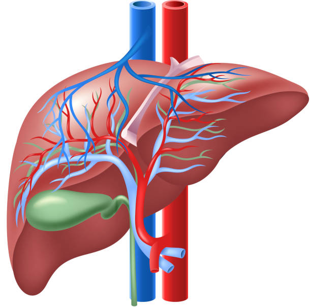
Illustration of Human Internal Liver and Gallbladder

Types of viruses vector illustration labeled drawings. Helical, polyhedral, spherical and complex structure models. Biology science research for epidemic and pandemic crisis public health protection.

Flat vector illustration of cutaway coronavirus internal structure showing Spike Glycoprotein, Hemagglutinin-esterase, RNA and N protein and Envelope. Close-up view of red COVID-19 with description.

Cytomegalovirus. CMV structure. Close-up of a virion anatomy. Magnified of virus particle that cause mononucleosis and pneumonia. Vector diagram

Structure of Hepatitis C virus. Virion anatomy. Infectious disease of the liver caused by HCV. Viral hepatitis. Vector diagram

Illustration of the human internal liver

Papillomaviruses (HPV) are non-enveloped, icosahedral double-stranded DNA viruses. Papilloma virion visualization includes capsid protein, histone, and DNA strand.

Close-up vector illustration of COVID-19 internal and external structure showing Spike Glycoprotein, Hemagglutinin, RNA and N protein and Envelope. Anatomy of red coronavirus with description.
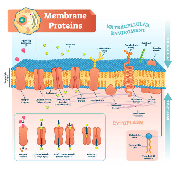
Membrane proteins labeled vector illustration. Detailed microscopic structure scheme. Anatomical diagram with receptor, open channel, closed gated and transport protein.

Cell anatomy. Cell structure and organelles Nucleus, Ribosomes, Endoplasmic reticulum, Golgi apparatus, mitochondrion, cytoplasm, lysosome. Close-up of lipid bilayer cell membrane. Vector poster. Isometric Flat illustration.

Colorful diagram of HIV virus particle structure with annotations on white background. Vector illustration
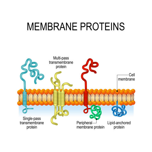
Membrane proteins. integral, and Peripheral membrane proteins, Single-pass, and Multi-pass transmembrane α-helix, Lipid-anchored protein. Vector illustration for biological, science and educational use

Microscopic real 3D model of the corona virus COVID-19. The image is a scientific interpretation of the virus with all relevant details : Spike Glycoproteins, Hemagglutinin-esterase, E- and M-Proteins and Envelope.

Major Mechanisms by which Molecules Cross the Cell Membrane, Simple Diffusion, Channel, and Pump / Carrier - Medical Vector Illustration

Phagocytosis - Process for nutrition in unicellular organisms, while in multicellular organisms it is found in specialized cells called phagocytes - Cell Medical Vector Illustration

Structure of Influenza virus. Close-up of a Virion anatomy. magnified of flu virus . Vector diagram

Plasma membrane (Fluid- Mosaic Membrane Model of cell membrane structure)

Flat vector illustration of COVID-19 external structure showing red spikes and envelope of virus capsid. Close-up view of coronavirus under microscope.

http://www.cumulocreative.com/istock/File Types.jpg
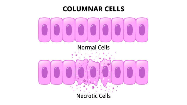
Columnar Cells - Normal Epithelial Cell - Necrotic Cell - Medical Vector Illustration

Cell Membrane Transport - Uniport, Cotransport (symport - antiport) of molecules through membranes - Histology - Medical Vector Illustration

AMH Anti-Müllerian hormone written on a notebook paper- vector illustration
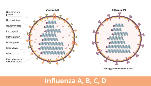
Four types of influenza virus cell, Influenza A and B (hemagglutinin and neuraminidase) and influenza C and D (hemagglutinin-esterase fusion).

Rod-shaped tobacco mosaic virus with a helical capsid surrounding RNA strand.

Flat vector illustration of doctors in masks tell about coronavirus internal structure on a tv or computer monitor. COVID-19 has Spikes, Glycoprotein, Envelope, RNA and protein. Pills, syringe around.

Flat vector illustration of doctors in masks show coronavirus internal structure on a smartphone or tablet. COVID-19 has Spikes, Envelope, RNA and N protein. There are pills, syringe around.

Facilitated Diffusion - Membrane Transport - Molecules Across Cell Membrane - Medical Vector Illustration

Colorful diagram of Hepatitis B virus particle structure with annotations on white background. Vector illustration

Liposome Structure of Phospholipid Molecule - Lipid Bilayer - Membrane Cell - Medical Vector Illustration

Herpes simplex virus particle structure on white background. Vector illustration

rabies virus structure. Anatomy of virion Rabies lyssavirus. structure. Anatomy of virion Rabies lyssavirus.

Structure of fibroblast cells. These cell are vital to the skin's strength and elasticity. The fibroblasts also synthesise the ground substance of the dermal matrix.

Fluid mosaic model vector illustration. Labeled cell membrane structure infographic. Educational scheme with phospholipids, protein, carbohydrates and cholesterol. Biological elastic functions graphic

Fluid mosaic model with cell membrane anatomical structure outline diagram. Labeled educational scheme with glycoprotein, integral protein, glycolipid and phospholipid vector illustration.

An RNA virus, a member of the family Togaviridae causes Chikungunya infection. Viral cell with RNA strand, glycoproteins, nucleocapsid and bilayer lipid membane.

Neuron, Neurone, or Nerve Cell - Anatomy and Histology nerve cell - Axon, Schwann Cell, Dendrite, Synapse - Vector Illustration

Mechanically Gated Ion Channels - Membrane Transport - Transmembrane Ion Channel Protein - Cell Biology - Medical Vector Illustration

Lipids in Membrane Structure - Lipid Bilayer - Cholesterol Structure Formula - Vector Medical Illustration

Receptor, enzyme, gated ion channel, cell-identity marker and cell-adhesion molecule. Chemical messenger breakdown and ions transportation.