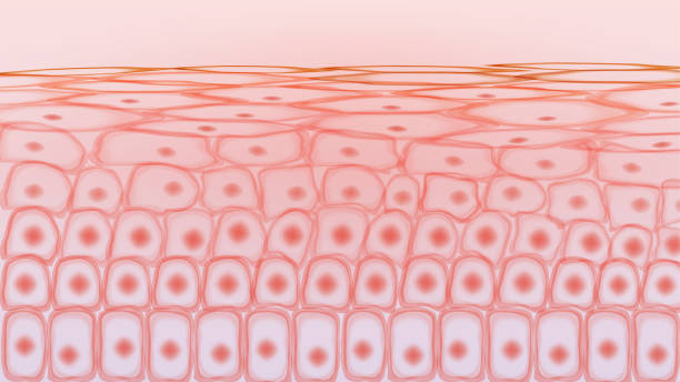
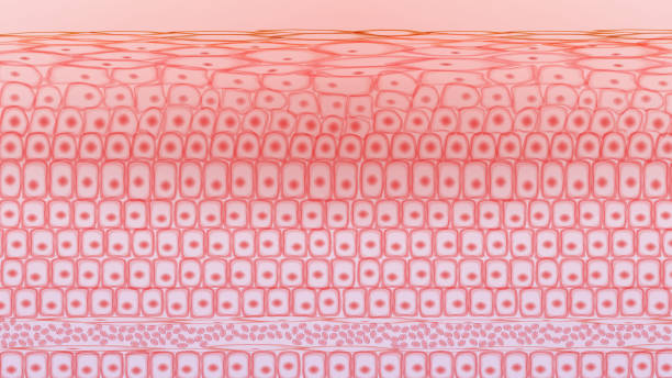
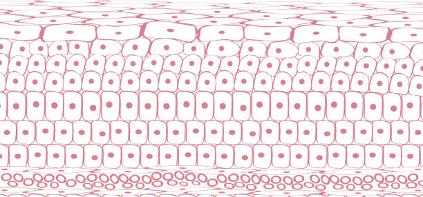
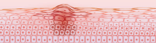
Cell division - scientific background
Browse 350+ human cell division stock illustrations and vector graphics available royalty-free, or search for human cell division icon to find more great stock images and vector art.
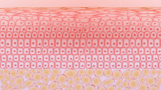
Cell division - scientific background
Hair loss treatment flat line icons set. Shampoo ph, dandruff, hair growth, keratin, conditioner bottle vector illustrations. Outline signs for beauty store. Pixel perfect 64x64. Editable Strokes.
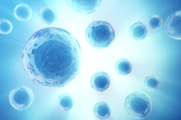
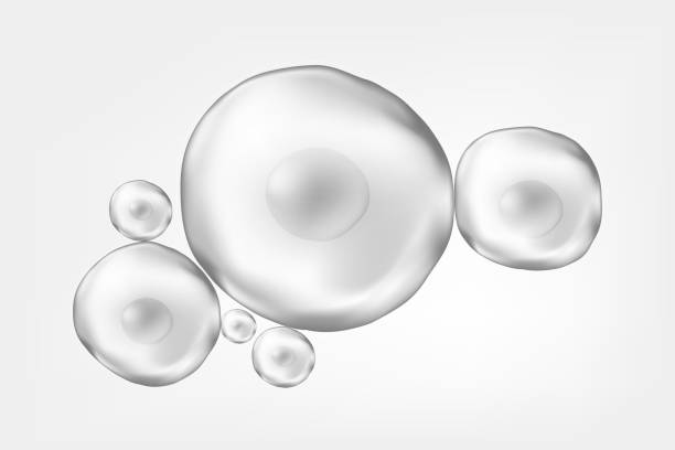
Human or animal cells on colorful background. Medicine scientific concept. 3d rendering.
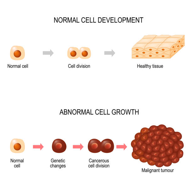
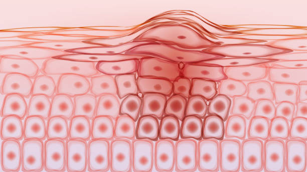
Cancer cells. illustration showing cancer disease development. Healthy tissue and Malignant tumour. Vector diagram for your design, educational, biological, science and medical use

induced pluripotent stem cell (iPS cell) and regenerative medicine, vector illustration
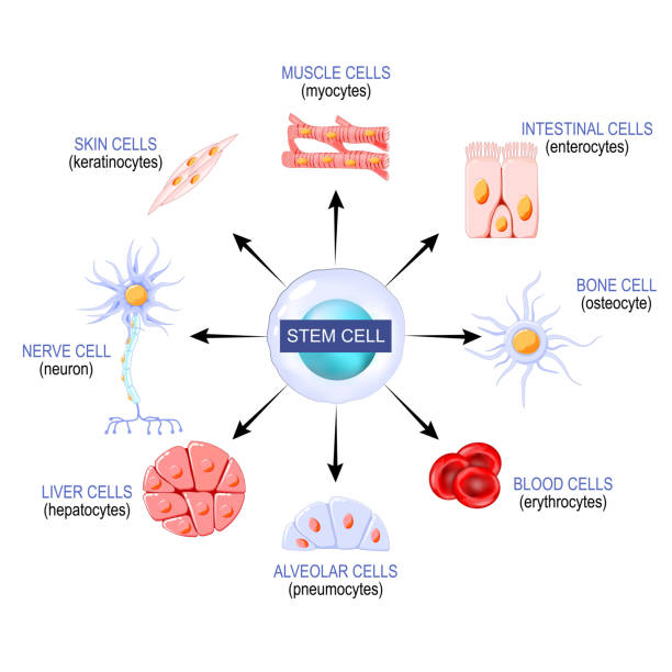
Stem cells from a blastocyst that can become any tissue in the body. for example: neuron, osteocyte, enterocytes, red blood cells, myocytes, hepatocytes, pneumocytes, and epithelial skin cells or keratinocytes. Using stem cells to treat disease. Vector poster
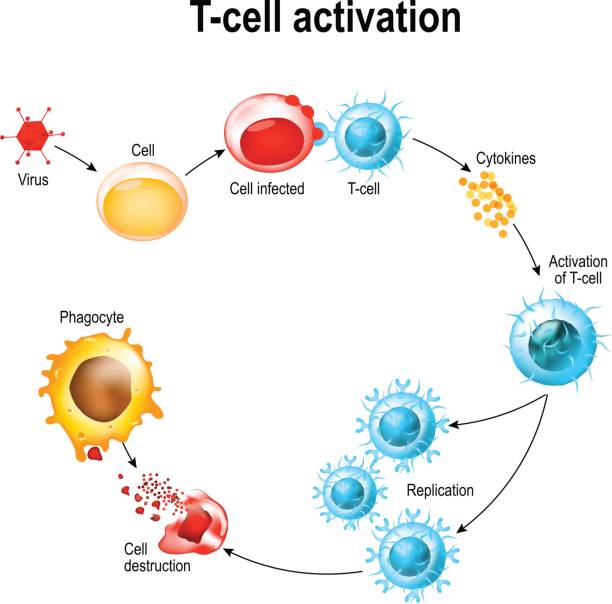
Activation of T-cell leukocytes. T-cell encounters its cognate antigen on the surface of an infected cell. T cells direct and regulate immune responses and attack infected or cancerous cells.

A telomere is a repeating sequence of double-stranded DNA located at the ends of chromosomes. Each time a cell divides, the telomeres become shorter. Eventually, the telomeres become so short that the cell can no longer divide.

Stem cell transplant. Stem cells divide and change into the different types of blood cells. Vector illustration

Human lung cancer under a magnifying glass. Polygonal design of interconnected lines and dots. Blue background.

cellular senescence. From Normal to Senescent cell. Telomere and DNA damaged, mitochondrial dysfunction are primary drivers of damage in aging. Anti-aging therapy. Senescence-associated secretory phenotype SASP. Anti aging medicine. vector poster

Cytotoxic T cell. T-cell regulate immune responses, release the perforin and granzymes, and attack infected or cancerous cells. Through the action of perforin, granzymes enter the cytoplasm of the target cell, and lead to apoptosis (cell death).
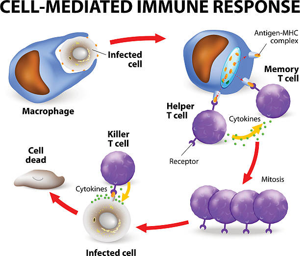
Cell-mediated immunity. T lymphocytes do not secrete antibodies. this response incorporates activated macrophages, natural killer cells, antigen-specific cytotoxic T-lymphocytes as well as release of cytokines.

Types of T-cell. Lymphoid cell. T lymphocytes: Naive, Regulatory, Memory, helper, and T-killer or Cytotoxic T cells. Vector illustration
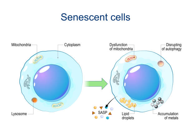
Senescent cells. Cellular senescence from Dysfunction of mitochondria, accumulation of metals, Disrupting of autophagy, Lipid droplets to release of Senescence-associated secretory phenotype SASP and chronic inflammation. DNA damage response. Aging cells. Vector diagram
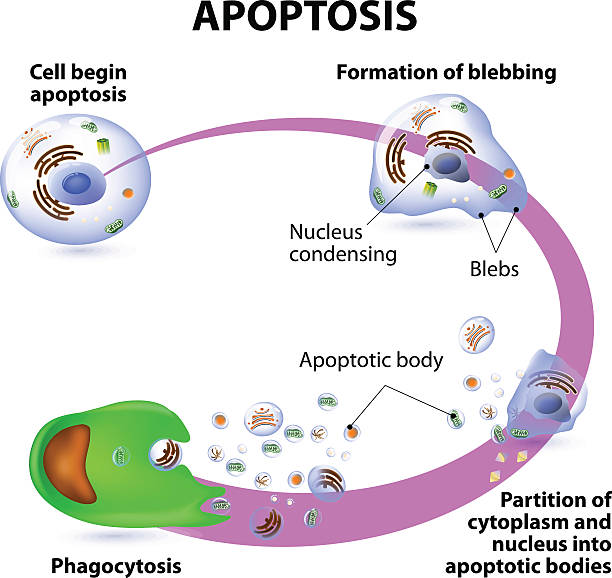
Apoptosis is the process of programmed cell death. Vector diagram
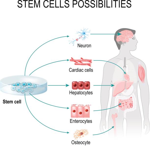
Stem cells possibilities. These cells can become any tissue in the body. Internal organs in the background of a male figure and a human cell (gepatocytes, osteocyte, cardiac, enterocytes, neuron).

Microscopic view of myelocyte human white blood cells in mitosis (cell division) found in the blood from a patient with chronic myelogenous leukemia. Vintage etching circa mid 19th century.
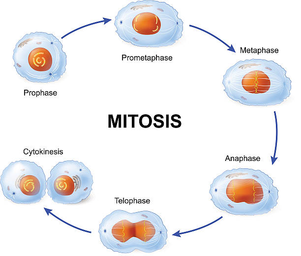
Cell division. Mitosis. Vector scheme
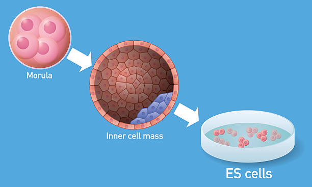
Embryonic stem cell (ES cell) and regenerative medicine, vector illustration

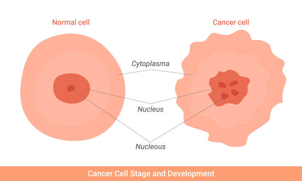
Cancer cell and normal cell. Healthy tissue and Malignant tumour. illustration showing cancer disease development. Vector diagram for your design, educational, biological, science and medical use

Cancer and cytotoxic T-cells. T lymphocyte kills cancer cells. T-cell (immune responses), release the perforin and granzymes, and attack cancerous cells. Through the action of perforin, granzymes enter the cytoplasm of the target cell, and lead to apoptosis (cell death

Cancer development from normal cell to Formation of tumor, Spreading cancer cells in blood flow, Invasion of other tissue, and formation of metastases. vector illustration

Cancer cell. illustration showing cancer disease development. Vector diagram for your design, educational, science and medical use

Cell division - scientific background

Stem cells under microscope. Biotechnology. Biology. "r"nMedical science background.

Chromosome of a Normal fibroblast and Cell senescence. Telomeres shorten with each round of replication. aging process. Vector poster

Embryonic stem cell (ES cell) and regenerative medicine, vector illustration

fraud triangle theory template infographic concept for slide presentation with pyramid stack box layer description 3 point list with flat style vector

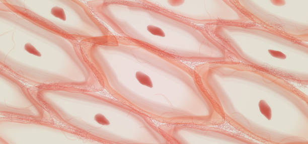
Chondrocytes is cells found in healthy cartilage. Chondrocyte lie in lacunae (spaces around cells).

Vector isolated illustration of malignant tumor in healthy tissue. Doctor in laboratory. Spreading of cancer cells, tumor development concept. Medical infographic for poster, science and medical use.
Hair Repair Concept Line Icon. Hair Protect, Nutrition, Growth with Keratin Treatment Black Outline Pictogram. Healthy and Growing Follicle Icon. Editable Stroke. Isolated Vector Illustration.

Stem cells under microscope. Biotechnology. Biology. Medical science background.

Cell division background. Vector

Stem cells under microscope. Biotechnology. Biology. Medical science background.