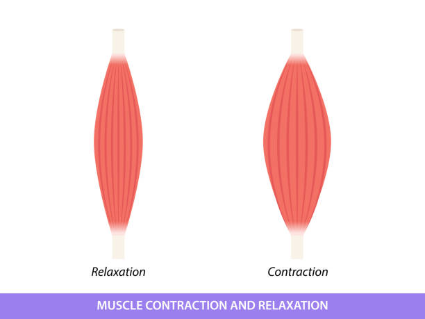
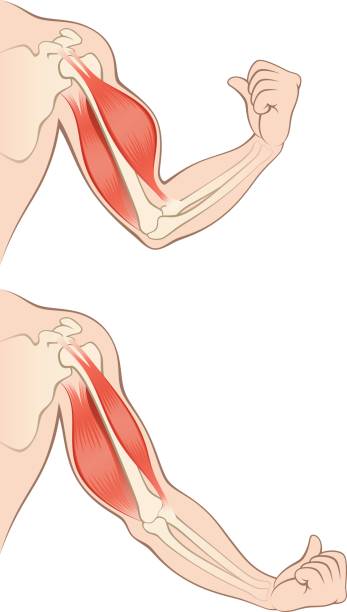
When a muscle gets shorter it has contracted and when a muscle gets longer it has relaxed. Most muscles come in antagonistic pairs. This means when one contracts the other must relax.
Browse 1,700+ muscle contraction stock illustrations and vector graphics available royalty-free, or search for skeletal muscles or muscle anatomy to find more great stock images and vector art.
When a muscle gets shorter it has contracted and when a muscle gets longer it has relaxed. Most muscles come in antagonistic pairs. This means when one contracts the other must relax.

Muscle contractions scheme with arm cross section and fitness weight lifting exercise movement. Concentric, eccentric and isometric contraction types diagram.
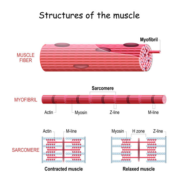
Structure Skeletal Muscle. myofibril with thin and thick filament. close up of a sarcomere. Muscles contract by sliding the myosin and actin filaments along each other. Biomedical Science. Mechanism of mechanical contraction
Tension muscles human hand on a white background

Extension and Flexion of Muscles. Biceps and triceps. Contracted and Relaxed. Agonist and Antagonist muscles. vector illustration

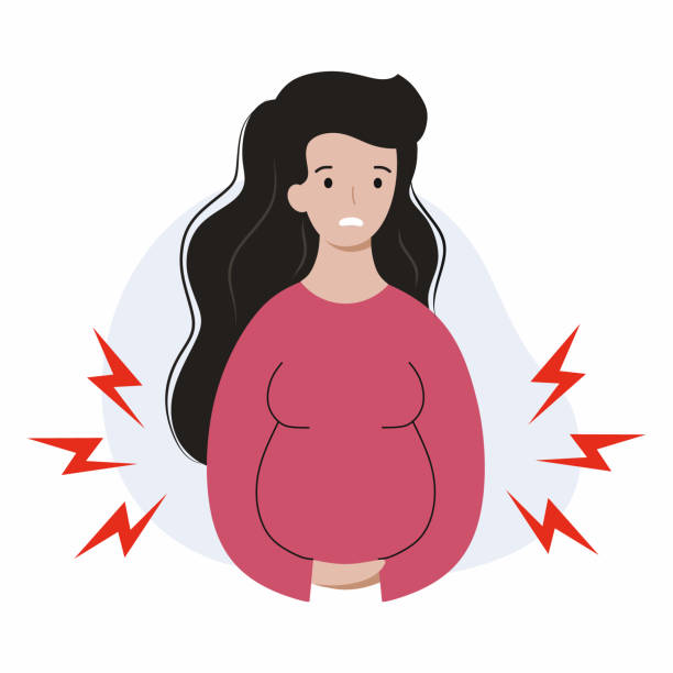
Childbirth contractions. Pregnant frightened african american woman suffering from labor pains. Real or false contractions? Travail pangs. Vector illustration.

Different illustrations for human heart. Simple images for human heart drawing. Outline and colored versions. For educational and scientific purpose.
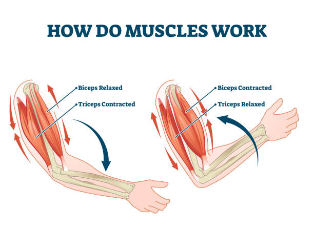
How do muscles work labeled principle explanation scheme vector illustration. Anatomical and physical movement process example with biceps relaxed and triceps contracted. Educational comparison graph.
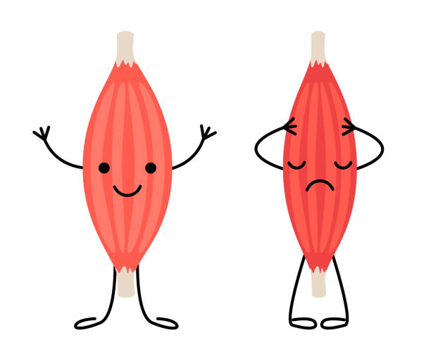
Healthy happy muscle and weak sad pain muscle character. Strong and frail tension fiber part body human. Skeletal muscle, inside tissue. Vector
Muscle contraction and relaxation isolated on white background. Muscle icon human internal organ anatomy healthcare medicine concept. Vector illustration.
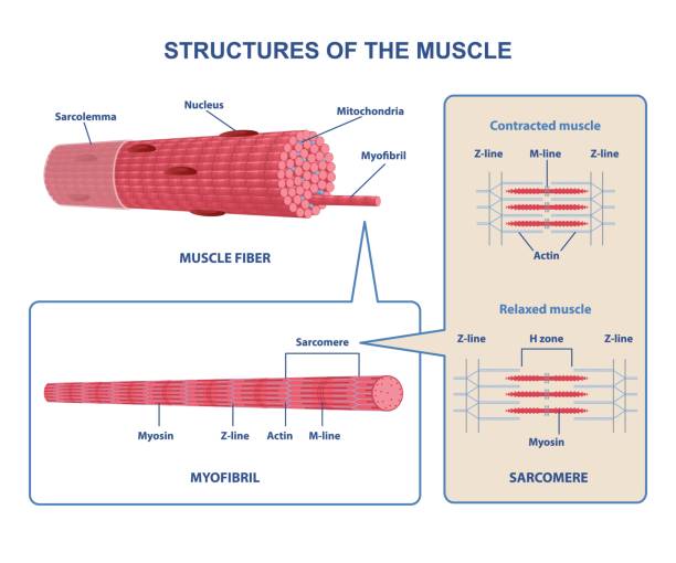
Colorful structure skeletal muscle scheme on white background. Muscles contract by sliding myosin and filaments along each other. Myofibril with thin and thick filament. Flat vector illustration
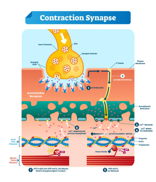
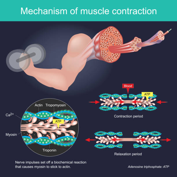
Contraction synapse vector illustration. Labeled closeup medical structure scheme. Diagram with full cycle of ACH release, action potential, troponin bonding and filament
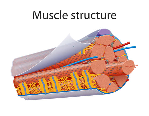
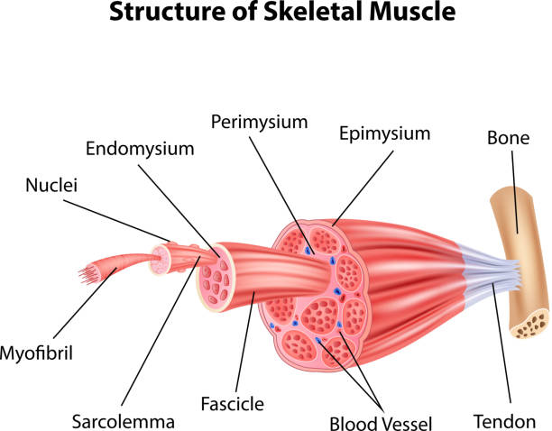
Skeletal muscles are organs of the vertebrate muscular system that are mostly attached by tendons to bones of the skeleton. The muscle cells of skeletal muscles are much longer than in the other types of muscle tissue, and are often known as muscle fibers

ATP muscle contraction cycle vector illustration labeled scheme. Educational diagram with muscle, fibers and cells. Structure of myofibril with thin and thick filament.
Muscle flat line icon. Vector outline illustration of human anatomy. Black thin linear pictogram for muscular system.

A synaptic connection between the terminal end of a motor nerve and a muscle. Presynaptic (nerve terminal), postsynaptic part, synaptic cleft.
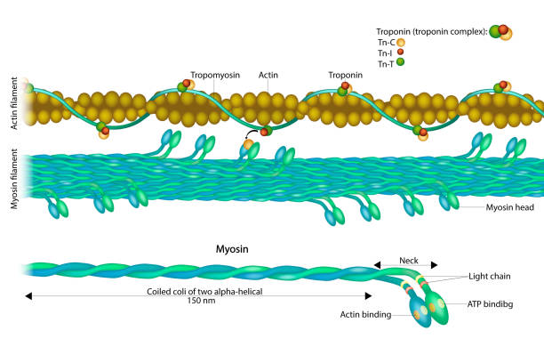
Actin filament and Myosin filament. Structure Myosin. Muscle Actin myosin interaction. Troponin or troponin complex.
Muscle icon. Muscle fiber. Muscle part. Editable vector.

Types of muscles on a white background

Troponin complex and three regulatory proteins structure outline diagram. Labeled educational scheme with tropomyosin and actin for muscle contraction vector illustration. Myocardial infarction marker

Muscle relaxation, stretching, and contraction. Close-up of a Skeletal muscle fiber. Isometric flat vector Illustration

Motor neuron. Structure and anatomy of a efferent neuron. Close-up of a Muscle fiber, and motoneuron with Dendrites, Synapse, Telodendria, Axon, Schwann cell. The axons carry signals from the spinal cord to muscles. Vector illustration
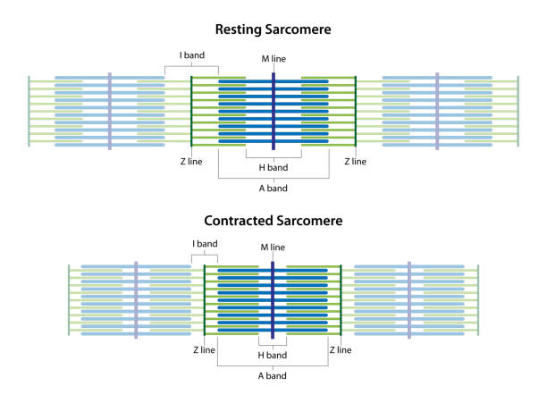
Sarcomeres in different functional stages: resting and contracted. Sarcomere showing the location of the I band, A band, H band, M line, and Z lines.

Painful arc test and physiopedia syndrome medical diagnosis outline diagram. Labeled educational position scheme with subacromial pain angle in hand movement vector illustration. Rotator cuff disorder
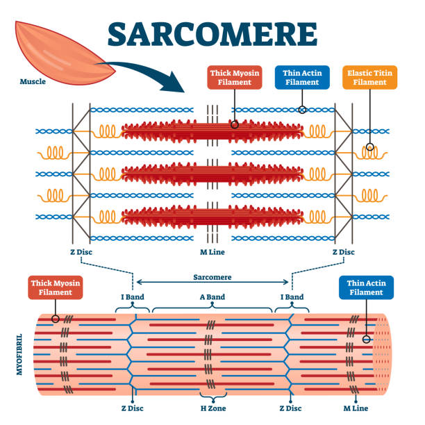
Sarcomere muscular biology scheme vector illustration. Myosin filaments, discs, lines and bands. Myofibril detailed labeled diagram. Sports educational health information. Muscular system anatomy.
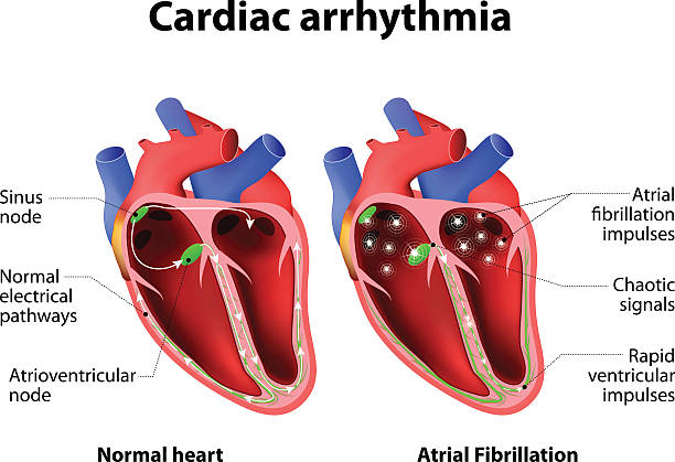
Cardiac arrhythmia. cardiac dysrhythmia or irregular heartbeat. Medical illustration

Different muscles in human body and muscular classification outline diagram. Labeled educational physiological parts scheme with anatomic skeletal, smooth and cardiac division vector illustration.
Types of muscle on a white background

An autoimmune disease of the neuromuscular junction when antibodies block or destroy nicotinic acetylcholine receptors (AChR) at the junction between the nerve and muscle.
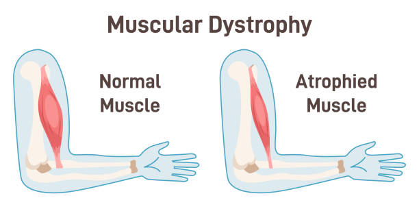
Muscular dystrophy. Healthy muscle versus atrophied one. Anatomical structure of the healthy human muscle and muscle with duchenne disorder. Flat vector illustration

Pregnant woman holds stomach, standing near cabinet, experiencing pain due to complications caused by fetal development disorders. Pregnant girl needs hospitalization to avoid miscarriage

Reflex reaction with knee stimulus test process explanation outline diagram. Labeled educational scheme with anatomical body reaction to impulse vector illustration. Receptors or sensory neuron check

Muscle contraction mechanism. Muscles work principle scheme. Anatomical structure and physical movement process example with contracted and relaxed biceps. Flat vector illustration

Muscle strain grading. Grade 1 is A minimal loss of strength. Grade 3 is a severe tear and complete loss of function. Vector illustration
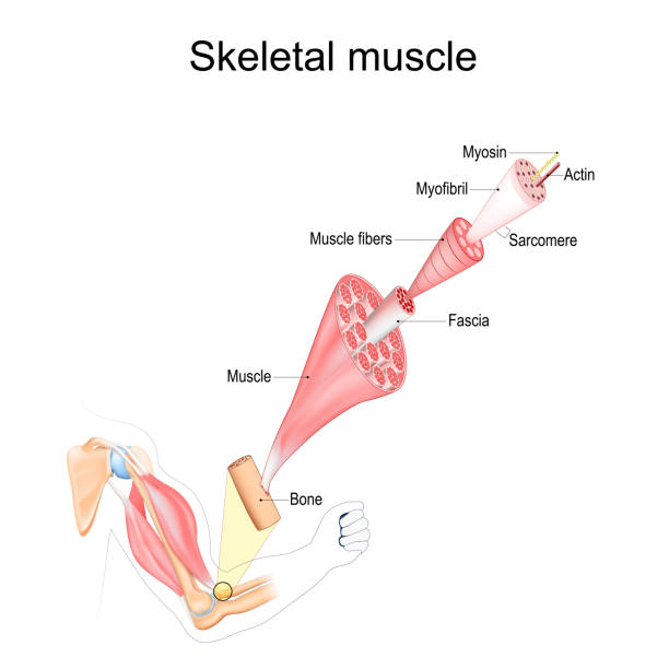
Skeletal Muscle anatomy. structure of Muscle fibers from Fascia and Tendon to Actin and Myosin. Vector poster

Isometric vector image on a blue background, a white sheet with the contract or business document and a pen for signing, the conclusion of contracts

Peristalsis, or wavelike contractions of the muscles in the outer walls of the digestive tract, carries the bolus by the esophagus. Peristalsis does not only occur in the esophagus. It continues through the stomach, small intestine, and large intestine. Vector diagram

Cervical effacement set. Cervix dilation during delivery from not effaced and dilated to fully effaced and totally dilated. Fetus in a womb during labor. Flat vector illustration

Breathing during labor. Vector infographic about the techniques of a breath of women in the different stages of pregnancy.
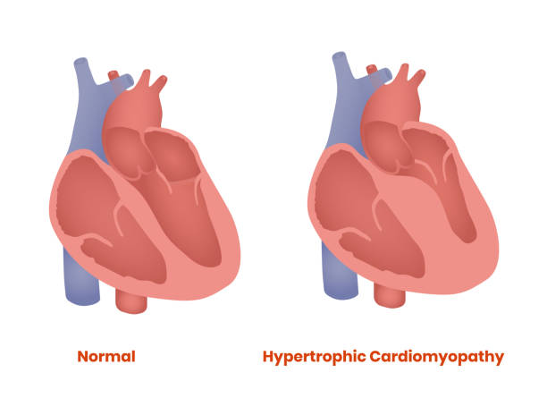
Hypertrophic Cardiomyopathy vs healthy heart vector illustration
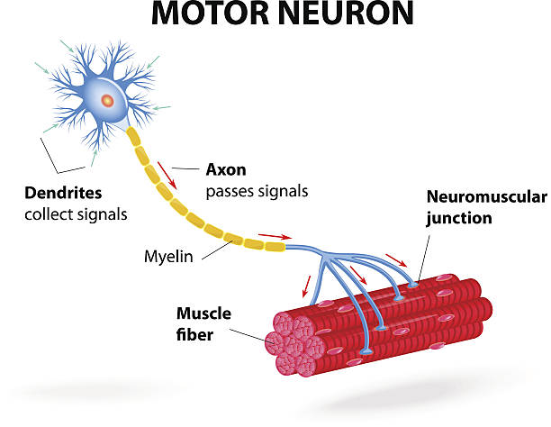
structure motor neuron. Vector diagram. Include dendrites, cell body with nucleus, axon, myelin sheath, nodes of Ranvier and motor end plates. The impulses are transmitted through the motor neuron in one direction
Vector gym icon set. Realistic 3d illustration isolated on white background.
Couple fitness Silhouette of a man / athlete and slender woman with dumbbells.
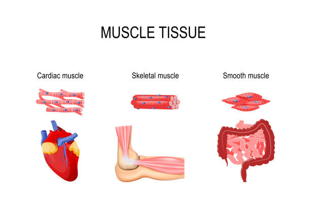
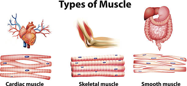
Types of muscle tissue. Skeletal muscle (elbow joint), smooth (gastrointestinal tract) and cardiac muscle (heart). Human internal organs and Muscle cells. vector illustration for medical, educational and science use
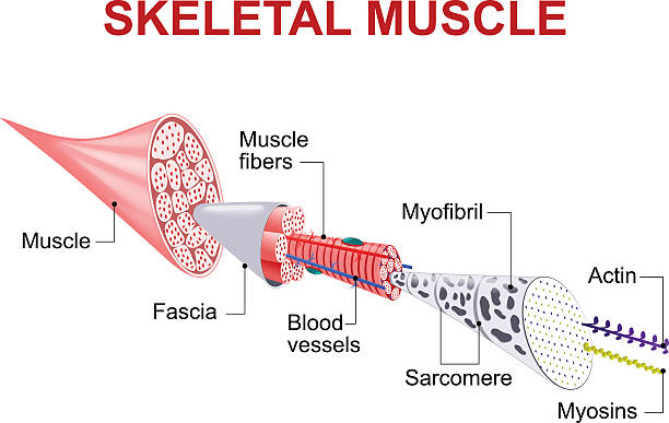
Each skeletal muscle fiber has many bundles of myofilaments. Each bundle is called a myofibril. This is what gives the muscle its striated appearance. The contractile units of the cells are called sarcomeres.

Skeletal muscle tissue structure. Skeleton with joints, cartilages and ligaments in the human body. Muscle fibers and connective tissue sheaths. Musculoskeletal anatomy flat vector medical anatomy.

Multiple red arrows falling across world map symbolize global synchronized economic decline. Vector illustration. Concept of global recession, market crash, economic contraction, trade collapse.
Tabloid Newspaper Design Template Vector. Images, Articles, Business Information. Daily Newspaper Journal Design. Illustration
© 2025 iStockphoto LP. The iStock design is a trademark of iStockphoto LP. Browse millions of high-quality stock photos, illustrations, and videos.
Do Not Sell or Share