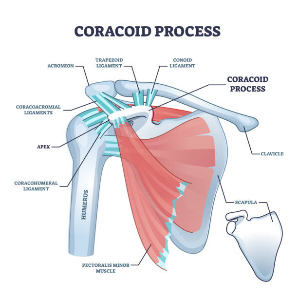
Coracoid process with anatomical osseous skeletal structure outline diagram. Labeled educational physiology scheme with shoulder bones, ligaments and muscle titles description vector illustration.
Browse 1,000+ where is the humerus bone pics stock illustrations and vector graphics available royalty-free, or start a new search to explore more great stock images and vector art.
Coracoid process with anatomical osseous skeletal structure outline diagram. Labeled educational physiology scheme with shoulder bones, ligaments and muscle titles description vector illustration.

Oblique muscles and human inner skeletal and muscular system outline diagram. Labeled educational external and internal obliquus abdominis parts description vector illustration. Anatomical scheme.

Pain in human arm. Lateral epicondylitis tennis elbow. Trauma or inflammation in hand. Muscular system and skeletal anatomical poster. Structure of muscle groups and bones isolated vector illustration

Skeleton chest bones showing heart shape. Heart rib cage in goth style illustration. Hand drawing illustration isolated on white background. Vector EPS 10

3D Illustration of Spinal cord a Part of Human Skeleton Anatomy

Ball of foot pain bones skeleton realistic illustration. Medial view. Anatomy of joints, human leg realistic black and yellow transparent skeleton. For medical orthopedic advertising. Vector illustration stock vector.

Shoulder bursitis inflammation. Inflamed bursa in the human body. Rotator cuff disease, pain and deformity. Anatomical musculoskeletal poster for clinic or hospital. Medical banner vector illustration

Arm parts poster with explanation in beautiful font, human body piece made of bones, humerus and ulna radius vector illustration isolated on blue

A stylized x-ray "front on view" of a person walking (2 styles - a dot and line version). The 12 red circular glow highlights indicate potential injury / pain areas.

A stylized x-ray "Birds-eye" view of a person running (2 styles - a dot and line version). The 11 red circular glow highlights indicate potential injury /pain areas.

Ulnar nerve entrapment. Cubital tunnel syndrome and Guyons canal syndrome. Pressure or pulling, stretching of the ulnar nerve in the elbow and wrist regions. Pain in arm anatomical vector illustration
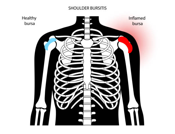
Shoulder bursitis inflammation. Inflamed bursa in the human body. Rotator cuff disease, pain and deformity. Anatomical musculoskeletal poster for clinic or hospital. Medical banner vector illustration

Shoulder joint and human ribs. Rear view. Polygonal design of interconnected lines and points. Blue background.

3D Illustration of Human Body Bone Joint Pains Anatomy (Foot Joints)

Palmaris longus skeletal and muscular body structure for human arm outline diagram. Labeled educational scheme with anatomical and medical hand inner parts physical description vector illustration.

Martin's natural history for the youth 1872

Man with a dislocation of the humerus at the shoulder

Illustration of the horse Skeletal System on a white background

human skeleton system outline isolated on white background . Didactic board of anatomy of human bony system

Osteosarcoma. Malignant bone tumor. Human leg with Bone cancer. Close-up of a knee joint and bone tissue with tumor. Vector illustration. Medical poster

Shoulder bursitis inflammation. Inflamed bursa in the human body. Rotator cuff disease, pain and deformity. Anatomical musculoskeletal poster for clinic or hospital. Medical banner vector illustration

Labeled human shoulder bone anatomical vector illustration diagram poster. Medical health care information.

Humerus, radius and ulna bone anatomical poster. Human skeleton concept. Hand bones and joints anatomy. Elbow and wrist. Medical flat vector illustration for clinic, hospital or education. X ray image

Female skeleton image. Vector illustration isolated on a dark grey background useful for creating medical and scientific materials. Anatomy, medicine and biology concept.

Lateral Epicondylitis known as Tennis Elbow - Stock Illustration as EPS 10 File

Human skeleton worksheet vector illustration. Blank educational bones scheme. Inner skeletal system practice lessons task template. Workbook topic material for school teachers anatomy or biology tests
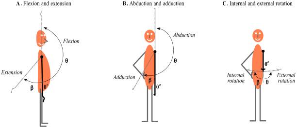
Shoulder movement: flexion, extension, abduction, adduction, external rotation and internal rotation

Tibia bone anatomy. Shinbone In human skeletal system diagram. Shankbone poster, Skeleton in male, female, baby, child and adult silhouettes. Cartilage and joints in body xray flat vector illustration

Anatomy of normal arm bone. Illustration about human body part in vector style.

Human anatomy scientific illustrations with latin/italian labels: elbow joint

The Talocrural joint or ankle joint, body weight and Gastrocnemius and soleus muscles form a second class lever

A stylized x-ray side view of a figure walking (2 styles - a dot and horizontal line version). The 19 red circular glow highlights indicate potential injury /pain areas.

Human shoulder anatomy with vector sketch of scapula and humerus bones, medicine and health care design. Shoulder skeleton diagram with head and deltoid tubercle of humerus, scapula skeletal structure

A vintage antique engraving illustration, of the skeleton of a bat, from the book Animal Kingdom With It's Wonders and Curiosities, published 1880.

Anatomical Illustration Highlighting Bones and Structure for Comprehensive Understanding.

A stylized x-ray semi rear view of a figure walking (2 styles - a dot and line version). The 21 red circular glow highlights indicate potential injury /pain areas.

A stylized x-ray view of a Male running - 3 quarter front view (2 styles - a dot and line version). The 10 red circular glow highlights indicate potential injury /pain areas.

Anatomical Illustration Highlighting Bones and Structure for Comprehensive Understanding.

Muscular dystrophy concept. Disease in human arm muscles. Muscular system and bones in male silhouette. Anatomical poster of biceps brachioradialis and brachialis. Medical vector illustration of hand.

Biceps, triceps, and extensor muscles anatomical poster. Human muscular system and skeleton parts. Bones and joints in male silhouette. Medical flat vector illustration of hand for clinic or hospital

A stylized x-ray "worm" view of a person running (2 styles - a dot and line version). The 13 red circular glow highlights indicate potential injury /pain areas.

"Some of the parts of the forearm shown include: superior ulnar collateral artery, ulnar nerve, inferior ulnar collateral artery,medial epicondyle, brachialis muscle, median nerve, flexor muscles, median artery, anterior interosseous artery,flexor carpi ulnaris muscle, tendons, flexor digitorum profundus muscle, radial nerve, radial collteral artery, deep radial nerve, common interrosseos artery, superficial radial nerve etc."


