
Images
Browse 410+ acl rehab stock photos and images available, or start a new search to explore more stock photos and images.


Young man undergoing ACL physical therapy during rehabilitation after knee injury
Our creative library is free of AI-generated contentChoose your visuals with confidence knowing our creative library is free from AI-generated content, so your searches only return safe, high-quality visuals you can trust.


Young woman after an ACL surgery, sitting on the couch and reading a book.


Male athlete with leg injury does leg lift exercise to strengthen leg

Young woman sitting on the couch examining her leg.


Young woman sitting at the table in the kitchen drinking tea and checking her phone.

The man is lying in hospital bed and take pictures of his injured knee.

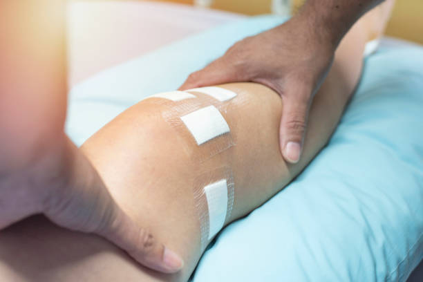
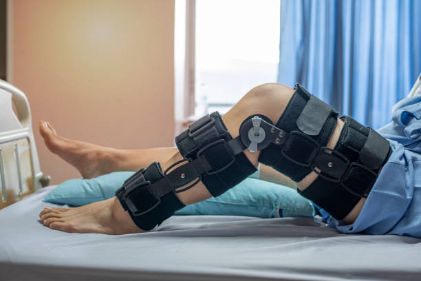
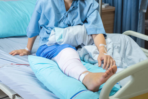
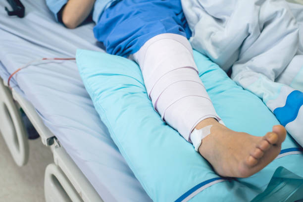
Human Leg with Patches and Orthopedic Brace After Anterior Cruciate Ligament Surgery: in Bed at Hospital.

Knee joint anatomy. Meniscus and ligaments of human knee. Joint structure. Head of tibia seen from above. Front aspect of knee. Poster. Vector illustration

Human Leg with Muscles Electrostimulation Device and Ice Bag during Rehabilitation Exercises on Bed After Knee Surgery.

Knee brace for ACL football knee injury.


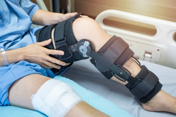
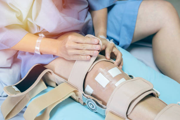
Young woman after an ACL surgery, sitting on the couch adjusting her knee orthosis.

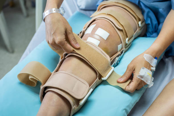
Young woman after an ACL surgery, putting on her knee orthosis.


Knee joint anatomy. Labeled of all bones. Isometric Flat vector illustration


Young woman standing in the kitchen looking out through the window and drinking her coffee.

Young man undergoing ACL physical therapy during rehabilitation after knee injury

Young woman sitting in the kitchen and drinking her tea.

Young woman sitting at the table in the kitchen drinking tea and checking her phone.

comprehensive knee examination of a patient in a hospital clinical setting, focused on orthopedic treatment and diagnosis


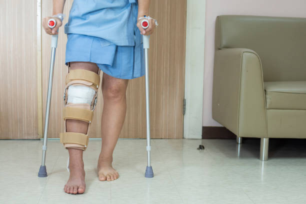
horizontal full length image of a caucasian young man trying to step out of the bathroom on his crutches with his knee heavily bandaged after knee surgery


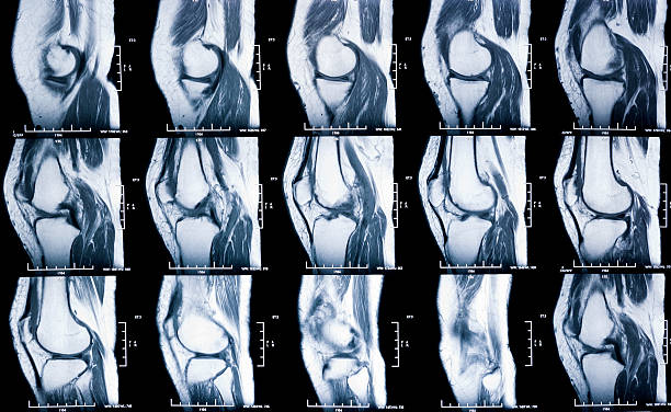
Knee X-ray and MRI befor and after arthroscopic surgery for Anterior cruciate ligament injury

Pensive young woman sitting on the couch looking at her leg, after an ACL surgery.


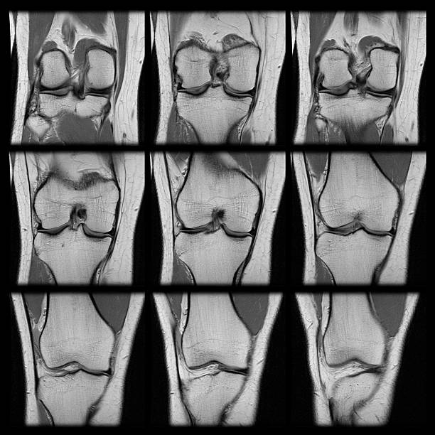
A series of nine MRI shots from one scan, showing a human female's left knee showing a torn ACL ligament.


Young woman in the kitchen waiting for her coffee to be ready.

Meniscus structure. Knee joint anatomy. Cartilage and Ligaments. Vector illustration


Anterior Cruciate Ligament Surgery on a dog

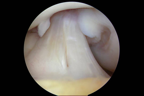
Arthroscopic view of the right human knee in about 45 degrees of flexion during an arthroscopic examination by an orthopaedic surgeon at a day-surgery center. The anterior cruciate ligament (ACL) consists of two major fibre bundles, namely the anteromedial (to the right) and posterolateral (to the left) bundles. When the knee is extended, the posterolateral bundle (PL) is tight and the anteromedial (AM) bundle is moderately lax. As the knee is flexed, the AM bundle tightens and the PL bundle relaxes. The AM bundle is the primary restraint against anterior tibial translation, whereas the PM bundle tends to stabilize the knee near full extension, and particularly against rotatory loads. Injury (tear or rupture) of the ACL is a common injury, especially in sports with rapid pivoting movements such as European team handball and soccer. A torn ACL may lead to giving-way episodes. To regain the ability to do sporting activities, an ACL reconstruction procedure is often warranted (using a graft secured close to the anatomic origins of the native ACL). The image was captured with a 4mm 30 degrees arthroscope. A copy of the same image with explaining text (labels) added onto the image, to aid a designer with limited medical knowledge, has been included in the same series.

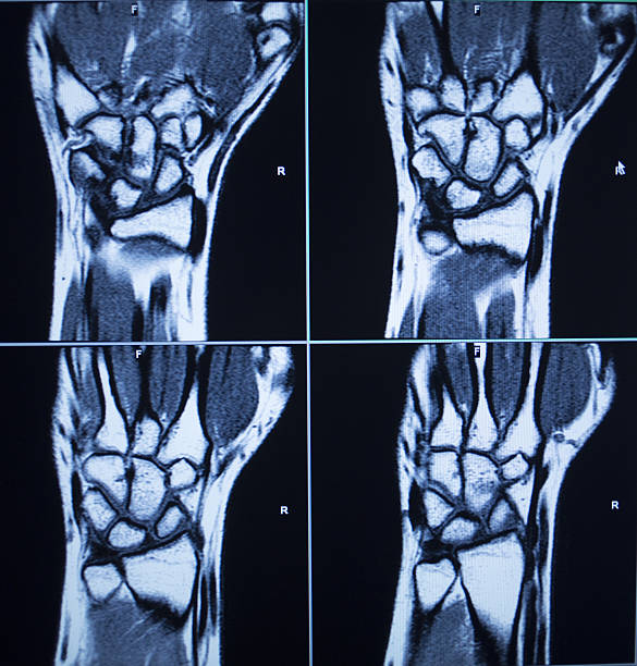
MRI magentic resonance imaging nuclear scanning scan test results wrists hands injury photo.


A vector illustration of the grades of ligament sprain

Surgeons perform knee arthroscopy with PCL reconstruction using arthroscope and tools in sterile orthopedic surgery setting

Young woman in the kitchen waiting for her coffee to be ready.
of7Next