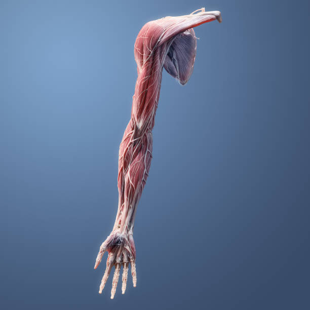
Full muscular, skeletal, nerve, vessel, ligament, tendon anatomy of the human upper extremity on blue background
Browse 30+ anterior forearm muscles stock photos and images available, or start a new search to explore more stock photos and images.
Full muscular, skeletal, nerve, vessel, ligament, tendon anatomy of the human upper extremity on blue background

Full 3D anatomy of the palmar side of the hand, wrist, and distal forearm including muscles, vessels, nerve, bone, ligaments, and tendons on blue background

Blue polygonal illustration of the elbow joint, lateral and medial view.

The upper limb is divided into three regions. These consist of the arm, located between the shoulder and elbow joints; the forearm, which is between the elbow and wrist joints; and the hand, which is located distal to the wrist. There are 30 bones in each upper limb.

This stock image highlights the serratus anterior muscle along the ribcage and spine, showcasing its structure and integration with the skeleton. Positioned between the ribs and scapula, the serratus anterior supports shoulder stability and movement. This stock image view emphasizes its attachment points on the ribs, spine, and upper limb, illustrating its key role in arm elevation, respiration, and core body strength.

"Some of the parts of the forearm shown include: superior ulnar collateral artery, ulnar nerve, inferior ulnar collateral artery,medial epicondyle, brachialis muscle, median nerve, flexor muscles, median artery, anterior interosseous artery,flexor carpi ulnaris muscle, tendons, flexor digitorum profundus muscle, radial nerve, radial collteral artery, deep radial nerve, common interrosseos artery, superficial radial nerve etc."

The upper limb is divided into three regions. These consist of the arm, located between the shoulder and elbow joints; the forearm, which is between the elbow and wrist joints; and the hand, which is located distal to the wrist. There are 30 bones in each upper limb.

Blue polygonal illustration of the elbow joint, lateral and medial view.

The pronator teres muscle is a long, round muscle that is located on the anterior aspect of the forearm.

he flexor carpi radialis muscle is a long, superficial muscle of the forearm that belongs to the anterior muscle group and lies in the first layer.

The muscles in the anterior compartment of the forearm are organised into three layers .

The muscles in the anterior compartment of the forearm are organised into three layers .

The muscles in the anterior compartment of the forearm are organised into three layers .

The muscles in the anterior compartment of the forearm are organised into three layers .

The pronator teres muscle is a long, round muscle that is located on the anterior aspect of the forearm.

he flexor carpi radialis muscle is a long, superficial muscle of the forearm that belongs to the anterior muscle group and lies in the first layer.

The muscles in the anterior compartment of the forearm are organised into three layers .

Serratus anterior Muscle Anatomy For Medical Concept 3D Illustration

Serratus anterior Muscle Anatomy For Medical Concept 3D Illustration

Serratus anterior Muscle Anatomy For Medical Concept 3D Illustration

The muscles in the anterior compartment of the forearm are organised into three layers .

3D Illustration of Human Skeleton System Upper Limbs Anatomy Anterior View

Serratus anterior Muscle Anatomy For Medical Concept 3D Illustration

The muscles in the anterior compartment of the forearm are organised into three layers .

Atlas of descriptive anatomy of the human body C. Bonamy - Paul Broca Victor Masson et Fils Paris 1866

The muscles in the anterior compartment of the forearm are organised into three layers .

The muscles of the upper limb can be divided into 6 different regions: pectoral, shoulder, upper arm, anterior forearm, posterior forearm, and the hand

The muscles in the anterior compartment of the forearm are organised into three layers .

Serratus anterior Muscle Anatomy For Medical Concept 3D Illustration

Illustration of a Arteria interossea anterior

Illustration of a Arteria interossea anterior

Illustration of a Arteria interossea anterior

The muscles of the upper limb can be divided into 6 different regions: pectoral, shoulder, upper arm, anterior forearm, posterior forearm, and the hand

"Some of the parts of the forearm shown include: superior ulnar collateral artery, ulnar nerve, inferior,ulnar collateral artery, medial epicondyle, brachialis muscle, median nerve, flexor muscles, median artery,nterior interosseous artery,flexor carpi ulnaris muscle, tendons, flexor digitorum profundus muscle, radial nerve, radial collteral artery,deep radial nerve, common interrosseos artery, superficial radial nerve etc."


