Bacteria cell wall illustration. Gram positive and gram negative cell wall differents. Isolated on white
Browse 510+ bacterial cellulose stock photos and images available, or start a new search to explore more stock photos and images.
Bacteria cell wall illustration. Gram positive and gram negative cell wall differents. Isolated on white

3d rendering of Gram-negative bacteria, have a thin peptidoglycan cell wall, which is surrounded by an outer membrane containing lipopolysaccharide

Gram negative bacteria do not retain the crystal violet stain. Gram positive bacteria give a positive result in the Gram stain test. comparison and difference type of bacterial cell wall. vector poster

Internal anatomy of the prokaryotic cell. Different types of bacteria. Comparison with the eukaryotic cell.

Types of bacterial cell wall. Gram-negative bacteria and Gram-negative bacteria. comparison, structure, and composition. Vector illustration

A lysosome is a membrane-bound organelle found in most animal cells and some plant cells. It is known as the "digestive bag" or "recycling center" of the cell due to its primary function of breaking down various biomolecules and cellular debris. Lysosomes are spherical vesicles surrounded by a single membrane. This membrane helps to maintain an acidic pH inside the lysosome, which is essential for the activity of its hydrolytic enzymes. Lysosomes are formed by the fusion of vesicles containing enzymes synthesized in the endoplasmic reticulum (ER) and modified in the Golgi apparatus. These enzymes include acid hydrolases, which are capable of breaking down, Proteins into amino acids, Nucleic acids into nucleotides, Carbohydrates into simple sugars, Lipids into fatty acids and glycerol, etc. Lysosomes digest materials taken up by the cell through endocytosis (engulfing of particles or molecules by the cell membrane) or phagocytosis (engulfing of large particles such as bacteria or cellular debris). Lysosomes are crucial for autophagy, a process where they degrade damaged organelles and macromolecules to recycle their components. This helps in maintaining cellular health and homeostasis. Lysosomes are involved in apoptosis (programmed cell death) by releasing enzymes that break down cellular components, leading to the orderly dismantling of the cell. Lysosomes in immune cells such as macrophages play a role in destroying engulfed pathogens by digesting them.

3D Isometric Flat Vector Conceptual Illustration of Archaebacteria, Anatomical Bacteria Structure

Bacterial multidrug efflux pumps (E-CmeB) expel antibiotics from inside the bacterium (bottom): ciprofloxacin (yellow), chloramphenicol (violet), ampicillin (red).

Internal anatomy of the prokaryotic cell.

Prokaryotic Cell Structure Chart, vector medical illustration, online education material. English translation text

Winogradsky column device method for soil science research outline diagram. Culturing microorganisms, bacteria and algae method vector illustration. Educational perpetual layer structure description.
Icon representing an isolated microscopic cell. Ideal for promotional and institutional materials
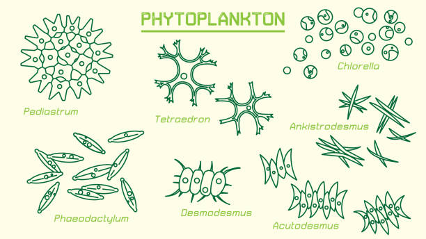
Phytoplankton (cyanobacteria and microalgae) can convert light energy and mineral nutrients into organic matter. They are responsible for the photosynthetic fixation of around 50×10^15 g C annually

Borrelia bacteria, Lyme disease, tick-borne illness. 3D medical illustration of Borrelia spirochetes. Borrelia bacteria cause Lyme disease, a bacterial infection. Microbiology, pathology, medicine
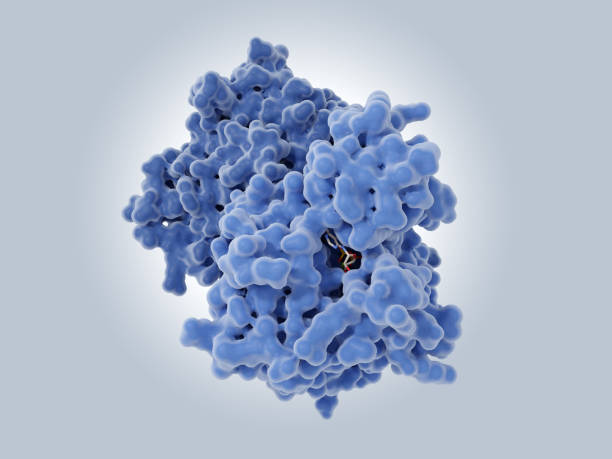
VanA synthesizes cell-wall precursors in bacteria, that are resistant to the antibiotic vancomycin. The cofactor ADP is visible in the active site. Source: PDB entry 1e4e.
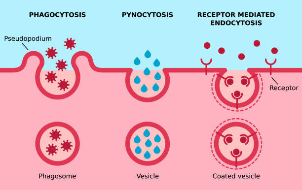
Three major types of endocytosis. Cell eating, cell drinking, receptors coated pit on cell membrane. Vector illustration.

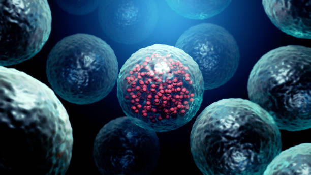
A detailed 3D illustration of stem cells positioned alongside a DNA helix structure. This conceptual image highlights the intersection of genetic material and cellular biology, emphasizing cutting-edge research in biotechnology and molecular science. Ideal for themes related to genetic engineering, medical diagnostics, and innovation in life sciences.

Structure of Mycoplasma cell. the bacterium is the causative agent of sexually transmitted diseases, pneumoniae, atypical pneumonia and other respiratory disorders. unaffected by many antibiotics. Cell parasitic or saprotrophic.
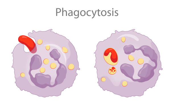
Phagocytes contain membranous sacs called lysosomes that contain various digestive enzymes, microbicidal chemicals, and toxic oxygen radicals. The lysosomes fuse with the phagosomes containing the ingested microbes and the microbes are destroyed

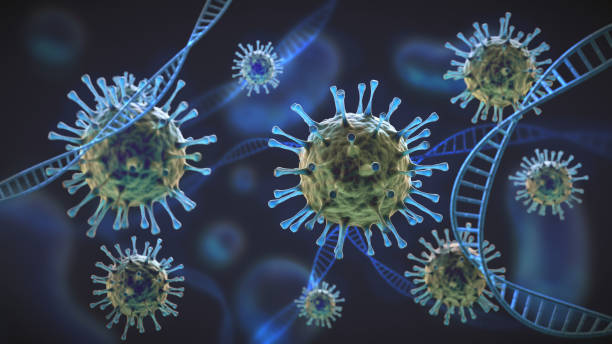
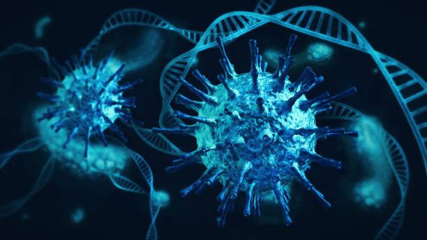
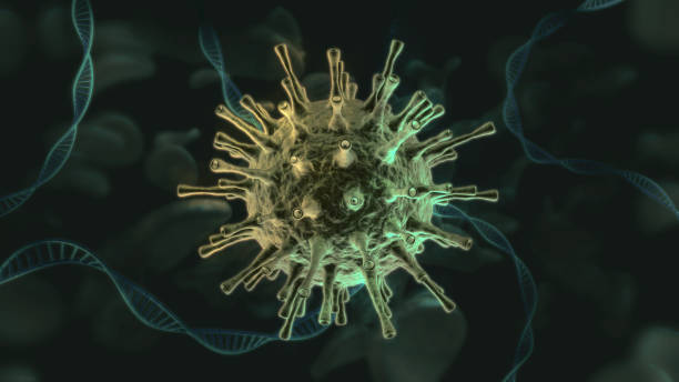
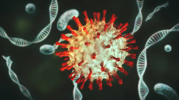
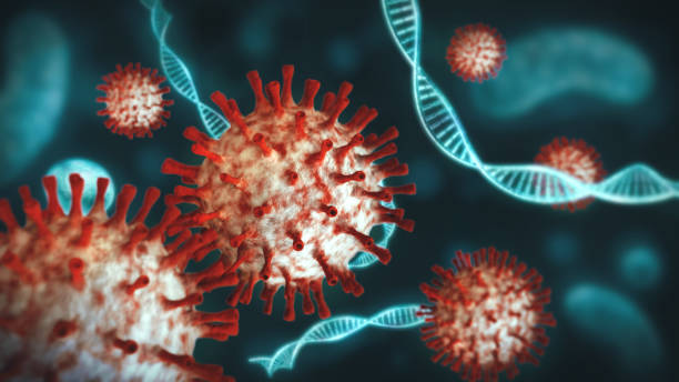
Bacteriophage Virus Cycle Phage replicating inside a pathogen as a virus with nucleic acid infecting bacteria as a virology symbol as a pathogen that attacks bacterial infections as a bacteriophages.
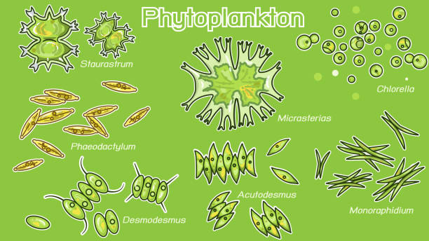
Phytoplankton are photosynthesizing microscopic protists and bacteria that inhabit the upper sunlit layer of marine and fresh water bodies of water on Earth.

Bacteriophages infecting bacterial cells
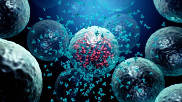
Human egg cell icon. Nucleus and membrane tissue under microscope. Medical design illustration.

Illustration of Escherichia coli bacteria with pink cell shapes and flagella, shown in a simplified cartoon style on a white background. Concept of microbiology. Vector illustration