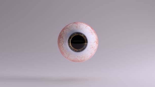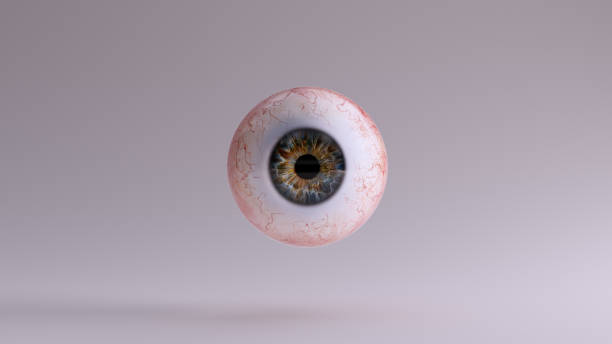
Human Eyeball 3d illustration 3d render
Browse 450+ ciliary-process stock photos and images available, or start a new search to explore more stock photos and images.
Human Eyeball 3d illustration 3d render

close-up of a lasik eye surgery.

Diabetic retinopathy. Retinal damage. Cross section of human eye. Diabetes. Close-up of a macula, optic disc, choroid, retina, sclera, and fovea. Medical condition. Microaneurysm of the small blood vessels. Vector poster
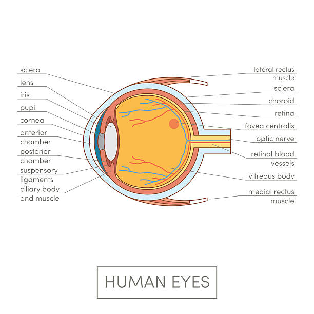
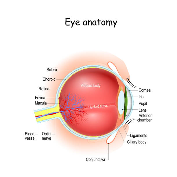
Human eye anatomy. Cartoon simple vector illustration for medical atlas or educational textbook. Cross-section of an eyes.

Eye anatomy. Cross section of a Human eyeball. Diagram for education and medical use. Vector illustration
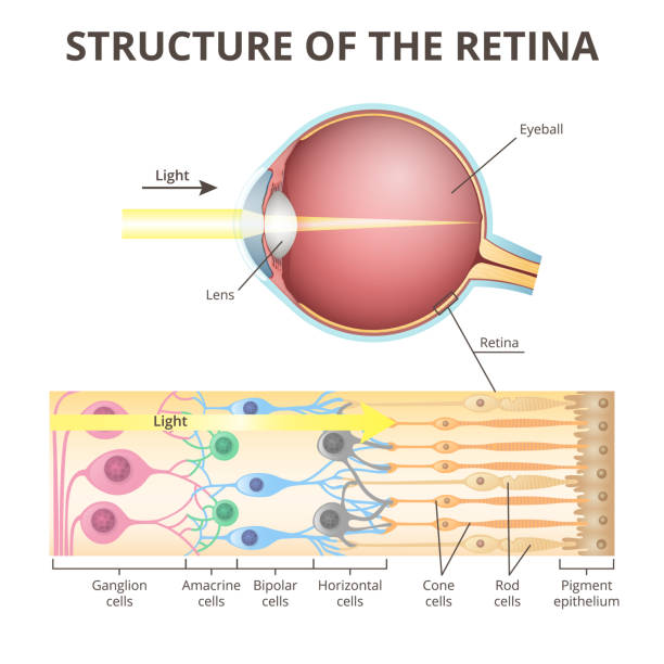
anatomy of the eyeball, functions and structure of the retina, projection of the image in the eye, the mechanism of visual perception

Macular degeneration. Age-related macular degeneration. Cross section of human eye. Close-up of a macula, optic disc, choroid, retina, sclera, and fovea. Medical condition. Image of the back of the eye showing Wet AMD and Dry macular degeneration. Vector poster
eye. Schematic diagram of the eye. human anatomy. labeled
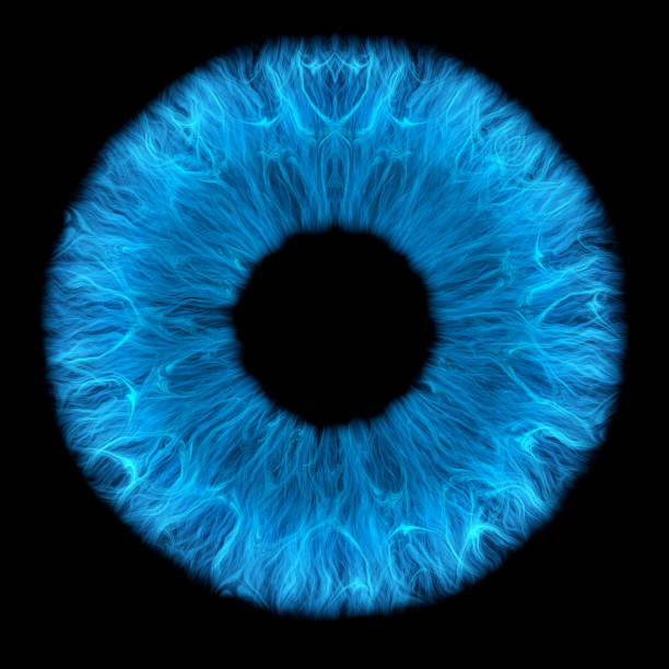
Isolated of blue human iris (3D Rendering)

Isolated of green human iris (3D Rendering)
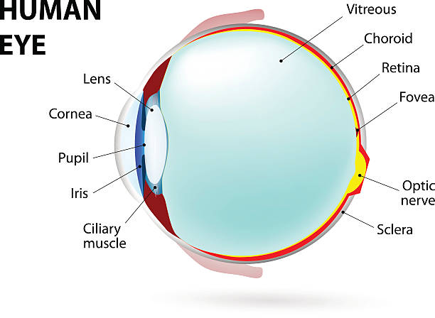
Eye anatomy and Physiology. How the Human Eye Works. Cross section of eyeball, eyelids, and Optic nerve. Details about visual system with retina, sclera, macula, fovea, and etc. vector Illustration.

anatomy of the eyeball, projection of the image in the eye

Human Eyeball with 75 Percent Dilated Pupil 3d illustration 3d render

anatomy of the human eyeball in section, detailed medical image on a white background

Eye anatomy diagram,vector illustration.

Pupillary light reflex PLR or photopupillary reflex. Schematic drawing of the pupillary light reflex pathway.

Anatomy of the Human Eye - one of the sense organs in human body

optical system of the eye and visual impairment, visual defect, myopia, eye structure

Black Human Eyeball with Red Iris 3d illustration 3d render

Diagram of human eyeball anatomy illustration

The human eye is an organ which reacts to light and pressure. As a sense organ, the mammalian eye allows vision. Human eyes help provide a three dimensional, moving image, normally coloured in daylight. Rod and cone cells in the retina allow conscious light perception and vision including color differentiation and the perception of depth.

Illustration of the eye - Translation: Schlemm's canal, corner angle, aqueous humor, pupil, anterior chamber, cornea, iris, posterior chamber, ciliary body, ciliary body, lens, vitreous body, external eye muscle, retina, choroid, sclera, central fossa (macula), optic nerve, optic nerve papilla

Eye accommodation infographic. Retina and sclera, ciliary muscle are contracted and relaxed, rounded and flattened lens. Objects at different distances and maintain clear images vector illustration

Motor nerves of the eye anatomy poster. Abducens, trochlear and oculomotor nerves in the human brain. Ciliary gland and muscle, coordinate eye position. Sensory and motor functions vector illustration

From First Book on Analytic, Physiology and Hygiene 1872

Felt pen Eyelash extension tool Element in the style of line art beauty theme on a white background. Doodle and scribble lash lifting rollers. Design Eyelashes Elements for medicine Lashes procedure

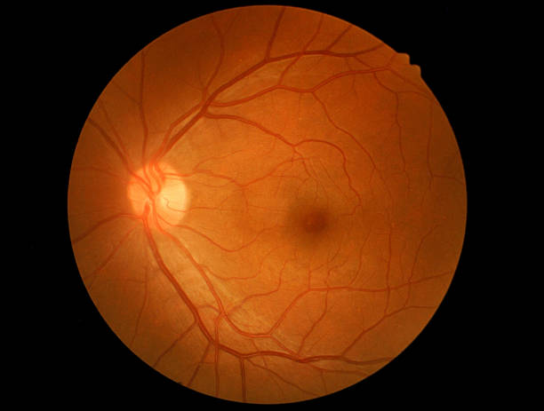
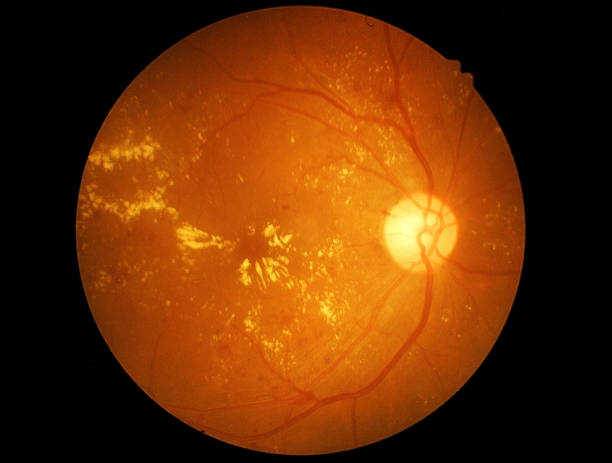
Human eye anatomy taking images with Mydriatic Retinal cameras.

The posterior chamber, trabecular meshwork, and eye drainage. Crucial role in removing intraocular fluid. Intraocular pressure. Eye health concept, glaucoma treatment research vector illustration

Bacterial interaction with respiratory mucus. Mucociliary transport. Respiratory pathogens release factors. Medical vector illustration
Extraction and processing of oil. Pixel art. Old school computer graphic style. Games elements.

Felt pen Curlers lifting tool Element in the style of line art beauty theme on a white background. Doodle and scribble blue applicator. Design Eyelash for medicine Lashes procedure