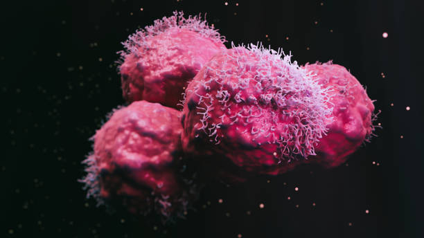
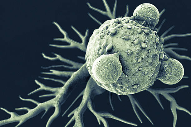
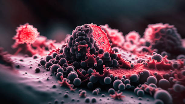
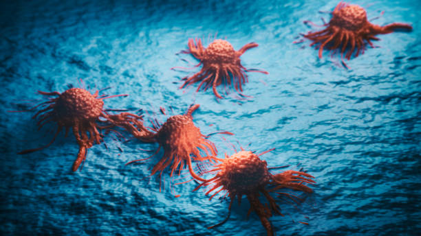
Cancer malignant cells - 3d rendered image, abstract enhanced scanning electron micrograph (SEM) of cancer malignant cells. Visual of overall shape of the cell's surface at a very high magnification. Medical research concept.
Browse 2,300+ electron microscope photography stock photos and images available, or start a new search to explore more stock photos and images.
Cancer malignant cells - 3d rendered image, abstract enhanced scanning electron micrograph (SEM) of cancer malignant cells. Visual of overall shape of the cell's surface at a very high magnification. Medical research concept.
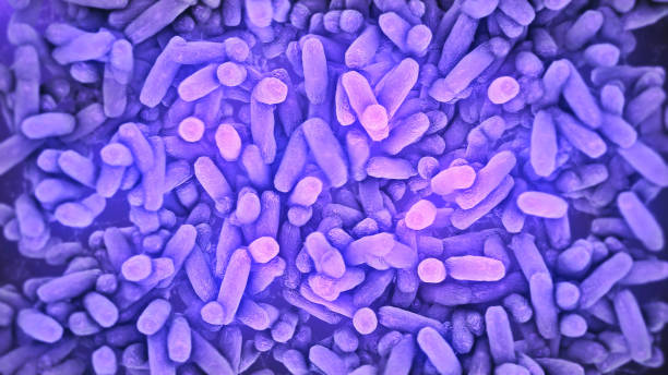
Bacteria Lactobacillus in human intestine,Beneficial healthy intestinal bacterium microflora,Probiotic bacterium

3D macro illustration of mold colonies (Mucor / Aspergillus-like fungi) growing in damp, humid conditions. Fungal growth on a neutral background; contamination, decay and microbial hazard. For scientific articles, medical infographics, health and hygiene warnings, microbiology research and environmental risk visuals
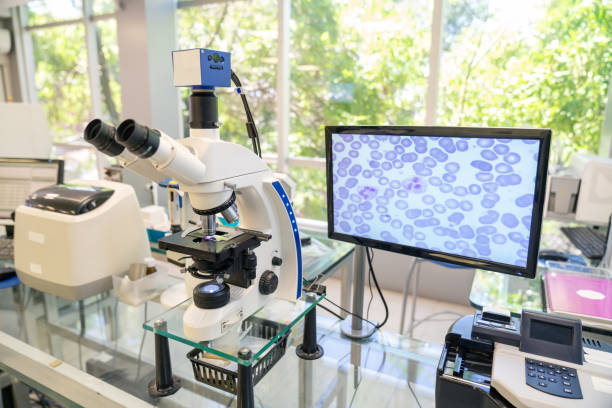
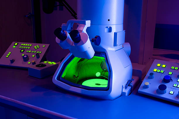
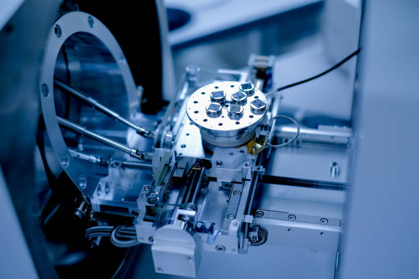
Modern microscope equipped with a digital camera, computer and monitor in a biological research laboratory. Modern microscope with digital imaging system

Cancer malignant cells - 3d rendered image, abstract enhanced scanning electron micrograph (SEM) of cancer malignant cells. Visual of overall shape of the cell's surface at a very high magnification. Medical research concept.
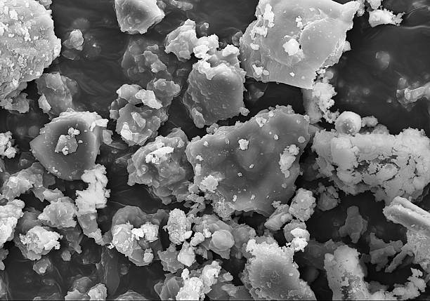
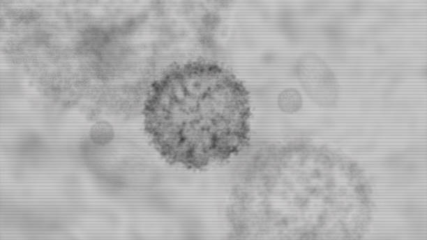
Technically, this image is not a photograph, since it was not originated by light ("photo") but by an electron beam: the image was captured by an Hitachi ultra-high-resolution Analytical FE Scanning Electron Microscope SU-70. it is a huge magnification of the common dust of industrial outdoor environments. Badly for us, this dust is rich in polluting and corrosive elements, such as chlorine and sulphur.

Cancer malignant cells - 3d rendered image, abstract enhanced scanning electron micrograph (SEM) of cancer malignant cells. Visual of overall shape of the cell's surface at a very high magnification. Medical research concept.
T lymphocytes attached to a cancer cell artwork.

Accurate Illustration of the novel COVID-19 "2019-nCoV" on white background. All details of the virus are in place, including E- and M-Proteins, virus Envelope, Hemagglutinin-esterase and Spike Glycoproteins.

3d illustration closeup of corona virus with DNA strands and blood cell antibodies on black background concept for immune response

Cancer cells vis - 3d rendered image, enhanced scanning electron micrograph (SEM) of cancer cell. Visual of overall shape of the cell's surface at a very high magnification. Medical research concept.
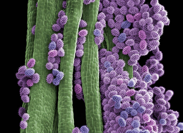
Pollen grains on Cosmos bipinnatus anther. Cosmos bipinnatus is a flowering herbaceous plant in the Asteraceae family, known commonly as garden cosmos or Mexican aster. This herb is a common plant of open fields, roadsides. The variably colored flowers also make it a popular ornamental garden plant. The anthers are the male reproductive parts that produce the pollen. Coloured scanning electron micrograph (SEM), magnified x150 when printed at 10cm wide.
The hydroid, Ectopleura larynx, protecting its sexual buds. The hydroid is a marine animal found attached to mussel shells, rocks and seaweed. Coloured scanning electron micrograph (SEM) magnified x32 when printed at 10cm wide.
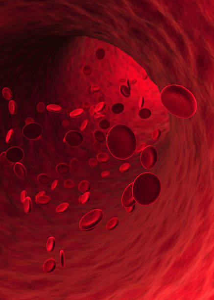
Red Blood Cells flowing in blood vessel. Shallow depth of field.
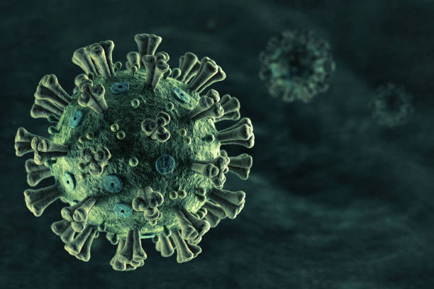
Accurate Illustration of the spreading novel COVID-19 "2019-nCoV". Green Background with copy space to the right. All details of the virus are in place, including E- and M-Proteins, virus Envelope, Hemagglutinin-esterase and Spike Glycoproteins.

Young woman working in printing factory. Printing Press

Cancer cell view - 3d rendered image, enhanced scanning electron micrograph (SEM) of cancer cell. Visual of overall shape of the cell's surface at a very high magnification. Medical research concept.

3d illustration closeup of corona virus with DNA strands on black background concept for strain mutation

Young woman working in printing factory. Printing Press

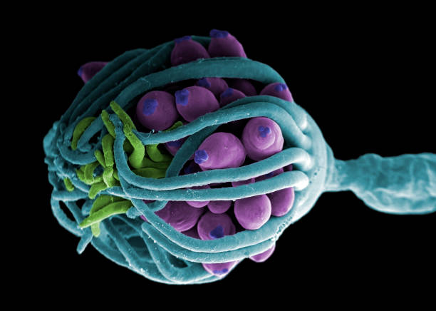
Molecules of cell nucleus in the interphase. Nucleolus (the granular structure), DNA being transcripted to mRNA (pink) by the RNA polymerase (green), and several enzymes: the endonuclease (violet), the DNA ligase (yellow) and the DNA topoisomerase (magenta)
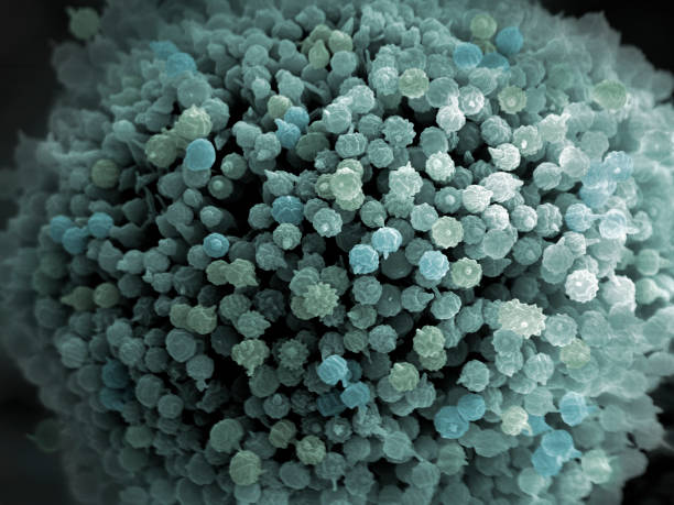
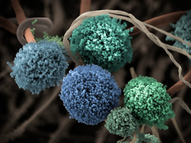
Aspergillus niger spores (reproductive cells). The fungus Aspergillus niger is a widely distributed saprophyte which grows on household dust, soil, and decaying vegetable matter, including stale food. A. niger is one of the most common causes of fungal ear infections and may result in the serious lung disease aspergillosis in people with compromised immune function or other lung diseases. A. niger is used for waste management and biotransformation in addition to other industrial uses, such as production of citric acid and extracellular enzymes. Coloured scanning electron micrograph (SEM), magnified x1500 when printed at 10cm wide.
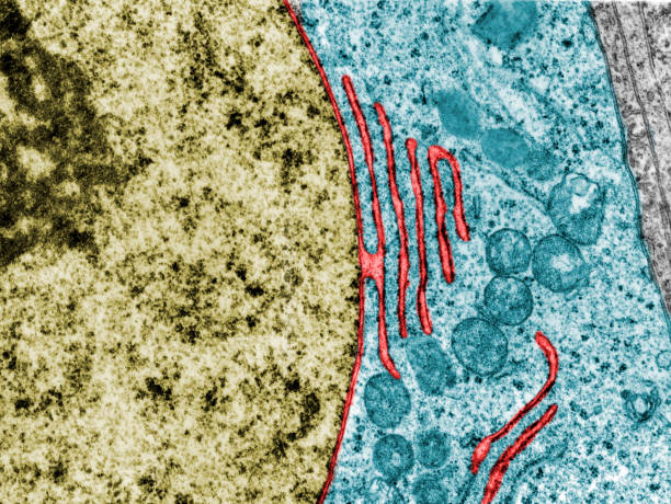
False colour transmission electron microscope micrograph showing a continuity between the nuclear envelope and a cistern of the rough endoplasmic reticulum (red). Nucleus (gold). Cytoplasm (blue)

Cancer cells vis - 3d rendered image, enhanced scanning electron micrograph (SEM) of cancer cell. Visual of overall shape of the cell's surface at a very high magnification. Medical research concept.
Cancer cells vis - 3d rendered image, enhanced scanning electron micrograph (SEM) of cancer cell. Visual of overall shape of the cell's surface at a very high magnification. Medical research concept.

Corona virus, lassa virus isolated needle medication mask object

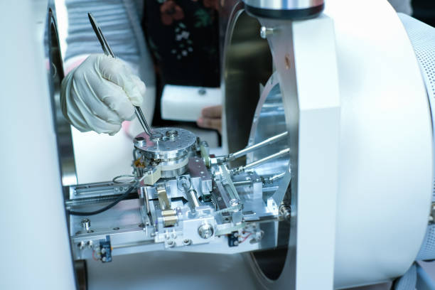
A technician looking into an Electron Microscope in a medical laboratory.

Illustration of Influenza Virus H1N1. Swine Flu.
Advance head up display of electron microscope scanning airborne virus outbreak showing the anatomy of the virus in close up details

Cancer cell view - 3d rendered image, enhanced scanning electron micrograph (SEM) of cancer cell. Visual of overall shape of the cell's surface at a very high magnification. Medical research concept.

Bacteria Lactobacillus in human intestine,Beneficial healthy intestinal bacterium microflora,Probiotic bacterium

Scientific molecular structures on dark blue background.
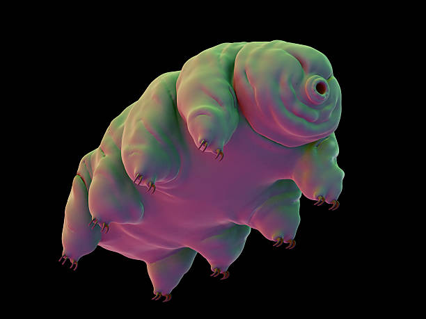
medically accurate illustration of a water bear
Cancer cells vis - 3d rendered image, enhanced scanning electron micrograph (SEM) of cancer cell. Visual of overall shape of the cell's surface at a very high magnification. Medical research concept.

Coloured transmission electron micrograph (TEM) showing the nucleus of a protein-secreting (serous) cell. The nuclear envelope is connected to rough endoplasmic reticulum cisterns (red). The large nucleolus with dense fibrillar (green) and granular (yellow) components, surrounded by nucleolus associated chromatin (dark blue) can be seen. Four secretory granules (light blue) can be seen in the cytoplasm.
Fruiting bodies (conidiophores) and hyphae of the fungus Aspergillus niger. A. niger is a widely distributed saprophyte which grows on household dust, soil, and decaying vegetable matter, including stale food. A. niger is one of the most common causes of fungal ear infections and may result in the serious lung disease aspergillosis in people with compromised immune function or other lung diseases. A. niger is used for waste management and biotransformation in addition to other industrial uses, such as production of citric acid and extracellular enzymes. Coloured scanning electron micrograph (SEM), magnified x415 when printed at 10cm wide.

Hologram Cancer cells view - 3d rendered image, enhanced scanning electron micrograph (SEM) of cancer cell. Visual of overall shape of the cell's surface at a very high magnification. Medical research concept.
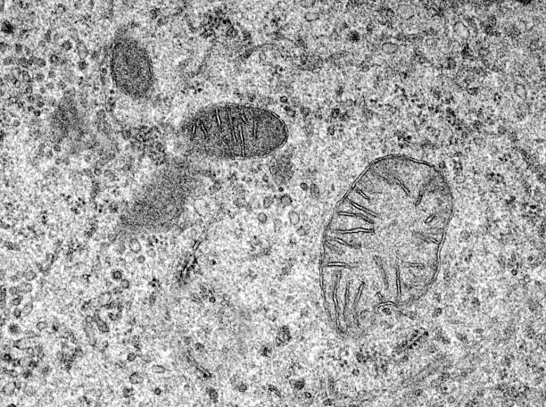
Transmition Electron microscopy of an epithelial cell where mitochondria of several sizes can be seen with the characteristic double membrane and internal cristaes. Mitochondria are believed to be of bacteria origin. It has its own genome and RNA and protein which are similar to those of bacteria.

Two young woman working in printing factory. Printing Press

Close-up Of Doctor Moving Ultrasound Probe On Pregnant Woman's Stomach In Hospital.

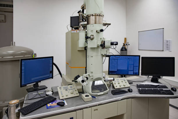
Optical electron microscope. Laboratory instrument, concept of science and microscopic research.

Cancer cells vis - 3d rendered image, enhanced scanning electron micrograph (SEM) of cancer cell. Visual of overall shape of the cell's surface at a very high magnification. Medical research concept.