Anatomy of foot and ankle with labeled medical location outline diagram. Labeled educational scheme with skeletal bones and muscles in latin names vector illustration. Biology and medicine study.
Browse 1,600+ foot anatomy diagram stock photos and images available, or start a new search to explore more stock photos and images.
Anatomy of foot and ankle with labeled medical location outline diagram. Labeled educational scheme with skeletal bones and muscles in latin names vector illustration. Biology and medicine study.
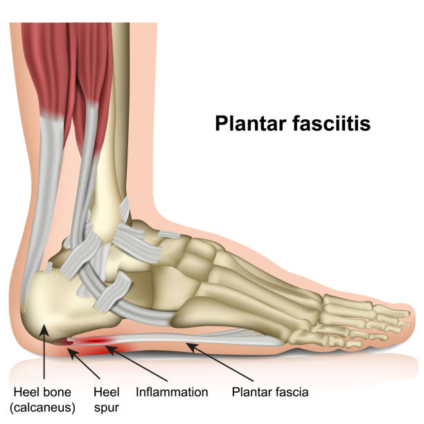
plantar fasciitis 3d medical vector illustration on white background eps 10

Diabetic foot medical diagram. Infographics with symptoms of diabetes and insulin resistance disease. Treatment and prevention. Cartoon flat vector illustration isolated on white background
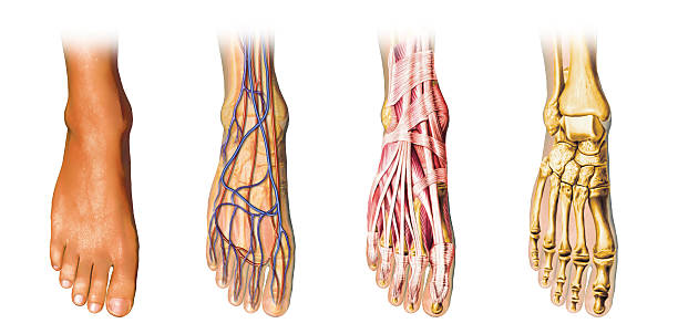
"Human foot anatomy cutaway representation, showing skin, veins and arterias, muscles, bones. With clipping path included."
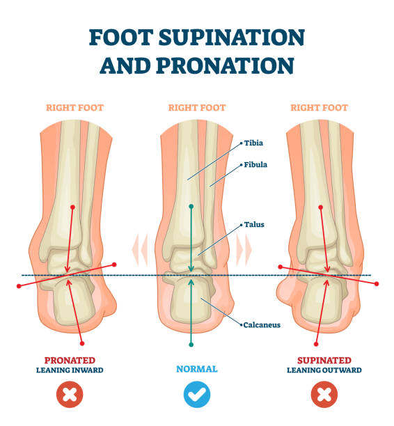
Foot supination and pronation vector illustration. Labeled medical scheme with incorrect leg joint movement. Educational diagram with pronated, normal and supinated compared examples with bone titles.

Achilles tendinopathy as injury to tendon in heel outline diagram. Labeled educational scheme with anatomical leg and foot skeleton and muscles vector illustration.Trauma and band of tissue problem.

Foot anatomy. Human foot with the name and description of all bones and sites. top view and side view. Arches of the feet. skeleton anatomy. Vector illustration

Lever systems in human body for neck, leg and arm movement outline diagram. Labeled educational scheme with biomechanics examples as effort, fulcrum or load force motion principle vector illustration.

Medical vector illustration of Hallux valgus types. Healthy foot, initial stage and pronounced deformation.
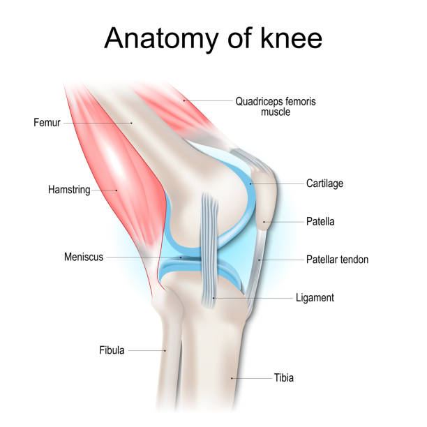
Knee anatomy. Structure of knee joint. Major parts of human leg. Bones, muscles, cartilage, tendon and Ligaments of knee. Side view. Vector poster
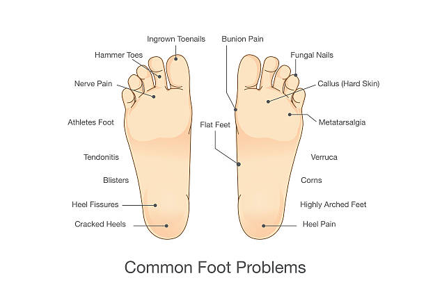
Common foot problems. Illustration about disease and abnormality of human foot.

Ankle fracture and right foot anterior trauma medical view outline diagram. Labeled educational scheme with anatomical comparison for healthy and fractured bones vector illustration. Leg tibia injury

Plate 133: Anatomy of the Vascular System Engraving Antique Illustration, Published 1851. Source: Original edition from my own archives. Copyright has expired on this artwork. Digitally restored.
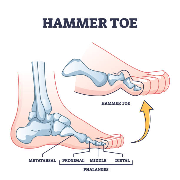
Hammer toe medical problem anatomy with foot phalanges deformation outline diagram. Labeled educational scheme with bent metatarsal, proximal, middle and distal bones deformity vector illustration.
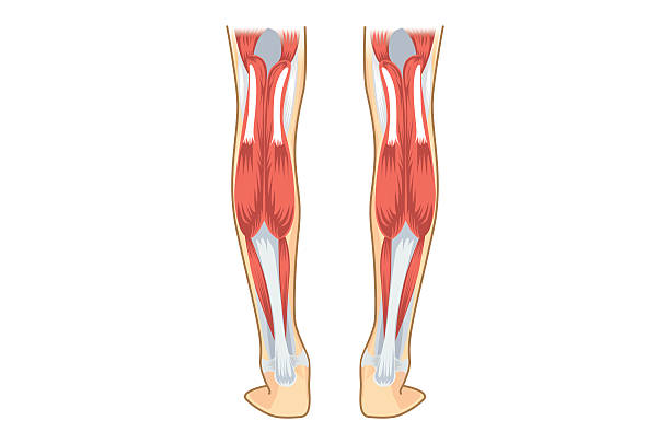
Calf Muscle of human. Illustration about human leg Anatomy.
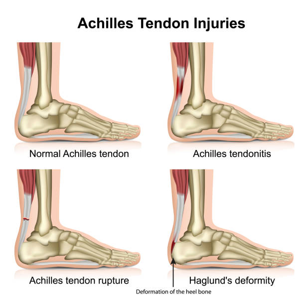
achilles tendon injures medical vector illustration isolated on white background with english description eps 10 infographic

Plantar fasciitis. Heel pain. Cross section of human leg. Structural breakdown of the plantar fascia. Microtrauma of thick connective tissue aponeurosis that supports the arch of the foot. Schematic illustration. Medical diagram. Detailed Vector poster.

Human Joints Pain concept as skeleton and muscle anatomy of the body with a group of sore joints as a painful injury or arthritis illness symbol for health care and medical symptoms as a symbol of Rheumatology and orthopedics and physical rehabilitation.

Engraved illustrations of The Anatomy of the Vascular System from Iconographic Encyclopedia of Science, Literature and Art, Published in 1851. Copyright has expired on this artwork. Digitally restored.

Foot problems are common in people with diabetes. They can happen over time when high blood sugar damages the nerves and blood vessels in the feet. The nerve damage, called diabetic neuropathy, can cause numbness, tingling, pain, or a loss of feeling in your feet. Diabetic blood vessel damage in the feet is a common complication associated with diabetes, particularly in individuals who have had the condition for a long time and/or have poorly controlled blood sugar levels. This condition is referred to as diabetic peripheral neuropathy (DPN) and diabetic peripheral vascular disease (PVD). It affects the blood vessels and nerves in the feet and lower extremities, leading to a range of problems.

Plate 122: Anatomy of the Bones Engraving Antique Illustration, Published 1851. Source: Original edition from my own archives. Copyright has expired on this artwork. Digitally restored.

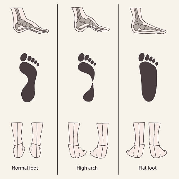
Foot deformation. Types pathologies of foot. Hollow, flat and normal foot. Medical infographic.

Plantar fasciitis vector illustration. Labeled human feet sport disorder diagram. Educational medical scheme with orthopedic leg disease. Painful plantar fascia inflammation and irritation infographic
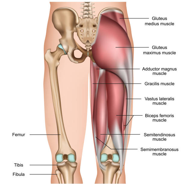
leg back muscles 3d medical vector illustration on white background eps 10
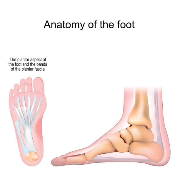
Anatomy of the foot. Bones and Plantar fascia. Realistic Vector illustration. bottom view and side view

Human foot bones anatomy with descriptions. Colored leg base parts structure. Educational diagram with human internal organ illustration. Talus, calcaneus, tarsals, cuboid, metatarsals foot parts.

Antique illustration (1898): Human foot

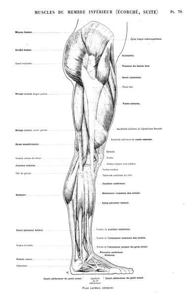
Human body muscles of the leg shown from the front and back anatomy or medical anatomical diagram illustration.

Peripheral neuropathy poster. Hands and feet highlighted areas of pain, numbness, and tingling. Diabetes or injury. Damaged nerves in the nervous system, myelin sheath deformity vector illustration

Human anatomy scientific illustrations with latin/italian labels: foot nerves

A vector illustration of the human skeleton made to look like a blueprint.

Human foot bones anatomy sketch design of vector orthopedics medicine. Skeleton leg ankle joints and toe phalanges, cuboid, metatarsal, navicular and cuneiform bones, hand drawn dorsal view of foot

Foot bones anatomy. Human skeleton. Horizontal medical poster. Medical education. Editable vector illustration isolated on light background. Graphic design for any purposes

Medical illustration showing different types of skin damage caused by friction, including callus, corn, and friction blister, with detailed cross sections of the affected areas

Plantar fascial fibromatosis. Plantar fibroma. Ledderhoses disease. Morbus Ledderhose. Skeleton of foot. Medial aspect of bones and plantar fascia with non-malignant thickening of connective tissue. Detailed Vector poster
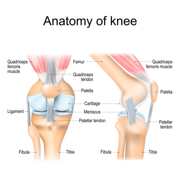
Knee anatomy. Structure of leg joint. Major parts. Vector poster with text label for medical education

BLACK AND WHITE ENGRAVING OF IVERTICAL SECTION OF THE FOOT AND LOWER LEG OF A HORSE. CIRCA 1882

Vector isolated illustration of human arterial and venous circulatory system in leg anatomy. Blood vessels diagram. Medical infographics for poster, educational, science and medical use.
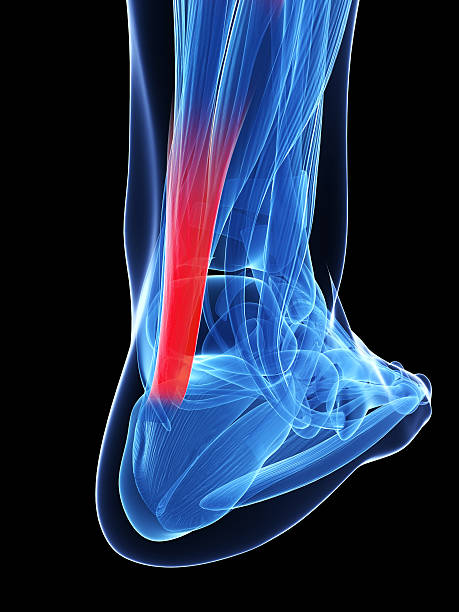
3d rendered illustration of the achilles tendon

Plate 124: Anatomy of the Ligaments and Muscles Engraving Antique Illustration, Published 1851. Source: Original edition from my own archives. Copyright has expired on this artwork. Digitally restored.

Medical infographic with orthopedic anatomy charts. Human silhouette in motion with marked spine, pelvis, knee, foot, shoulder, elbow, hand bones and joints. Orthopedics medical group design
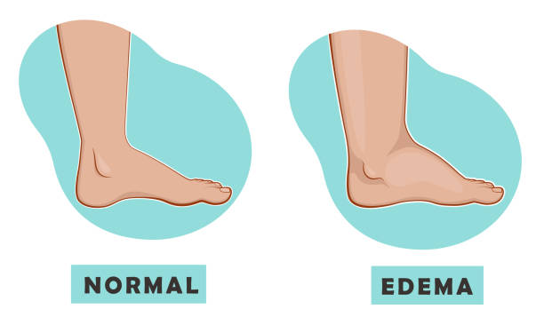
A swollen foot and ankle and a normal foot. Vector illustration of the disease
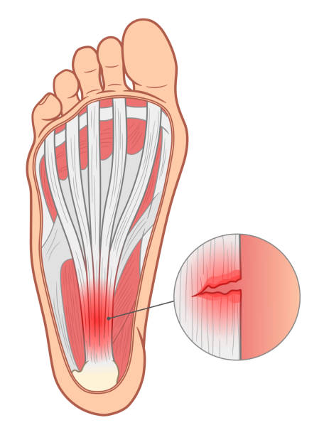
Plantar fasciitis medical vector illustration.
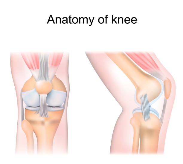
knee anatomy. side and front view. Cross section of the joint showing the main parts: femur, fibula, articular capsule, menisci, muscles and ligaments. vector illustration
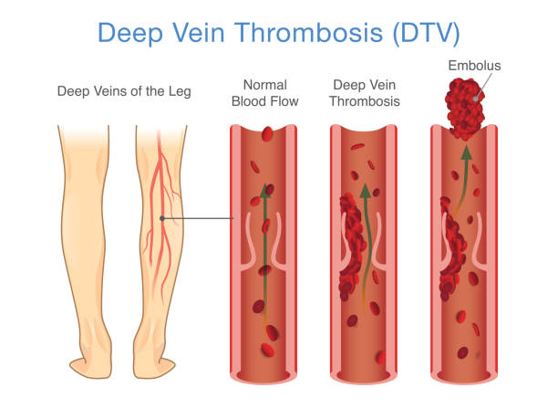
Medical Diagram of Deep Vein Thrombosis at leg area. Illustration about abnormally of blood floow.

Human ankle icon for clinic. Normal foot joints and bones anatomy in leg silhouette. Skeleton x ray medical poster. Orthopedic or chiropractic treatment. Anatomical logo concept. vector illustration.

Skeletal system with body skeleton structure and anatomy outline diagram. Labeled educational medical physiology with skull, spine, ribs, hand and leg bones vector illustration. Biological human model

Joints Set icons like x-ray image. Disease and disorder in shoulder, knee, ankle, wrist and pelvis. Arthritis, Osteoporosis, Rheumatism, inflammation, fracture. Joints and Body parts with pain rings. infographics elements. Medical poster. Flat vector illustration. Xrays examination

Peripheral artery disease PAD vector illustration. Labeled medical structure scheme with healthy or narrowed plaque in vessels comparison. Anatomical damaged peripheral organ and blocked blood example
Leg vector in front view. Illustration about human legs composition.
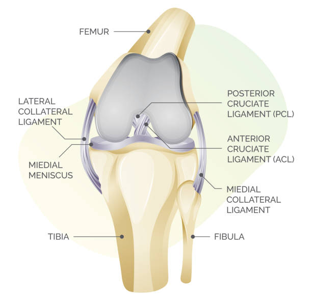
Knee Cartilage Anatomy - Illustration as EPS 10 File
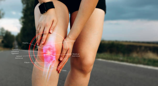
Human Knee joint anatomical diagram, medical scheme. Educational information template