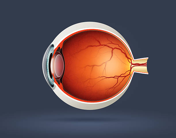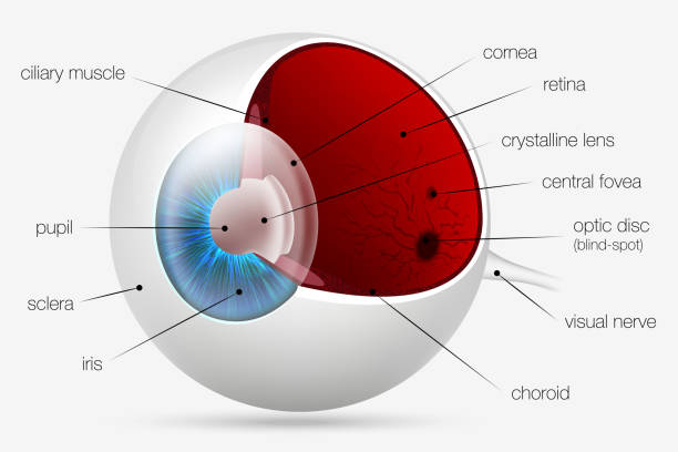
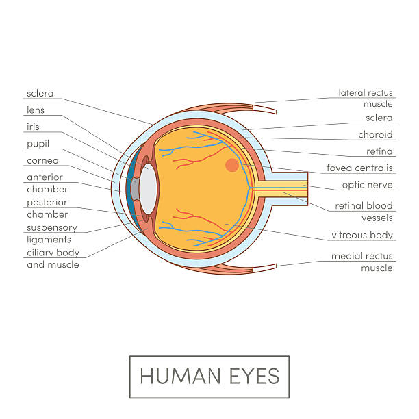
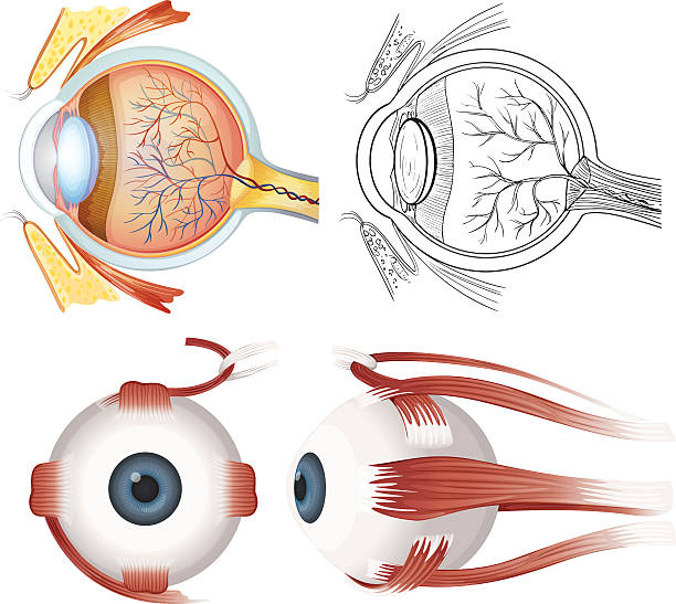
Components of human eye. Illustration about Anatomy and Physiology.
Components of human eye. Illustration about Anatomy and Physiology.
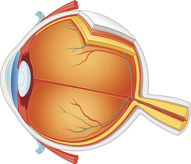
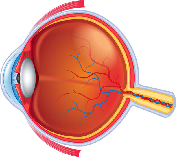
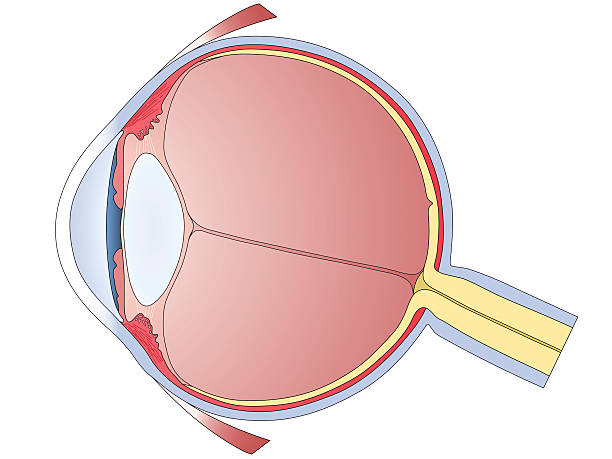
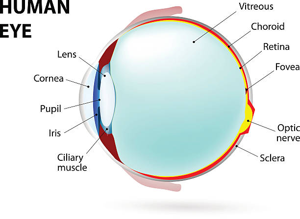
Structure of human eye. In side view.
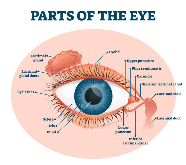
Parts of the eye, labeled vector illustration diagram. Educational beauty and nursing information. Eyelid, eyelashes, pupil, lacrimal gland and other anatomical parts. Healthy visual sensory organ.

Eye anatomy. Rod cells and cone cells. The arrangement of retinal cells is shown in a cross section. Vector diagram for your design, educational, biological, science and medical use
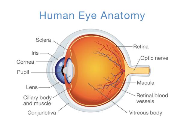
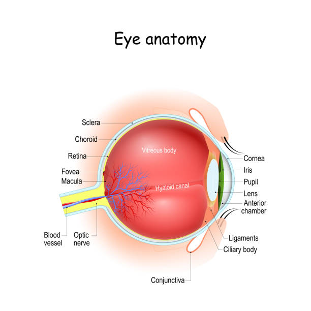
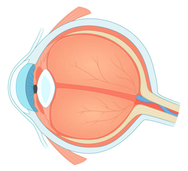
Human eye anatomy. Cartoon simple vector illustration for medical atlas or educational textbook. Cross-section of an eyes.

Digital artificial intelligence blue human eye formed by fiber optics. Futuristic technology concept 3d Illustration render.
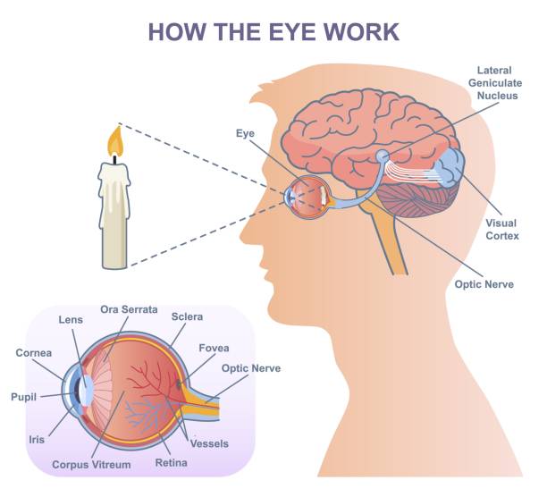
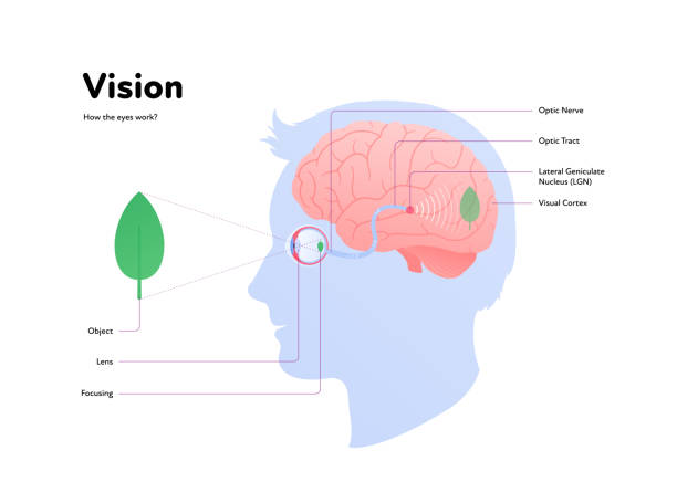
Medical diagram of eye work. Anatomical infographics with structure of visual system, connection with brain and function of eyes. Cartoon flat vector illustration isolated on white background

Human eyeball icon. Human eye structure. Vector illustration.
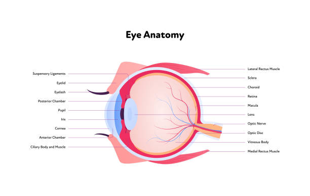
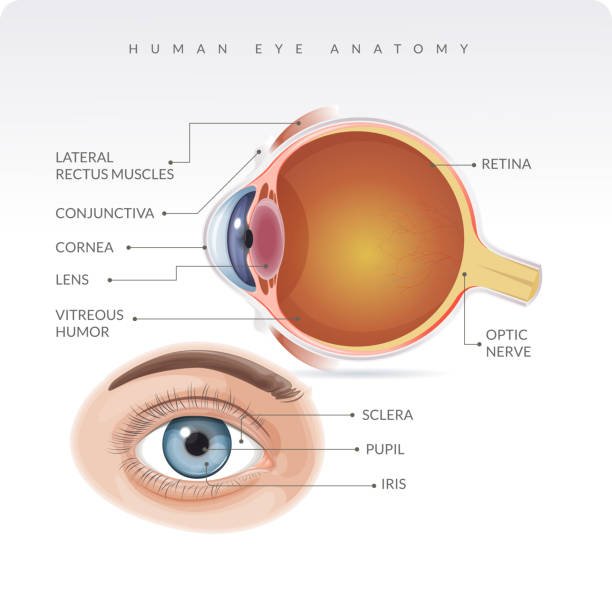
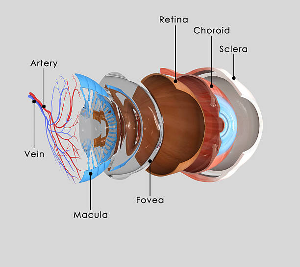
Eye anatomy and Physiology. How the Human Eye Works. Cross section of eyeball, eyelids, and Optic nerve. Details about visual system with retina, sclera, macula, fovea, and etc. vector Illustration.
Eye anatomy isolated on white photo-realistic vector illustration

How the eye works medical scheme poster, elegant and minimal vector illustration, eye - brain labeled structure diagram. Stylized and artistic medical design poster.Health care educational infographic

A beautiful illustration of human eye anatomy vector design
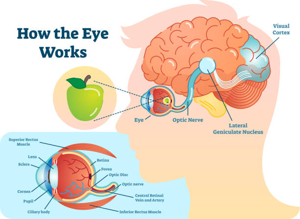
How eye work medical illustration, eye - brain diagram, eye structure and connection with brains. Vector EPS10
A human eye diagram in sagittal section. Each anatomical component is on a separate layer and accurately named.
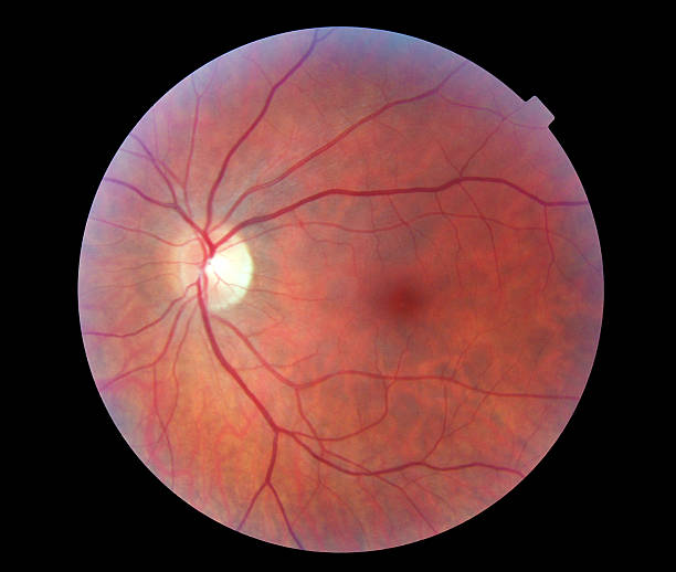
A digital image of a human retina, taken using professional ophthalmological equipmentRelated images:
Eye inner structure. Eye cross section. Ophthalmology medical vector illustration.
Anatomy of the human eye on a white background

Eye anatome concept. Structure of human body. Graphic elements for medical websites. Study, education. Physiological sight infographic banner with side and front view. Cartoon flat vector illustration
Human Eye Anatomy - Stock Illustration as EPS 10 File

Vector Illustration Of Human eye anatomy, right eye viewed from above

Eyeball infographic. Cartoon of eyeball vector infographic for web design
They may not be painful and you may not see changes in your vision until the disease has become very advanced. Vector graphic.

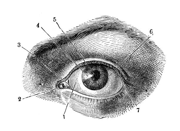
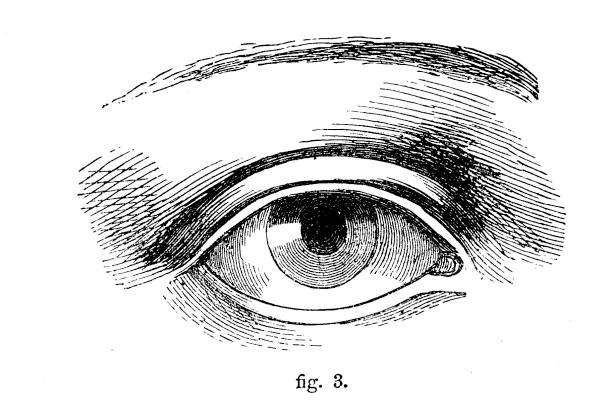
Engraving from "Meyers Konversations-Lexikon"; Author: Carl Joseph Meyer (1796-1856). The Meyers Konversations-Lexikon was a German encyclopedia. The first Edition appeared in October 1839. This image is in the public domain. Photographed and edited by J. C. Rosemann.

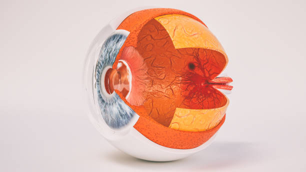
sectional structure of the human eyeball, detailed medical image, illustration of localization in the body
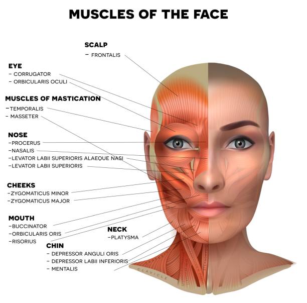
Facial and neck muscles of the female, half of the face muscles and half skin, each muscle with name on it, detailed bright anatomy isolated on a white background

Illustration of the human eye anatomy on the white background.

Structure of the eye, parts of the eye. Retina, macula, blind spot, optic nerve, cones, rods, vitreous humor, ciliary body, lens, pupil, aqueous humor, cornea, iris, sclera, choroid.
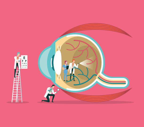
Team of doctors diagnose human eye

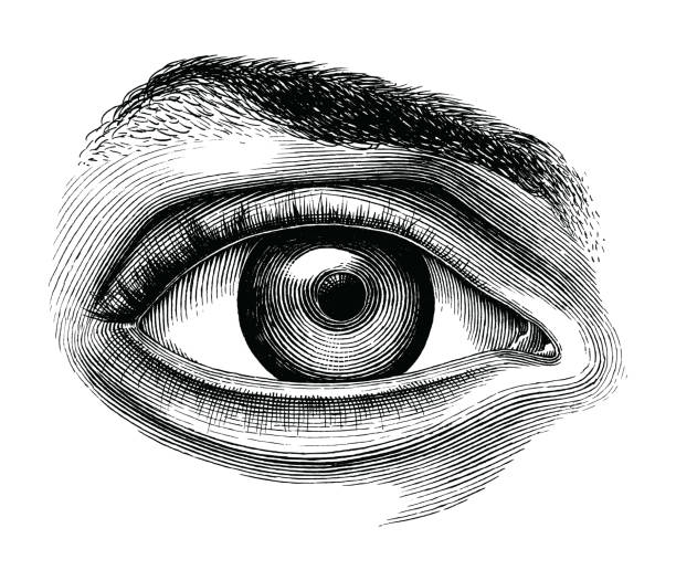
Anatomy of the Eye Engraving Antique Illustration, Published 1851. Source: Original edition from my own archives. Copyright has expired on this artwork. Digitally restored.
Taken from the Self-Aid Cyclopedia by Robert Scott Burn printed 1860. Chapter on Shading techniques. Front few pages missing from my own book hence image of the dedication on the hard cover dated 1877 and an image of the book for sale showing year of publishing.
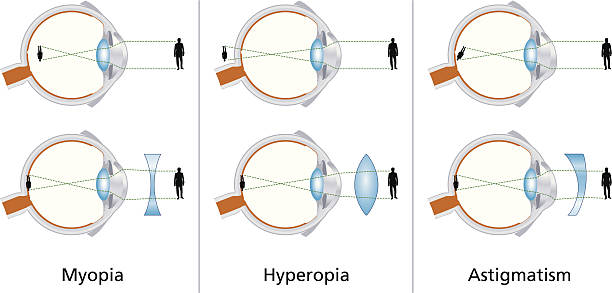
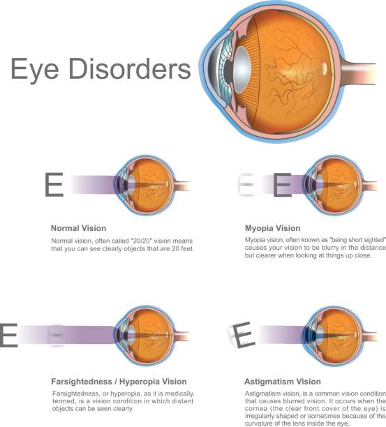
Illustration of the three visual defects Myopia, Hyperopia and Astigmatism and how to correct it with biconcave and biconvex lenses - with glasses or contact lenses.

The human eye is an organ that reacts to light and has several purposes. As a conscious sense organ, the mammalian eye allows vision.
The inner layer of the eye, or retina, is similar to film in a camera. It receives light from an image we are looking at, and converts that light into electrical impulses which are sent through the fibers of the optic nerve to the brain.

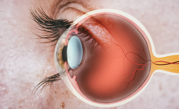
Realistic human eye hologram, cross sectional cut, isolate side view on black background. Healthcare concept, vision, catheract, ostegmatism, laser eye surgery. 3D illustration, 3D render.

Eye anatomy - inner structure isolated on white

Detailed illustration of the anatomy and structure of the human eye. The picture shows the iris, pupil, lens, retina, optic nerve, and other significant structures of the eye.
Eye icon isolated on white background

Eye, Human Body Part, Lens - Optical Instrument, Human Eye, Medicine

Anatomy of the human eye in front view. External View. Schematic diagram. detailed illustration

Structure of the human eye. Polygonal design of lines and dots. Blue background.

anatomy of the human eyeball in section, detailed medical image on a white background
eye. Schematic diagram of the eye. human anatomy. labeled

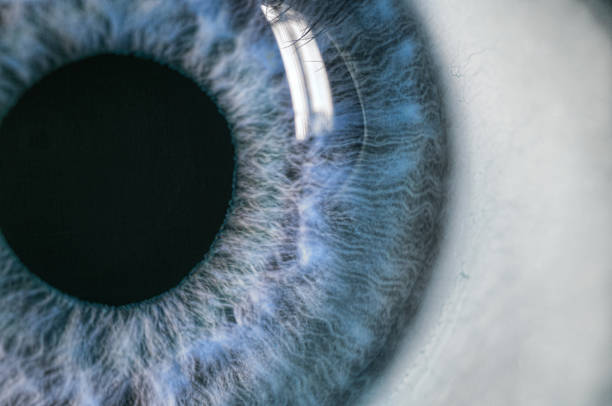
Description: Male Blue Colored Eye With Long Lashes Close Up. Structural Anatomy. Human Iris Macro Detail.