Human Knee joint anatomical diagram, medical scheme. Educational information template
Browse 100+ lateral knee anatomy pictures stock photos and images available, or start a new search to explore more stock photos and images.
Human Knee joint anatomical diagram, medical scheme. Educational information template

Doctor shows anatomy of structure of human knee joint

Human Knee joint anatomical diagram, medical scheme. Educational information template

Doctor shows anatomy of structure of human knee joint
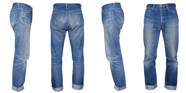
Blue jeans that I wore, I took a picture with a model wearing it and cut it out

Legs in gray sweatpants. Taken from front, back, left and right

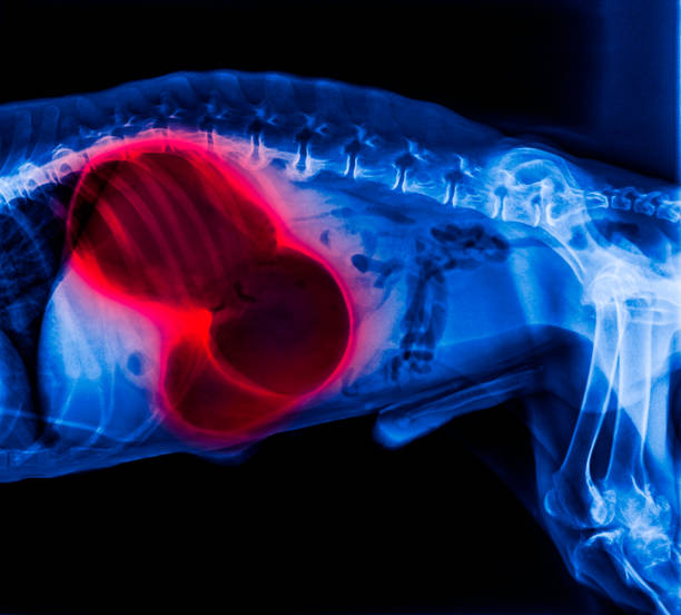
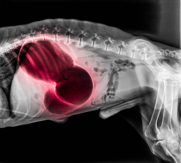
Comparison between normal human knee ( left image ) and osteoarthritis knee ( right image ) . Lateral view .

Comparison between normal human knee ( left image ) and osteoarthritis knee ( right image ) . Lateral view .

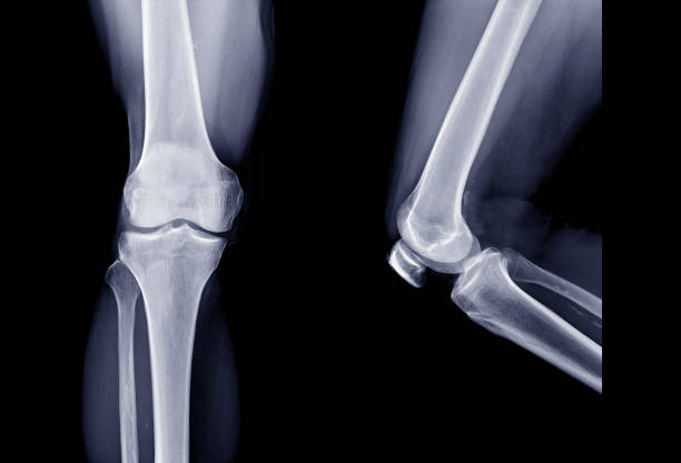
X-ray image of broken knee, lateral view, show broken knee.

X-ray image of broken knee, AP view, show broken knee

X-ray image of knee, lateral view, show fracture of patella

X-ray image of knee joint, AP and lateral view, show fracture of patella.

X-ray image of knee joint, AP view, show fracture of patella.

X-ray image of knee joint, AP and lateral view, show fracture of patella.

X- ray image of knee joint, AP view, show fracture of patella

X-ray image of knee,lateral view, show broken patella

Doctor shows anatomy of structure of human knee joint

Small joint effusion is seen, no fracture line or dislocation is noted, joint space is preserved.

Screws fixation at distal femur and proximal tibia, retained draining tube

Hands of a doctor are holding film x-ray knee joints. knee joint inflammation, Osteoarthritis knee. knee joints radiography concept.

This 3d illustration shows the vastus lateralis muscles on skeleton on a white background

plain x ray on knee joint showing joint space narrowing and Subchondral Sclerosis on medial compartment (thickening of bone that happens in joints affected by osteoarthritis), knee osteoarthritis, selective focus

Illustration of a View of a Lateral Region of knee

Medical model of the knee tendon and muscle anatomy. Skeleton leg concept

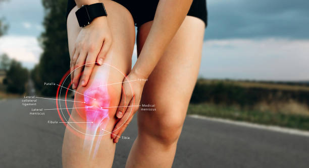
Explore the intricacies of pain and injury in the Gastrocnemius Muscles. This detailed visualization delves into the anatomy of the leg muscles, offering insights into common injuries, treatment options, and preventive measures. Ideal for medical education, fitness training, and rehabilitation resources.

Explore the intricacies of pain and injury in the Gastrocnemius Muscles. This detailed visualization delves into the anatomy of the leg muscles, offering insights into common injuries, treatment options, and preventive measures. Ideal for medical education, fitness training, and rehabilitation resources.

Plain X ray of the right knee shows apparent joint osteoarthritis according to Kellgren and Lawrence system for classification of osteoarthritis with definite osteophytes and join space narrowing

Plain X ray of the right knee shows apparent joint osteoarthritis according to Kellgren and Lawrence system for classification of osteoarthritis with definite osteophytes and join space narrowing

Plain X ray of the right knee shows apparent joint osteoarthritis according to Kellgren and Lawrence system for classification of osteoarthritis with definite osteophytes and join space narrowing