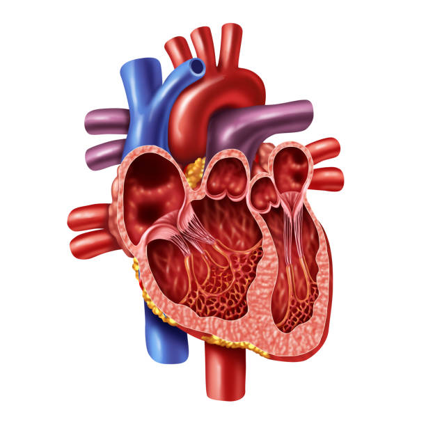
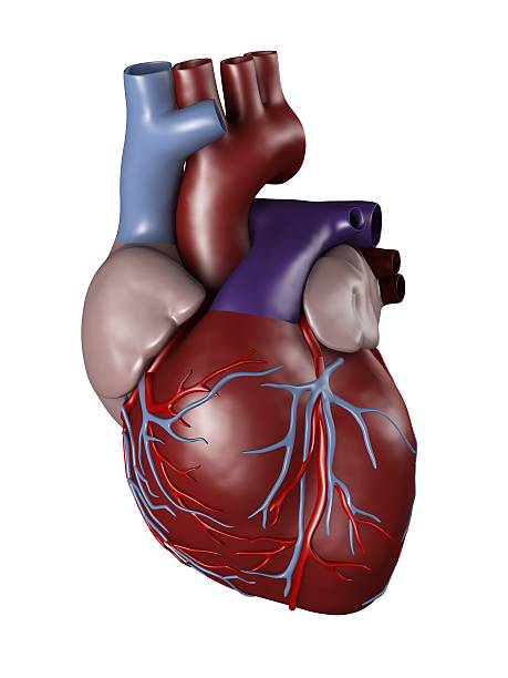
Human heart inner anatomy concept with valves from a healthy body isolated on white background as a medical health care symbol of an inner cardiovascular organ in a 3D illustration style.
Browse 2,300+ left atrium photos stock photos and images available, or start a new search to explore more stock photos and images.
Human heart inner anatomy concept with valves from a healthy body isolated on white background as a medical health care symbol of an inner cardiovascular organ in a 3D illustration style.

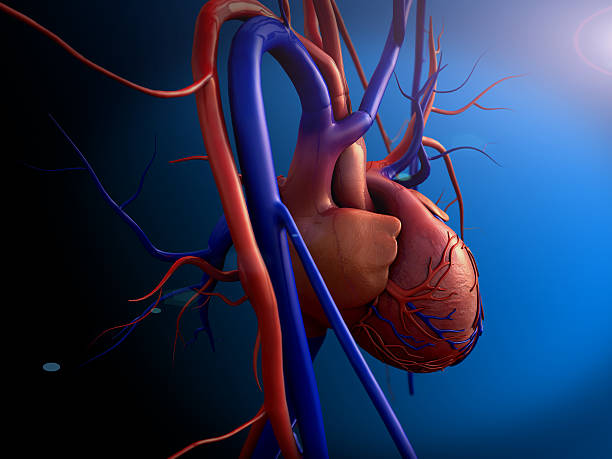
Human body with heart, with aorta, pulmonary trunk, veins, left ventricle, right ventricle, left atrium, right atrium, superior vena cava, inferior vena cava and artery, on black background. Great to be used in medicine works and health.
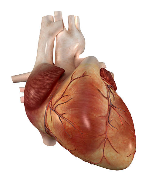
"Human heart, with aorta, pulmonary trunk, veins, left ventricle, right ventricle, left atrium, right atrium, superior vena cava, inferior vena cava and artery, on white background. Great to be used in medicine works and health."
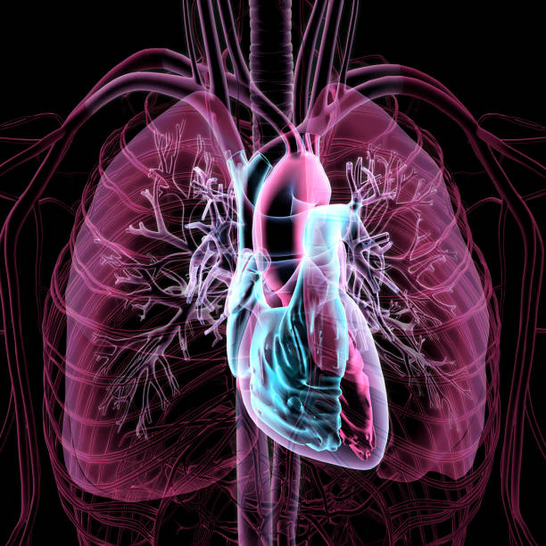
Stylized magenta and blue transparent CG image of human anatomy, showing the neck and chest area, heart, lungs, major arteries and veins isolated on black background.

"Torso of a man highlighting the heart, with aorta, pulmonary trunk, veins, left ventricle, right ventricle, left atrium, right atrium, superior vena cava, inferior vena cava and artery. Isolated on black background. Great to be used in medicine works and health."
CG image of human anatomy, showing the neck and chest area, heart, lungs, major arteries and veins isolated on black background.

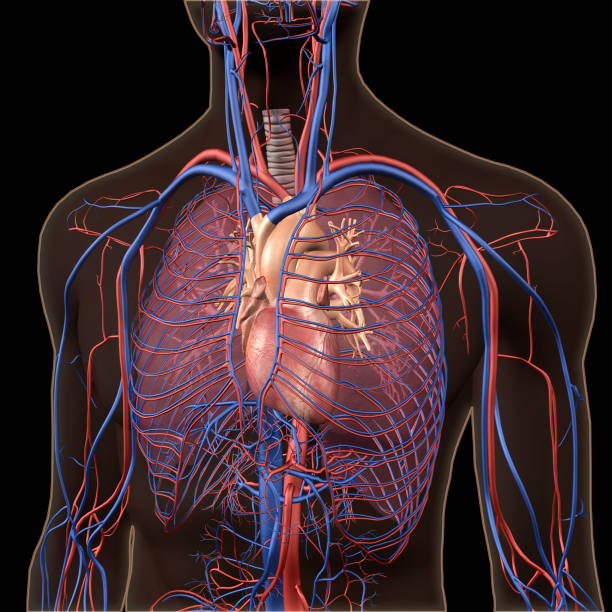
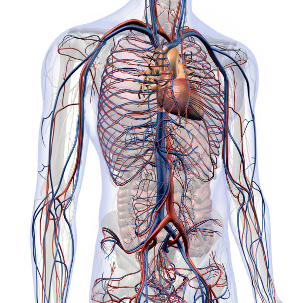
CG image of man's chest and abdominal areas showing the human circulatory system, heart, major arteries and veins isolated on black background.
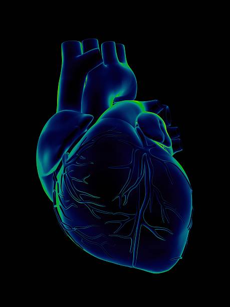
Human heart, with aorta, pulmonary trunk, veins, left ventricle, right ventricle, left atrium, right atrium, superior vena cava, inferior vena cava and artery, on black background. Great to be used in medicine works and health.
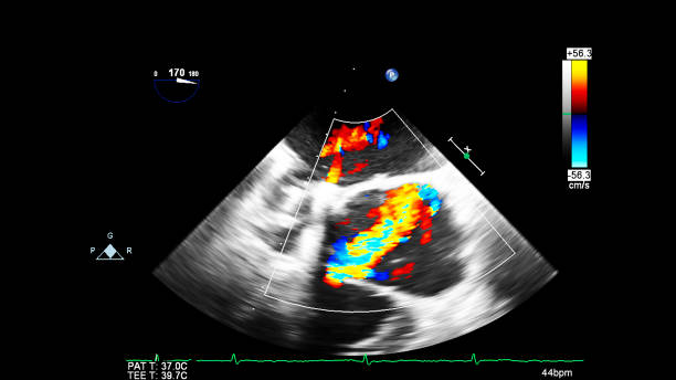
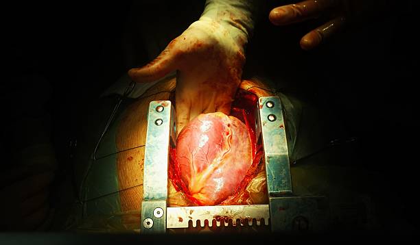
Image of the heart during transesophageal ultrasound with Doppler mode.
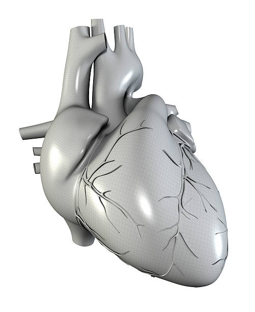
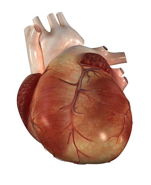
Heart model w/clipping path, Human heart model, Full clipping path included, Human heart for medical study, Human Heart Anatomy

Human heart anatomy close up diagram concept from a healthy body isolated on white background as a medical health care symbol of an inner cardiovascular organ.
"Human heart, with aorta, pulmonary trunk, veins, left ventricle, right ventricle, left atrium, right atrium, superior vena cava, inferior vena cava and artery, on white background. Great to be used in medicine works and health."

human heart, Heart model w/clipping path, Human heart model, Full clipping path included, Human heart for medical study, Human Heart Anatomy

Fetal echocardiography is a diagnostic method for congenital heart defects.
CG image of man's chest and abdominal areas showing the human circulatory system, heart, major arteries and veins isolated on white background.

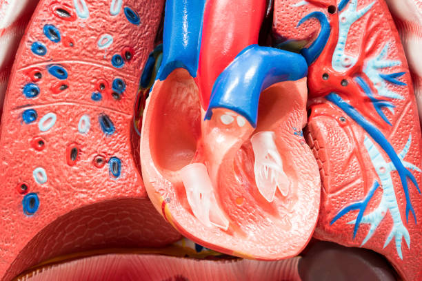
Cross section CG image of human heart, showing interior of the heart valves

Image of the heart in gray-scale mode during transesophageal ultrasound.

Image of the heart during transesophageal ultrasound with Doppler mode.

Image of the heart during transesophageal ultrasound with Doppler mode.
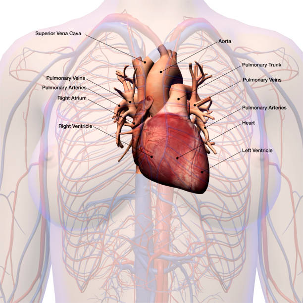
CG image of human heart isolated with arteries and veins within female chest area with anatomical labeling.
"Human heart, with aorta, pulmonary trunk, veins, left ventricle, right ventricle, left atrium, right atrium, superior vena cava, inferior vena cava and artery, on white background. Great to be used in medicine works and health."
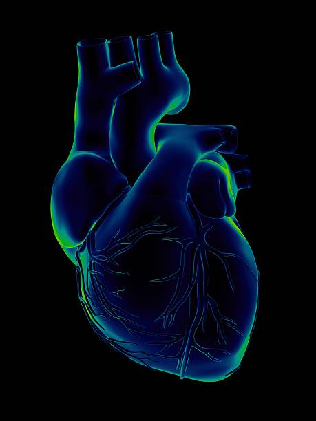
"Human heart, with aorta, pulmonary trunk, veins, left ventricle, right ventricle, left atrium, right atrium, superior vena cava, inferior vena cava and artery, on black background. Great to be used in medicine works and health."
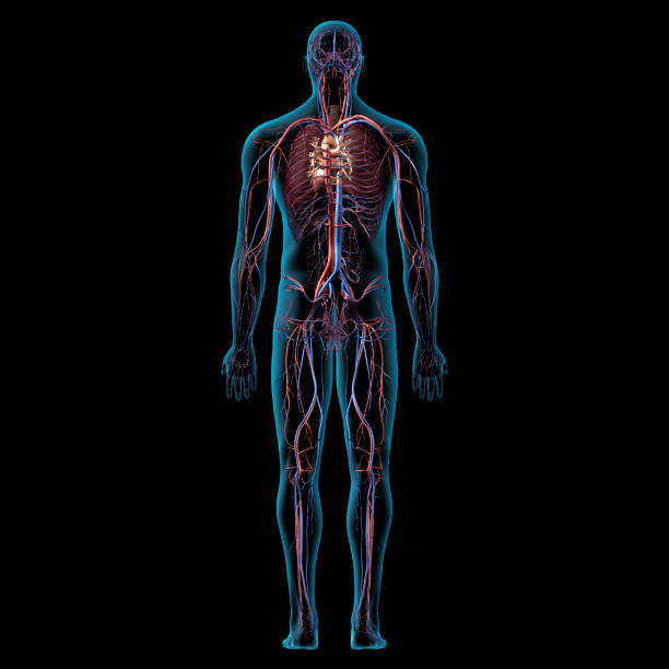
Human Cardiovascular System, Male Posterior View, No Genitalia, on Black Background, Computer generated image.

Fetal echocardiography is a diagnostic method for congenital heart defects.

Cross section CG image of human heart, showing interior of the heart valves

Cross section CG image of human heart, showing interior of the heart valves

Image of the heart in gray-scale mode during transesophageal ultrasound.

Image of the heart in gray-scale mode during transesophageal ultrasound.

Image of the heart during transesophageal ultrasound with Doppler mode.

Computer generated image of human heart, major arteries and veins with labeled anatomy, isolated on white background

CG image of human heart isolated, female chest area, with anatomical labeling.
"Human heart, with aorta, pulmonary trunk, veins, left ventricle, right ventricle, left atrium, right atrium, superior vena cava, inferior vena cava and artery, on white background. Great to be used in medicine works and health."

Image of the heart in gray-scale mode during transesophageal ultrasound.

Human body with heart, with aorta, pulmonary trunk, veins, left ventricle, right ventricle, left atrium, right atrium, superior vena cava, inferior vena cava and artery, on black background. Great to be used in medicine works and health.

Human body with heart, with aorta, pulmonary trunk, veins, left ventricle, right ventricle, left atrium, right atrium, superior vena cava, inferior vena cava and artery, on black background. Great to be used in medicine works and health.

Image of the heart during transesophageal ultrasound with Doppler mode.

Fetal echocardiography is a diagnostic method for congenital heart defects.

Heart model w/clipping path, Human heart model, Full clipping path included, Human heart for medical study, Human Heart Anatomy

Image of the heart during transesophageal ultrasound with Doppler mode.

CG image of man's chest and abdominal areas showing the human circulatory system, heart, major arteries and veins front view isolated on white background.

Fetal echocardiography is a diagnostic method for congenital heart defects.

Image of the heart in gray-scale mode during transesophageal ultrasound.
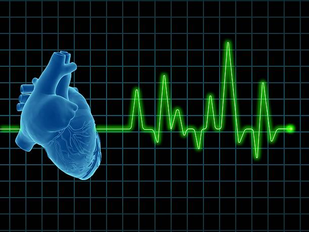
Image of a electrocardiogram (ECG / EKG), with human heart on screen. Great to be used in medicine works and health.

Cross section CG image of human heart, showing interior of the heart valves

Image of the heart in gray-scale mode during transesophageal ultrasound.

Image of the heart during transesophageal ultrasound with Doppler mode.

Fetal echocardiography is a diagnostic method for congenital heart defects.

Fetal echocardiography is a diagnostic method for congenital heart defects.

Image of the heart during transesophageal ultrasound with Doppler mode.

Image of the heart in gray-scale mode during transesophageal ultrasound.