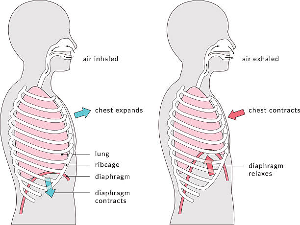
Breathing - a labelled diagram showing the mechanics of breathing. When inhaling, the diaphragm contracts and the lungs expand, pushing the chest upwards. When exhaling the diaphragm relaxes and the lungs contract, moving the chest back down.
Browse 60+ lungs in the rib cage side view stock photos and images available, or start a new search to explore more stock photos and images.
Breathing - a labelled diagram showing the mechanics of breathing. When inhaling, the diaphragm contracts and the lungs expand, pushing the chest upwards. When exhaling the diaphragm relaxes and the lungs contract, moving the chest back down.
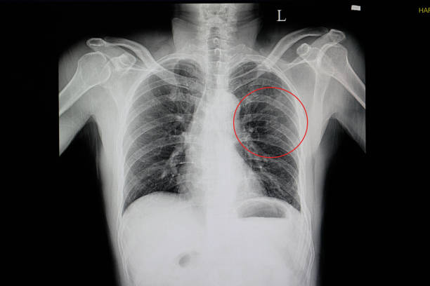
a chest x-ray of a blunt chest wall injuried patient showing fractured ribs 5, 6, 7, 8 on the left side(red cycle) with some pulmonary contusion
Set of human anatomy line vector icons. Editable stroke.
Set of human anatomy line vector icons. Editable stroke.

Normal human chest on computed tomography images

Female doctor examining lung x-ray image

A lateral projection x-rays of lungs and heart shadow, the chest shows a deformity called "pectus excavatum" (or sunken / funnel chest)

Chest X-Ray image showing human chest from the side, high dynamic range. Spine, ribs, heart and other organs are clearly visible.

Breathing. Exhalation and Inspiration. Side view of a human body with Diaphragm and Lungs cavity. vector illustration isolated on white background.

a chest x-ray film of a patient with cardiomegaly with left side heart failure, pulmonary edema, and pericadial effusion

blue rendition of lateral female chest X-ray

X-ray of a chest in a woman. This medical imaging technique is often used for screening for cancer and tuberculosis.

This is a 3d illustration of the side view of the human lungs

Normal anterior and lateral roentgenograms of the human chest

"Lateral Chest X Ray of a Female 34 years old showing ribs, spine, clavicles, heart, lungs and upper torso organs with Breast Silhouette."

Medical themes: Chest X- Ray that shows broken ribs

Medical X-ray of French Bulldog dog chest showing lung anatomy and veterinary diagnostic detail

side view of human thorax with lungs on X-ray image

The thorax or chest is a part of the anatomy of humans and various other animals located between the neck and the abdomen

Medical themes: Right chest CAT- SCAN of patient with broken ribs

The thorax or chest is a part of the anatomy of humans and various other animals located between the neck and the abdomen. The thorax includes the thoracic cavity and the thoracic wall. It contains organs including the heart, lungs, and thymus gland, as well as muscles and various other internal structures. Many diseases may affect the chest, and one of the most common symptoms is chest pain.

Lateral Projection of chest in a one year old child. Showing cardiothymic silhouette is within normal limits. The lung fields are free of pulmonary infiltrates or pleural effusions. Moderate peribronchial thickening is noted diffusely. No evidence of pneumonia.

Side view x-ray of human chest and lungs, healthy concept

Asian doctor and his colleagues looking at the chest x-ray

Chest x-ray photo of woman, with lungs, thoracic spine and thoracic cage. Side view

Medical themes: Lateral View of chest X- Ray that shows broken ribs

Doctor radiologically analyzing the chest with x-ray profile.Hand of a doctor analyzing and pointing with a pen to the affected area

Two doctors discussing a patient's chest x-rays wearing hospital scrubs

Human skull with blood vessels and internal organs. Polygonal model of the human skeleton. 3D. Side view. Vector illustration.

X-ray, Scoliosis, Human Chest and Head

a chest x-ray of a patient with pneumonia with left lower lung infiltration

a chest x-ray of a patient with pneumonia with left lower lung infiltration

Side view X-ray of a chest in a healthy man. This medical imaging technique is often used for screening for cancer and tuberculosis.

Medical X-Ray Image Of Human Chest MRI for diagnosis and prevention of diseases

Diverse group of medical doctors looking at and examining a patient's x-rays together

The thorax or chest is a part of the anatomy of humans and various other animals located between the neck and the abdomen. The thorax includes the thoracic cavity and the thoracic wall. It contains organs including the heart, lungs, and thymus gland, as well as muscles and various other internal structures. Many diseases may affect the chest, and one of the most common symptoms is chest pain.

X-ray of a chest in a woman. This medical imaging technique is often used for screening for cancer and tuberculosis.

CT- Scan showing a rib fracture. Medical themes

Diverse group of medical doctors looking at and examining a patient's x-rays together

Color image of a woman chest (side view) based on roentgenogram


