
Illustrattion of the foot muscles and tendons on a white background
Browse 30+ medial malleolus stock photos and images available, or start a new search to explore more stock photos and images.
Illustrattion of the foot muscles and tendons on a white background

Ankle joint fracture Type A medical vector illustration on white background eps 10
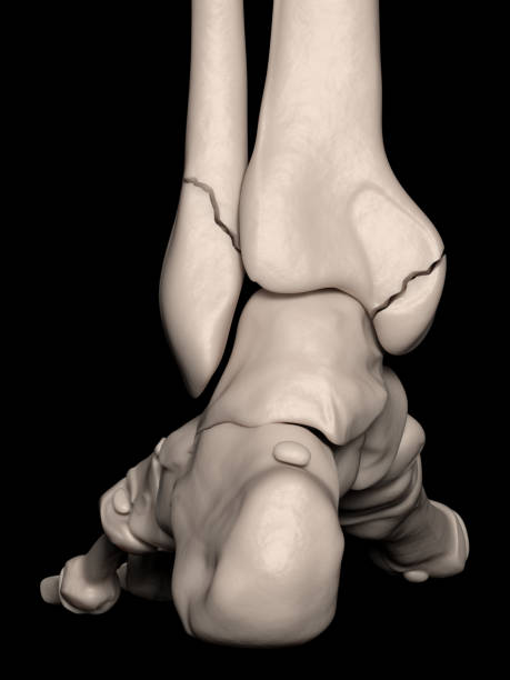
Digital medical illustration depicting a bimalleolar fracture (lateral + medial) (broken ankle). Posterior (rear) view.

Ankle joint fracture Type c medical vector illustration on white background eps 10

Medial view of foot bones Diagram. Anatomy

Digital medical illustration depicting a medial malleolus fracture (broken ankle). Posterior (rear) view.

A right foot with drawing of medial malleolus and ligamentum deltoideum. 50Mpx image.

X-ray foot after operation fix screws in medial malleolus tibia.Medical concept.

"Shown are the tibialis posterior muscle, medial crest of fibula, flexor digitorum longus muscle, tibialis posterior muscle, flexor hallucis longus muscle, tendon of flexor digitorum longus muscle, medial malleolus, tendon of tibialis posterior muscle, flexor retinaculum, calcaneal tendon, fibularis longus muscle, tibia, tendon of flexor hallucis longus, superior fibular retinaculum, calcaneal tuberosity, popliteus muscle, soleus muscle."

A right foot with drawing of medial malleolus and ligamentum deltoideum. 50Mpx image.

The flexor retinaculum of the foot extends from the medial malleolus above, to the calcaneus below 3d illustration

The flexor retinaculum of the foot extends from the medial malleolus above, to the calcaneus below 3d illustration

X-ray foot after operation fix screws in medial malleolus tibia.Medical concept.

The flexor retinaculum of the foot extends from the medial malleolus above, to the calcaneus below 3d illustration

The flexor retinaculum of the foot extends from the medial malleolus above, to the calcaneus below 3d illustration

The flexor retinaculum of the foot extends from the medial malleolus above, to the calcaneus below 3d illustration

The flexor retinaculum of the foot extends from the medial malleolus above, to the calcaneus below 3d illustration

The flexor retinaculum of the foot extends from the medial malleolus above, to the calcaneus below 3d illustration

The flexor retinaculum of the foot extends from the medial malleolus above, to the calcaneus below 3d illustration

The flexor retinaculum of the foot extends from the medial malleolus above, to the calcaneus below 3d illustration

The flexor retinaculum of the foot extends from the medial malleolus above, to the calcaneus below 3d illustration

X-ray foot after operation fix screws in medial malleolus tibia.Medical concept.

The flexor retinaculum of the foot extends from the medial malleolus above, to the calcaneus below 3d illustration

The flexor retinaculum of the foot extends from the medial malleolus above, to the calcaneus below 3d illustration

The flexor retinaculum of the foot extends from the medial malleolus above, to the calcaneus below 3d illustration

Straight-on view of a human tibia anatomical model showing the tibial tuberosity and medial malleolus for anatomy instruction.

Oblique close-up of the distal end of the tibia bone model showing the medial malleolus used in ankle joint articulation.

The flexor retinaculum of the foot extends from the medial malleolus above, to the calcaneus below 3d illustration

X-ray (projectional radiograph) of the foot of a 64-year-old female, by dorsoplantar projection

The flexor retinaculum of the foot extends from the medial malleolus above, to the calcaneus below 3d illustration

X-ray foot after operation fix screws in medial malleolus tibia.Medical concept.

The flexor retinaculum of the foot extends from the medial malleolus above, to the calcaneus below 3d illustration

Southeast Asian, Myanmar old womans foot of lateral view. Skin creases, loosen skin and veins show aging. Isolated on cement background.

Xray film of an ankle of a patient showing medial malleolus fracture.


