
Trapezius muscle labeled medical anatomy structure scheme vector illustration. Educational diagram with upper, middle and lower parts with acromion, humerus and scapula bones location on human body.
Browse 150+ pics of humerus labeled diagram stock photos and images available, or start a new search to explore more stock photos and images.
Trapezius muscle labeled medical anatomy structure scheme vector illustration. Educational diagram with upper, middle and lower parts with acromion, humerus and scapula bones location on human body.
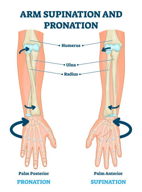
Arm supination and pronation vector illustration. Labeled anatomical scheme. Medical diagram with inner bones and joints. Compared palm posterior and anterior. Hand rotation movement biological terms.
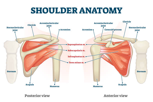
Shoulder anatomy vector illustration. Labeled inner skeleton and muscle structure scheme. Physiological educational posterior or anterior view with bones titles and location. Healthy organ description

Latissimus dorsi muscles the largest muscles in the upper body isolated within the shoulder area of human skeletal system in a rear posterior view on a white background.
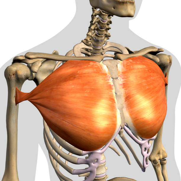
Pectoralis major muscles isolated within the chest and shoulder area of human skeletal system in a frontal anterior view on a white background.
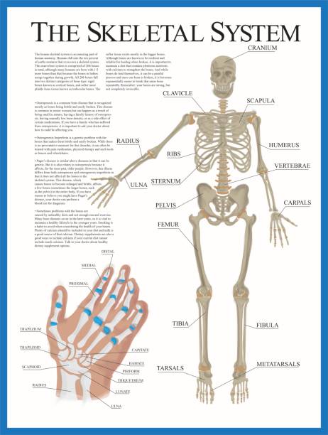
Human skeletal system poster containing detailed information about the skeletal structure. The poster contains a detailed illustration of the human hand.

Pectoralis major muscle as human chest muscular anatomy outline diagram. Labeled educational medical scheme with skeletal system and musculature in human body breast and ribs area vector illustration.
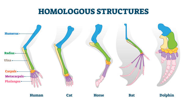
Homologous structures vector illustration. Biological species example scheme. Labeled structural diagram with bone titles. Humerus, ulna and carpals in various creature skeletons from common ancestry.

Skeletal system with body skeleton structure and anatomy outline diagram. Labeled educational medical physiology with skull, spine, ribs, hand and leg bones vector illustration. Biological human model

Shoulder dislocation. humerus bone trauma, Sports injuries, or Weak shoulder muscles. Human arm anatomy. Bones and joint of the Shoulder, and hand. Vector illustration

Clavicle fracture anatomy and broken shoulder collarbone outline diagram. Labeled educational scheme with anatomical hand and scapula skeletal joint vector illustration. Acromion and coracoid process.
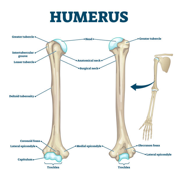
Humerus bone labeled vector illustration diagram. Long bone type in the upper arm. Skeleton anatomy scheme with greater tubercle, deltoid tuberosity, medial epicondyle, trochlea and other parts.

Bursitis vector illustration. Labeled bursae synovial inflammation scheme. Bone and tendon illness and disease diagnosis. Educational chronic problem symptoms, causes and anatomical structure diagram.

Ulnar collateral ligament or UCL with anatomical structure outline diagram. Labeled educational elbow inner parts with xray view vector illustration. Posterior, intermediate and anterior bundle scheme
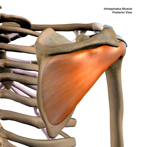
Infraspinatus muscle isolated within the shoulder area of human skeletal system in a rear posterior view on a white background.

Plantigrade, Digitigrade and Unguligrade comparison vector illustration. Educational labeled structure scheme with human, dog and pig legs collection. Bone skeleton parts with location explanation.

Bones types of Human skeleton: Flat, Long, Short, Sesamoid and Irregular bone. Classification of bones by shape.

Shoulder subluxation as partial dislocated arm joint problem outline diagram. Labeled educational medical scheme with body skeletal anatomy and dislocated bones vector illustration. Upper body trauma.
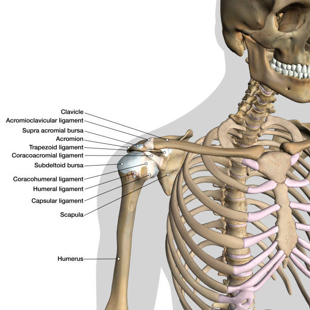
Labeled human anatomy diagram of man's shoulder ligaments, connective tissue and skeleton anterior view on a white background.

Biceps brachii muscles isolated within the human skeletal system and connective tissue in a frontal anterior view labeled on a white background.
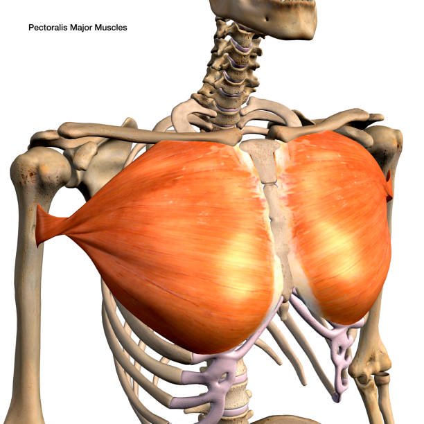
Pectoralis major muscle isolated within the chest and shoulder area of human skeletal system in a frontal anterior view on a white background.
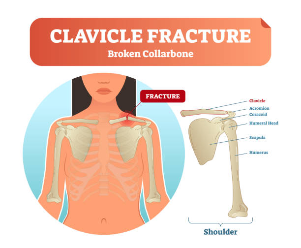
Clavicle fracture with broken collarbone vector illustration. Medical and anatomical labeled scheme with clavicle fracture, acromion coracoid, humeral head, scapula and humerus. Xray after injury.

Biceps tear vector illustration. Labeled medical scheme with humerus, muscle, radius and ulna isolated closeup. Anatomical diagram with human arm, elbow and shoulder. Pain, damage and injury reason.

Distal radius fracture and broken arm bone types anatomy outline diagram. Labeled educational scheme with extra articular nondisplaced and displaced radius and ulna comparison vector illustration.

Labeled human anatomy diagram of male shoulder area with biceps isolated within the skeletal system frontal anterior view on a white background.

Anatomy of elbow with lateral, posterior or anterior view vector illustration. Educational labeled scheme with skeleton bone structure description. Healthy body parts example for physiology handout.
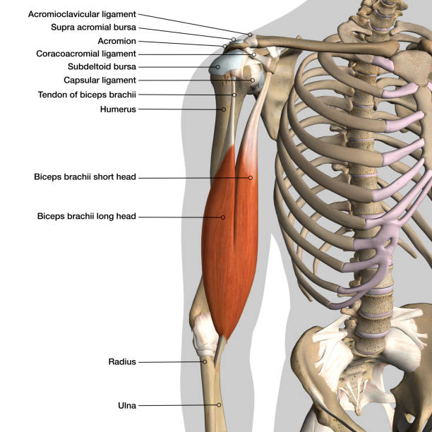
Labeled human anatomy diagram of male shoulder ligaments, connective tissue and biceps muscles isolated within the skeletal system frontal anterior view on a white background.

Pectoralis major muscles isolated within the chest and shoulder area of human skeletal system in a frontal anterior view on a black background.

Labeled human anatomy of male lower arm brachioradialis muscles isolated within the skeletal system bones in a frontal view on a white background.

Biceps Arm Muscle with Skeleton. Human Hand Muscle Tension on White Background with Male Silhouette Bones and Joints.

Human anatomy scientific illustrations with latin/italian labels: elbow joint

Labeled human anatomy of male forearm Flexor Carpi Ulnaris muscles isolated within the skeletal system bones in a frontal view on a white background.

Labeled human anatomy diagram of man's shoulder bones, triceps muscles and connective tissue in a posterior view on a black background.

Labeled human shoulder bone anatomical vector illustration diagram poster. Medical health care information.

Labeled human anatomy diagram of man's shoulder ligaments, connective tissue, biceps muscle and skeleton in an anterior view on a black background.

Extensor carpi ulnaris muscle for arm and hand wrist movement outline diagram. Labeled educational fusiform muscular system in lateral part of posterior forearm vector illustration. Skeletal bones.
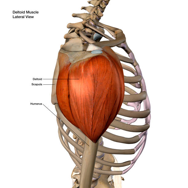
Deltoid muscle isolated within the shoulder area of human skeletal system in a lateral side view labeled on a white background.
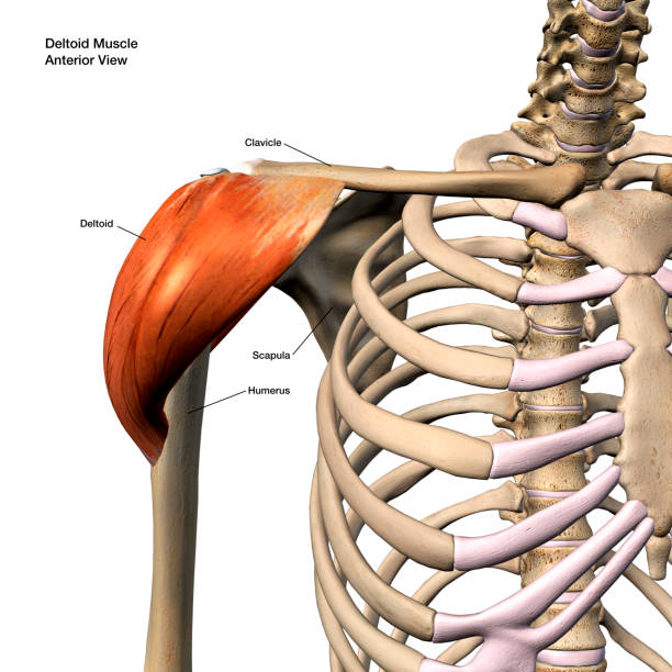
Deltoid muscle isolated within the shoulder area of human skeletal system in a frontal anterior view labeled on a white background.

Shoulder joint illustrated on white. Vector Illustration

Deltoid muscle isolated within the shoulder area of human skeletal system in a lateral side view on a white background.

Labeled human anatomy diagram of male shoulder ligaments, connective tissue and biceps isolated within the skeletal system frontal anterior view on a black background.

Human anatomy scientific illustrations with latin/italian labels: Humerus

Educational vector illustration of a bent human arm displaying the internal bone and muscle structure. The image highlights anatomical components including the humerus, radius, ulna, and surrounding muscles in a simplified medical style. Designed with clean lines and soft colors, this illustration is suitable for healthcare, anatomy education, medical presentations, biology learning materials, and fitness-related concepts.
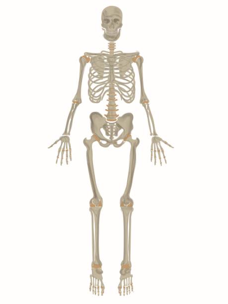
Poster of complete human skeleton.

Biceps brachii muscles isolated within the human skeletal system and connective tissue in a frontal anterior view labeled on a white background.

Latissimus dorsi muscle the largest muscle in the upper body isolated within the shoulder area of human skeletal system in a rear posterior view on a white background.

Pectoralis major muscle diagram. Anatomy of human chest with musculature in ribs area. Educational labeled infographic with medical skeletal system and sternum. Cartoon flat vector illustration

Elbow ligaments with medical medial or lateral xray structure outline diagram. Labeled educational anatomical joint capsule, radius and ulna parts as inner medicine explanation vector illustration.

Labeled human anatomy of male forearm Extensor Digitorum muscles isolated within the skeletal system bones in a rear view on a white background.
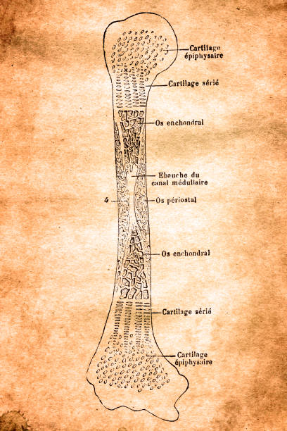
Illustration of a Humerus section

Labeled human anatomy of male lower arm Extensor Digiti Minimi muscles isolated within the skeletal system bones in a rear view on a white background.


