Similar to the gap junctions found in animal cells, the plasmodesmata, which penetrate both the primary and secondary cell walls, allow certain molecules to pass directly from one cell to another and are important in cellular communication
Browse 120+ plasmodesmata stock photos and images available, or search for smooth endoplasmic reticulum or rough endoplasmic reticulum to find more great stock photos and pictures.
Similar to the gap junctions found in animal cells, the plasmodesmata, which penetrate both the primary and secondary cell walls, allow certain molecules to pass directly from one cell to another and are important in cellular communication
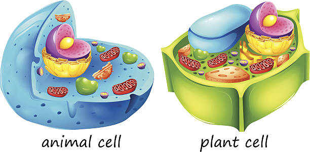
lllustration of the animal and plant cells on a white background
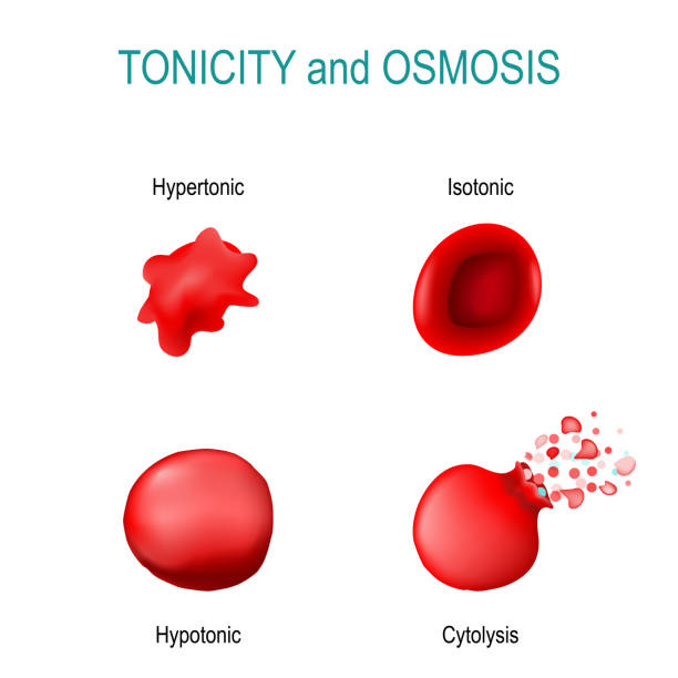
Tonicity is a measure of the osmotic pressure in red blood cells. isotonic (concentration of solutes outside the cell is equal to the concentration of solutes inside the cell); hypertonic (water to flow out of the cell); hypotonic(water diffuses into

Animal cells are generally smaller than plant cells. Animal cells range from 10 to 30 micrometers in length, while plant cells range from 10 and 100 micrometers in length. Animal cells come in various sizes and tend to have round or irregular shapes. Plant cells are more similar in size and are typically rectangular or cube shaped.
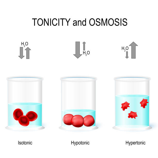
Isotonic, Hypotonic and Hypertonic solutions effects on animal cells. Tonicity and osmosis. This diagram shows the effects of hypertonic, hypotonic and istonic solutions to red blood cells. Vector illustration for biological, medical, science use

Root hair cell collecting mineral nutrients and water from soil, biological labeled plant system diagram. Vector illustration educational cross section scheme. Cytoplasm, nucleus and other elements.

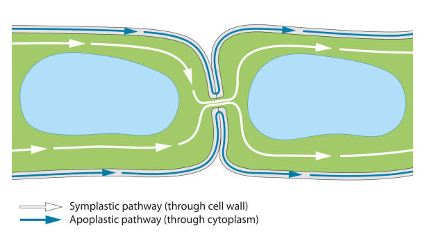
Plasmodesmata plant cells diagram, vector illustration. Educational microscopic labeled cross section scheme. Cell wall protein transport pathways. Agricultural science education and farming research.

Plant cell structure cross-section with Green cell wall, membranes, and chloroplasts, purple nucleus, orange endoplasmatic reticulum and ribosomes, blue vacuole, pink Golgi body and red mitochondrion.

Illustration of a plant cell

Illustration of the plant cells on a white background

Aquatic plant cell under microscope view

Cartoon vector illustration of structure of plant cell. Illustration showing the plant cell anatomy

Gap junction as anatomical intercellular connection structure outline diagram. Labeled educational multitude cell types space description with connexon monomer and plasma membranes vector illustration

Plasmodium vivax in thin film under microscopy, Malaria disease. P. vivax, Malaria parasite in red blood cells.

Different biology cells on a white background

Plasmodesmata are small channels that directly connect the cytoplasm of neighboring plant cells to each other, establishing living bridges between cells

"Microscopic photo of a professionally prepared slide demonstrating the cellular structure of the object.NOTE: Shallow DOF, uneven focus and chromatic aberration are inherent in microscopy, and what appears as dust is actually in the sample.See all my"

Plant cell structure, cross section, with legend. Schematic diagram of the components of plant cells, photosynthetic eukaryotes, with technical terms in english. Isolated illustration over white.

Plasmodesma arrangements in plant cells. Vintage etching circa 19th century.

We find the CIRs to adopt a novel structure, consisting of eight α-helices, divided into two lobes. Despite their different architectures, the CIRs share properties with other proteins from the surfaces of Plasmodium-infected erythrocytes

"Microscopic photo of a professionally prepared slide demonstrating the cellular structure of the object.NOTE: Shallow DOF, uneven focus and chromatic aberration are inherent in microscopy, and what appears as dust is actually in the sample.See all my"

"Microscopic photo of a professionally prepared slide demonstrating the cellular structure of the object.NOTE: Shallow DOF, uneven focus and chromatic aberration are inherent in microscopy, and what appears as dust is actually in the sample.See all my"

aquatic plant (Vallisneria gigantea) under the microscope showing chloroplasts and cell walls - optical microscope x400 magnification

aquatic plant (Vallisneria gigantea) under the microscope showing chloroplasts, cell walls and hairs - optical microscope x100 magnification

Aquatic plant cell under microscope view

Colorful vector illustration of plant cell structure with labeled organelles, highlighting cellular components and functions in a clear layout

Pitted cell walls of basal cells of the wavy broom moss, Dicranum polysetum, with polarization at 200x.


