Illustration of the anatomy of the male reproductive system on a white background
Browse 1,000+ pubic bone pic stock photos and images available, or start a new search to explore more stock photos and images.
Illustration of the anatomy of the male reproductive system on a white background
The external oblique muscle is one of the outermost abdominal muscles, extending from the lower half of the ribs around and down to the pelvis 3d illustration

The iliopsoas muscle refers to the joined psoas major and the iliacus muscles .
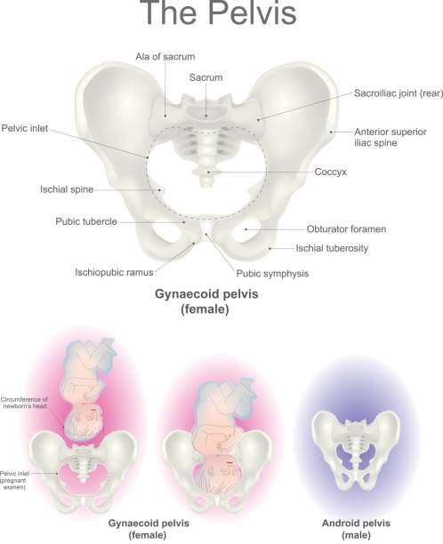
The pelvic region is the headquarters for reproductive organs and the end of the line for the digestive tract. It is also home to a collection of bones known as the pelvis, where the femur the largest bones in the body meets to form the leg. Vector graphic.

The erector spinae or spinal erectors is a set of muscles that straighten and rotate the back 3d illustration
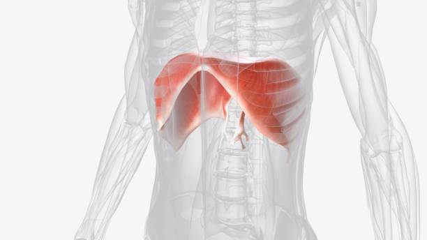
The diaphragm is a muscle that helps you inhale and exhale 3d illustration

The multifidus muscle is an important stabilizer of the lumbar spine .

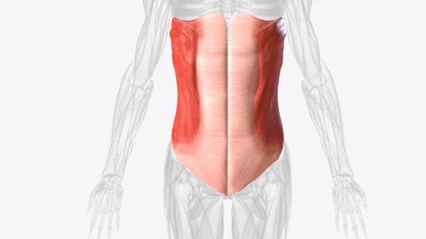
The abdominal muscles are the muscles forming the abdominal walls, the abdomen being the portion of the trunk connecting the thorax and pelvis

The pudendal nerve provides most of the movement and sensations .

The transversus abdominis (TrA) muscle is the deepest of the 6 abdominal muscles .

The triceps, or triceps brachii is a large muscle on the back of the upper limb of many vertebrates.

The transversus abdominis (TrA) muscle is the deepest of the 6 abdominal muscles .

The lateral pterygoid muscle (or external pterygoid muscle) is a muscle of mastication.

The flexor hallucis longus muscle (FHL) attaches to the plantar surface of phalanx of the great toe and is responsible for flexing that toe.

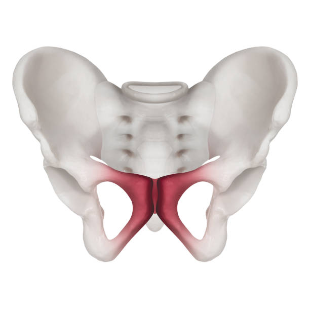
The pelvic floor muscles span the bottom of the pelvis and support the pelvic organs (bladder and bowel, and uterus (womb) in women)

The suprahyoid muscles are a group of four muscles located superior to the hyoid bone of the neck .

The gluteus maximus is the main extensor muscle of the hip in humans .

The erector spinae or spinal erectors is a set of muscles that straighten and rotate the back 3d illustration

The transversus abdominis (TrA) muscle is the deepest of the 6 abdominal muscles .

pelvis x-ray image of after childbirth women shows distasis of simphysis pubis called symphysiolysis

The suboccipital muscles are a group of four muscles located inferior to the external occipital prominence of the skull .

The muscles in the back are the trapezius, rhomboids, latissimus dorsi, erector spinae, multifidus, and quadratus lumborum.

The axial connective tissue system is a fiber continuum of the lung that maintains alveolar surface area during changes in lung volume

Deep plantar muscles of right foot 3d illustration

The buccinator compresses the cheeks against the teeth and is used in acts such as blowing .

The dorsal interossei muscles are muscles that abduct the second, third and fourth digits.

The infraspinatus is a thick triangular muscle that occupies much of the infraspinous fossa of the scapula .

The deltoid is responsible for elevating the arm in the scapular plane and its contraction in doing this also elevates the humeral head.

Eyeball, spheroidal structure containing sense receptors for vision, found in all vertebrates and constructed much like a simple camera .

The diaphragm, located below the lungs, is the major muscle of respiration .

The diaphragm, located below the lungs, is the major muscle of respiration .

The lumbricals are deep muscles of the hand that flex the metacarpophalangeal joints .

The biceps brachii (BB), commonly know as the biceps, is a large, thick muscle on the ventral portion of the upper arm .

Corrugator supercilii is a paired muscle found deep to the medial end of each eyebrow 3d illustration

Innermost intercostals comprise the third and deepest layer of intercostal muscles .

The suboccipital muscles are a group of four muscles located inferior to the external occipital prominence of the skull .

The serratus posterior inferior is a muscle within the intermediate compartment of the back.

The obturator externus is a muscle of the medial compartment of the thigh .

The flexor pollicis longus (FPL) muscle is one of the three deep flexors of the volar compartment of the forearm .

Pronator quadratus is a square-shaped muscle on the distal forearm that acts to pronate the hand .

The brachialis (brachialis anticus), also known as the Teichmann muscle, is a muscle in the upper arm that flexes the elbow.

Fibularis tertius muscle, also called peroneus tertius, is located on the lower lateral aspect of the leg .

Gracilis is a thin, flat, long muscle that attaches to the coxal bone and tibia.

The brachialis (brachialis anticus), also known as the Teichmann muscle, is a muscle in the upper arm that flexes the elbow.