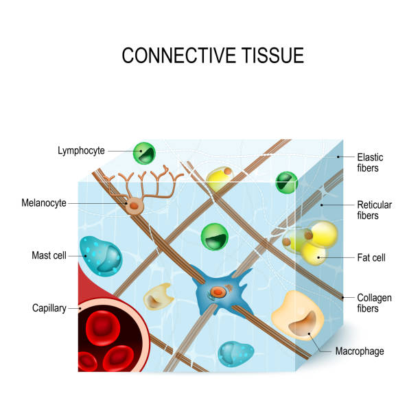
connective tissue that supports, binds, or separates more specialized tissues and organs of the body. Illustration showing a section of connective tissue with capillary, cells (lymphocyte, fat, melanocyte, macrophages, mast cell) and fibers (elastic, collagen, reticular). Human anatomy