Human Skeleton Anatomy.Vertebral Column of Human Body Anatomy infograpic diagram including all vertebra cervical thoracic lumbar sacral and coccygeal
Browse 1,800+ vertebrae diagram stock photos and images available, or start a new search to explore more stock photos and images.
Human Skeleton Anatomy.Vertebral Column of Human Body Anatomy infograpic diagram including all vertebra cervical thoracic lumbar sacral and coccygeal

A retro style diagram of the human spine showing the side view with different regions and vertebrae labelled. This is an editable EPS 10 vector illustration with CMYK color space.

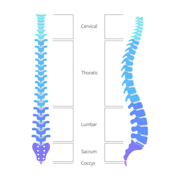
Spinal cord. sections of vertebral column: cervical, thoracic, and lumber spine, sacrum and coccyx. Human silhouette with backbone, intervertebral discs, hip bones and joints. Vector diagram for medical, educational and science use

Facet joints anatomy with bone capsule and cavity closeup outline diagram. Labeled educational medical explanation with body of vertebra, intervertebral disc and spinous process vector illustration.
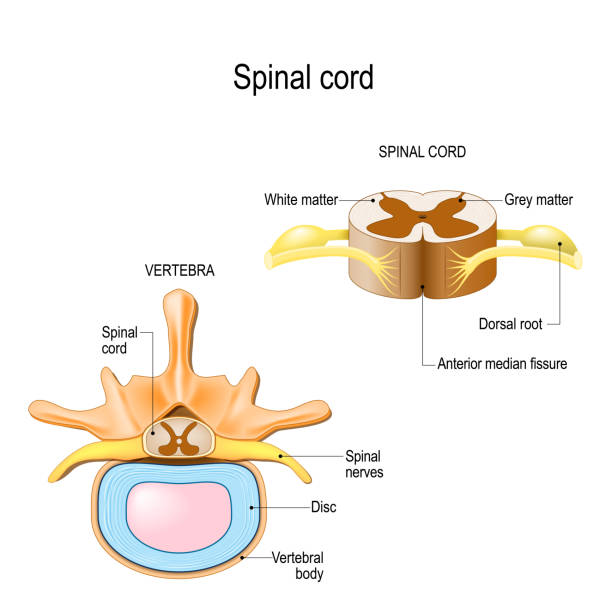
section of the human vertebral column and cross-section of spinal cord. Central nervous system. Vector illustration for medical, biological, and educational use

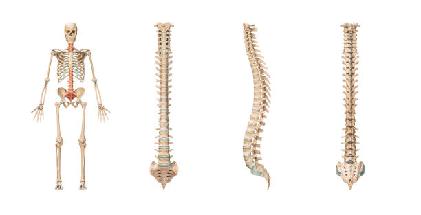
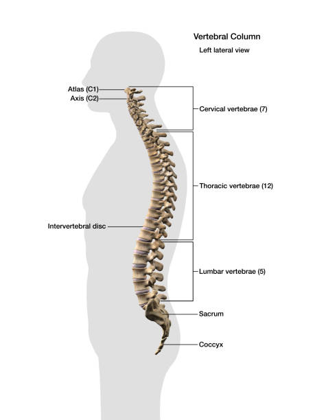
The vertebral column, also known as the backbone or spine. The human vertebral column and its regions Coccyx, Sacrum, Lumbar, Thoracic, Cervical. Lateral Anatomy of a vertebra
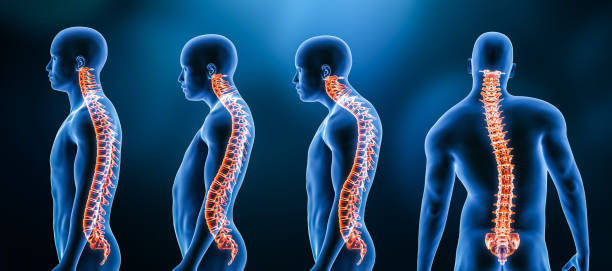
Human spine structure vector illustration. Backbone and vertebral column anatomy with section names. Scoliosis concept and symbol of spinal surgery. Back and lateral view isolated. Medical banner
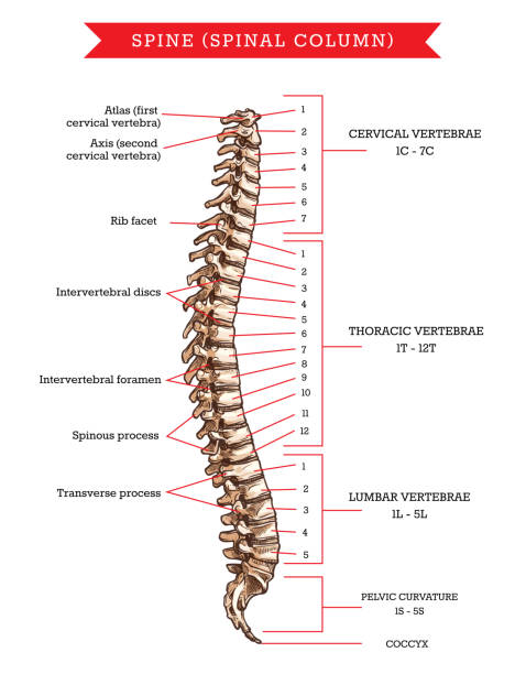
Human spine bones anatomy, vector sketch of skeleton backbone or vertebral column. Cervical, thoracic and lumbar vertebrae, pelvic curvature and coccyx, rib facet, intervertebral discs and foramen
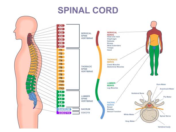
Medical diagram of spinal cord. Anatomical infographic with different part of spine, nervous system and vertebrae. Cervical, thoracic, lumbar, sacral and coccyx spine. Cartoon flat vector illustration

Medical illustration depicting types of spina bifida: hidden, meningocele, and myelomeningocele with detailed spinal anatomy.

Intervertebral disc structure from Nucleus pulposus and Annulus fibrosus to Endplates. Vertebra anatomy. Spinal Column. Vector illustration

Skeletal system with body skeleton structure and anatomy outline diagram. Labeled educational medical physiology with skull, spine, ribs, hand and leg bones vector illustration. Biological human model

A detailed blueprint of a human spine showing the side view with different regions and vertebrae labelled. This is an editable EPS 10 vector illustration with CMYK color space.

Spine osteoarthritis anatomical vector illustration diagram, educational medical scheme information.

Degenerative disc disease with spine and vertebra trauma outline diagram. Labeled educational normal intervertebral, degenerated, bulging, thinning and herniated problem example vector illustration.

Vertebral column: cervical, thoracic and lumbar spine, sacrum and coccyx. Numbering order of the vertebrae of the human spinal column. Vector diagram for medical use
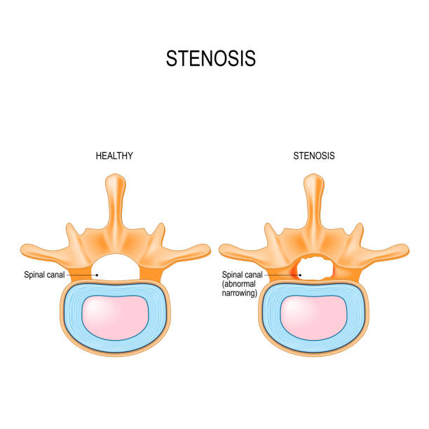
Lumbar Spinal Stenosis is an abnormal narrowing in spinal canal. section of the human vertebral column and cross-section of spinal cord. Vector illustration for medical, biological, and educational use

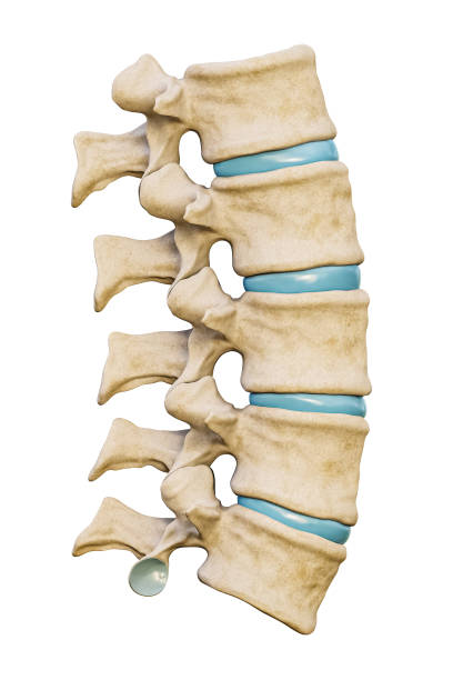
Anatomical model of spinal column, vector illustration on white background

Paraspinal muscles as erector spinae or back muscular system outline diagram. Labeled educational vertebrae movement and support anatomy vector illustration. Spinal and torso backview detailed model.

Spine anatomy with detailed back bone medical structure outline diagram. Labeled educational scheme with spinous process, lamina, vertebral canal, thoracic and lumbar body parts vector illustration.

Herniated spinal disc - Degenerative, Protrusion, Extrusion, Sequestration - vector patient-friendly diagram, hand drawn. Infographic of stages hernia of intervertebral disk
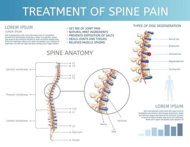
Vector Illustration Treatment of Spine Pain. Spine Anatomy. Square Flat Banner on White Background. Graphic Spinal Diagram Showing Cervical Vertebrae Below Pectoral and Lumbar Spine.

Cervical spondylosis problem compared with healthy spine outline diagram. Labeled educational scheme with human backbone disk bulging, bone spurs and narrowed space explanation vector illustration.
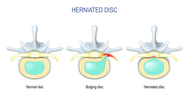
Spinal disc herniation. Difference Between Bulging disc and Herniated Disc. Vector illustration

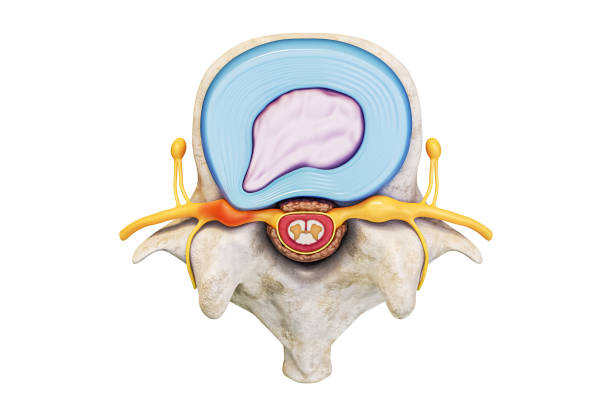
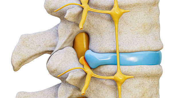
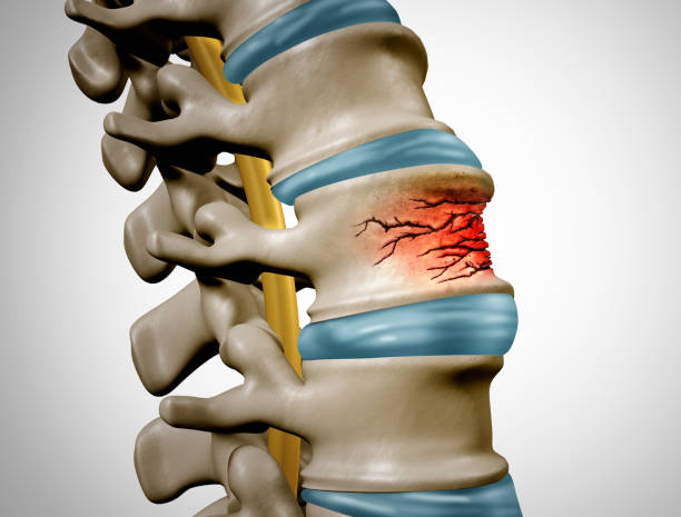
The intervertebral disc is a fibrocartilaginous structure that joint the vertebral bodies of the spinal column and provides it resistance and protection to the effect of axial loads

Thoracic vertebrae location and medical structure description outline diagram. Labeled educational scheme with anatomical backbone parts and detailed superior or lateral bone view vector illustration

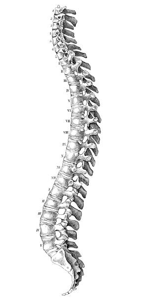
Victorian print of the Ligaments of the Vertebrae, 19th Century
Computer generated image of spinal column inside gray outline of man, with anatomical labeling, side view on white background.

Vector Medical illustration of the Spine, diagram of the human spinal column, vertebral sections, vertebra.

Lumbar puncture procedure. Spinal nerve roots. Spinal Tap Procedure. close-up of Vertebral Column. Syringe needle inserted into Epidural space to Collect Cerebrospinal Fluid. Vector illustration for medical use.

The lumbar plexus. Nervous plexus in the lumbar region of the body. Vector diagram.

A diagram of a human female spine showing a side view of the vertebra of the spinal cord within the the body. This is an editable EPS 10 vector illustration with CMYK color space.

Thoracic spine pain as soft tissue muscle inflammation outline diagram. Labeled educational medical backbone skeletal trauma as painful problem vector illustration. Chronic vertebrae posture condition
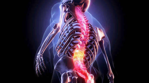
Spine-Part of Human Skeleton,Pain in spine,Pain concept,
Human anatomy scientific illustrations with latin/italian labels: Spine
Traumatic Spine Fracture and vertebral injury medical concept as a human anatomy spinal column with a broken burst vertebra due to compression or other osteoporosis back disease as a 3D illustration.
![3d illustration of Skull With Spinal Cord Anatomy The vertebral column, also known as the backbone or spine, is part of the axial skeleton. The vertebral column is the defining characteristic of a vertebrate, in which the notochord (a flexible rod of uniform composition) found in all chordates has been replaced by a segmented series of bones—vertebrae separated by intervertebral discs.[1] The vertebral column houses the spinal canal, a cavity that encloses and protects the spinal cord. vertebrae diagram stock pictures, royalty-free photos & images](https://media.istockphoto.com/id/967686594/photo/3d-illustration-of-skull-with-spinal-cord-anatomy.jpg?s=612x612&w=0&k=20&c=4CIT3kNCI9jg_MQNS2HdIkmYj51ozSPlNvEeqS6BHJ4=)
The vertebral column, also known as the backbone or spine, is part of the axial skeleton. The vertebral column is the defining characteristic of a vertebrate, in which the notochord (a flexible rod of uniform composition) found in all chordates has been replaced by a segmented series of bones—vertebrae separated by intervertebral discs.[1] The vertebral column houses the spinal canal, a cavity that encloses and protects the spinal cord.

The human spine column. Colorful vector illustration with useful information isolated on a dark grey background. Front view. Medicine, anatomy and biology concept.
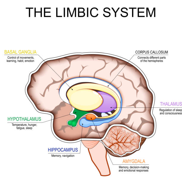
Limbic system. Cross section of a human brain. Mammillary body, basal ganglia, pituitary gland, amygdala, hippocampus, thalamus, cingulate gyrus, corpus callosum, hypothalamus. Part of limbic system for Emotion regulation, Memory formation, Behavioral responses, Autonomic functions and Fight-or-flight response. Detailed vector poster. Schematic diagram

Spinal cord compression with tumor and vertebrae condition outline diagram. Painful back cause with medical problem explanation vector illustration. Skeleton pathology and unhealthy skeletal position

Male human anatomy Rotatores Spinae back muscles in isolation on the skeletal system from a posterior view on a black background.

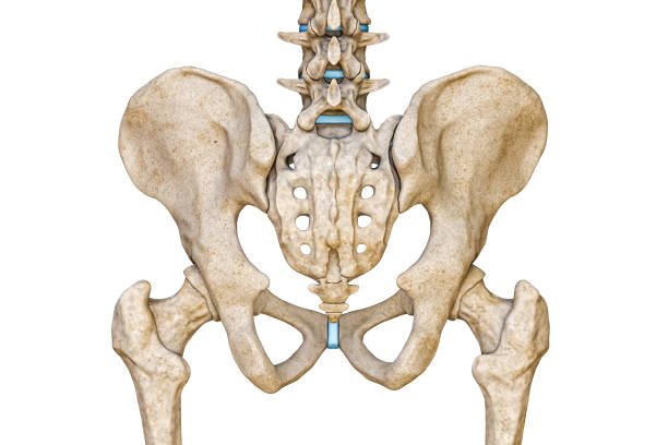
Lumbar vertebrae and sacrum skeletal bones of the human spinal column including the anterior longitudinal ligament of the spine. Labeled medical illustration on a white background.
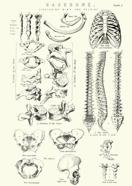
Vintage engraving of the human Backbone including Ribs and Pelvis. 19th Century

A diagram of the human spine showing the side view with different regions and vertebrae labelled. This is an editable EPS 10 vector illustration with CMYK color space.

Ankylosing spondylitis as inflammatory spine bone disease outline diagram. Labeled educational anatomical comparison with healthy and damaged vertebrae vector illustration. Fused skeletal back parts.