Images
X Ray Tarsal Ankle And Foot Pictures, Images and Stock Photos
Browse 650+ x ray tarsal ankle and foot stock photos and images available, or start a new search to explore more stock photos and images.
Most popular
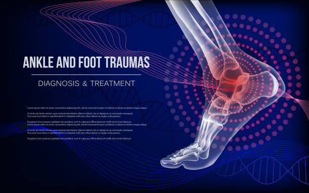
Ankle sore joints concept. Realistic bones of foot skeleton of human leg. Horizontal dark blue banner for ankle and foot joints traumas advertising, medical publications. Vector illustration stock.


Heel spur, injury to the plantar ligament of the leg. Polygonal design of interconnected lines and points. Blue background.

Horizontal light blue banner with ankle and foot joints traumas concept. For advertising, medical publications in social media. Realistic bones of foot skeleton of human leg. Vector illustration stock vector.
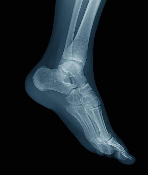
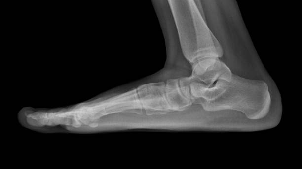
Digital X-ray image of a young foot, oblique view. normal.
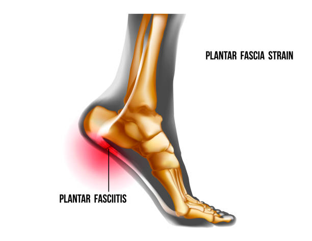
Plantar fasciitis inflammation and ruptures strain. Bones ot Foot pain realistic illustration. Medial view. Anatomy of joints, human leg black and yellow transparente skeleton. For medical orthopedic advertising. Vector illustration stock vector.
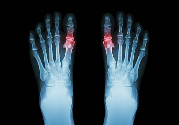
Gout , Rheumatoid arthritis ( Film x-ray both foot and arthritis at first metatarsophalangeal joint ) ( Medicine and Science background )
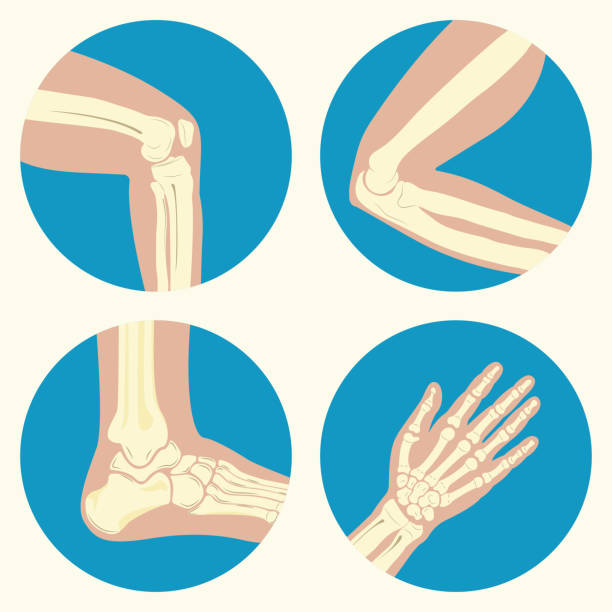
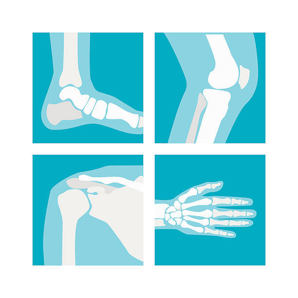
Set of human joints, knee joint, elbow joint, ankle joint, wrist, emblem or sign of medical diagnostic center or clinic, flat design, vector
Cartoon Human Joints Set Health Care Medical Diagnostic X-ray. Flat Design Style Vector illustration


White horizontal continuous line drawing concept banner about rheumatoid arthritis. Linear bones joints of foot. For advertising, medical publications in social media. Vector illustration.

Set of human joints, knee joint, elbow joint, ankle joint, wrist, skeletal spinal bone structure of Human Spine, emblem or sign of medical diagnostic center or clinic, flat vector illustration.


Doctor orthopedist on a computer shows a heel spur on an x-ray of the foot

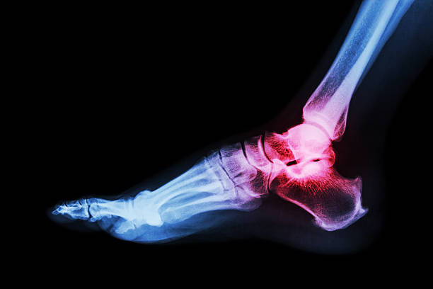
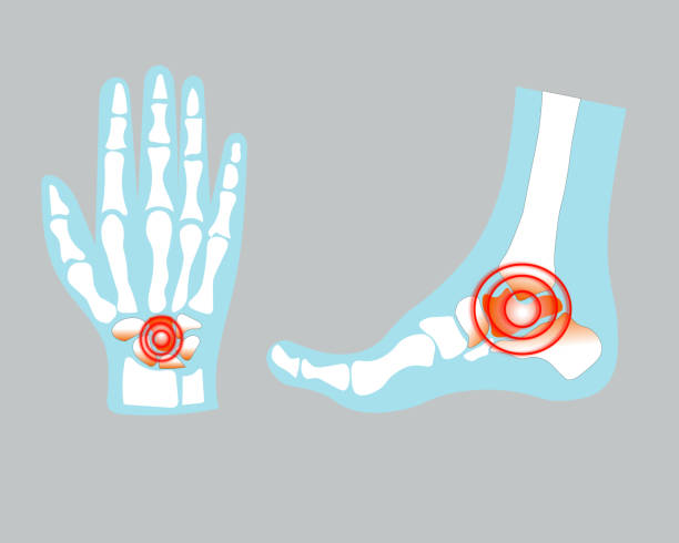
Arthritis of ankle . X-ray of foot . Lateral view . Invert color style . Gout or Rheumatoid concept .

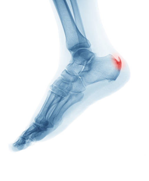
Lateral view of heel of human foot in X-ray (blue on black background), with pains on hell and Calcaneus bone.
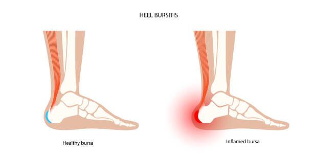
Heel bursitis inflammation. Inflamed bursa in human ankle. Achilles tendon and foot disease, pain and deformity. Diagnosis and treatment. Anatomical musculoskeletal poster, medical vector illustration

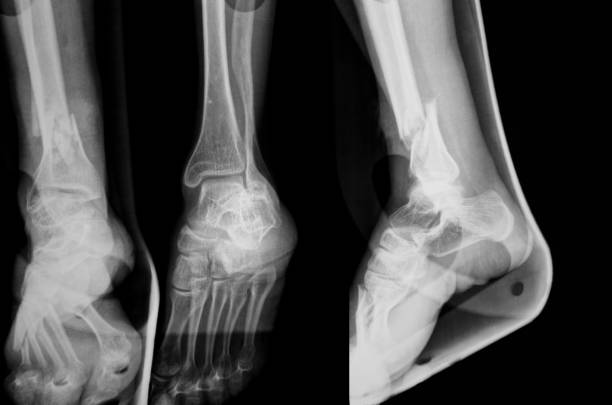
Fracture shaft of fibula bone ( leg bone ) . X-ray of leg ( 2 position : side and front view )

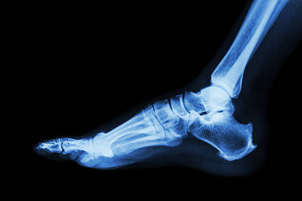
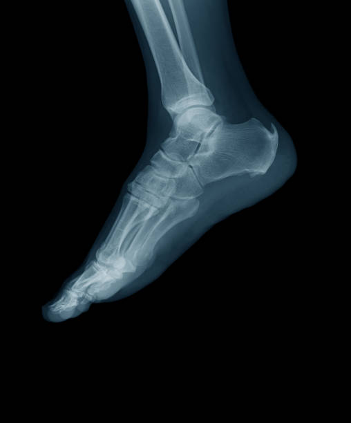
X-ray normal human foot . Lateral view . Invert color style .
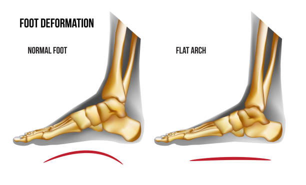
Anatomy flat foot arch. Bones the of foot medial view. Realistic skeleton of human leg. For advertising or medical publications. Vector illustration stock vector.


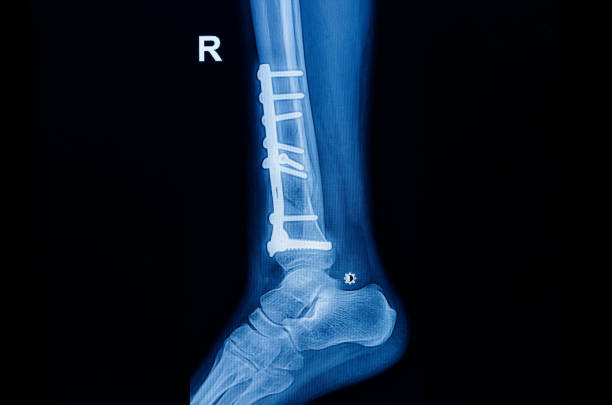
lateral view of heel of human foot in X-ray (blue on black background), after surgery to repair multiple fractures of the heel of Calcaneus bone very important, with tube and screw to fix bones.


Vector realistic dark navy blue x ray of skeleton of foot. Human leg bones. Anatomy of joints. Medial view. For advertising or medical publications. Illustration stock vector.

Skeletal foot - injuryd talus bone. Xray view. Medically accurate 3D illustration

bone of the hand and foot with arm and leg, anatomy, internal organs body part orthopedic health care, vector illustration cartoon flat character design clip art

Supinated foot, arch deformation, bottom and back view. Foot weight distribution. For medical orthopedic advertising. Vector illustration stock vector.

Set of xray of human skeletal, human joints, knee joint, elbow joint, ankle joint, wrist, skeletal spinal bone structure of Human Spine, emblem or sign of medical diagnostic center or clinic, flat vector illustration.

Gouty arthritis . film x-ray of human foot and arthritis at first metatarsophalangeal Joint . 2 position ( front and side view )


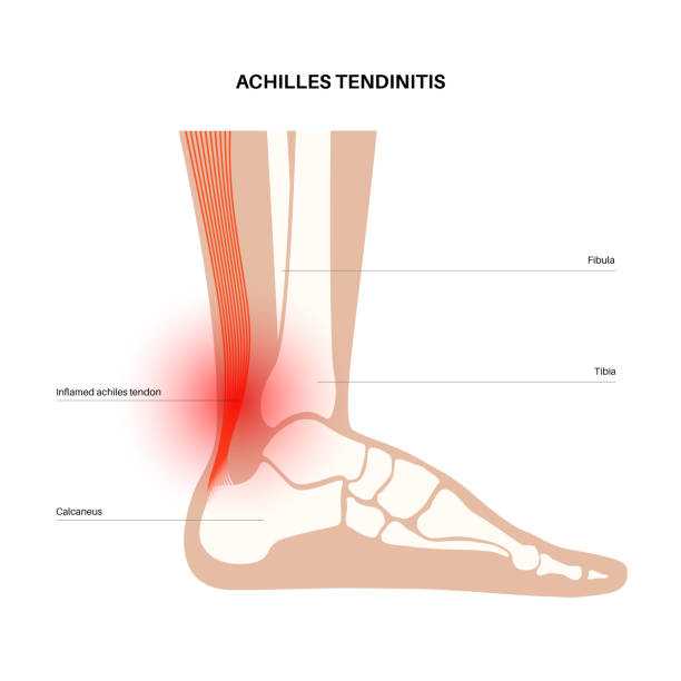
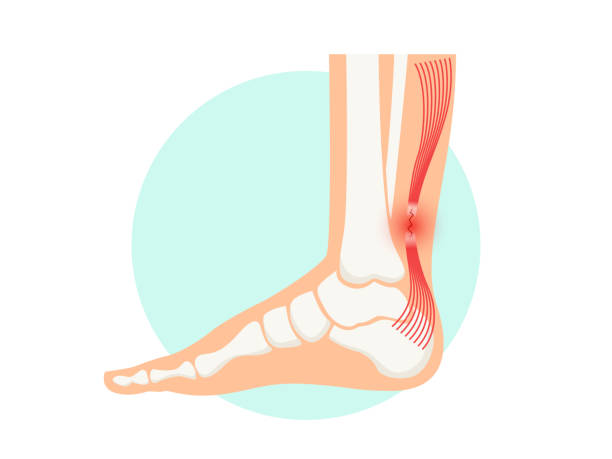
Achilles tendinitis anatomical poster. Ankle injury, ligament sprain and tear problems. Pain in the human muscular system. Tendinosis and podiatry, trauma in foot joints flat vector illustration.


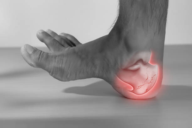
A man has a heel injury because his heel bone is broken.
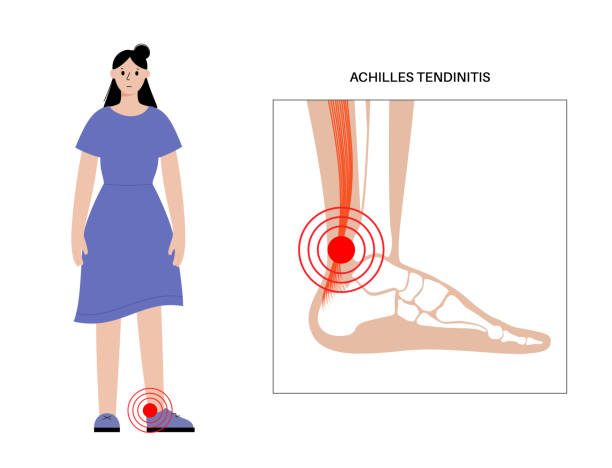
Achilles tendinitis anatomical poster. Ankle injury, ligament sprain and tear problems. Pain in the human muscular system. Tendinosis and podiatry, trauma in foot joints flat vector illustration.

Set of human knee and elbow, ankle and shoulder joints, wrist and spine and tooth, anatomy icons, emblem or sign of medical diagnostic center or clinic, flat design, vector


Set of xray of human,human joints,knee joint,elbow joint, ankle joint, wrist, skeletal spinal bone structure of Human Spine, emblem or sign of medical diagnostic center ,flat vector illustration.
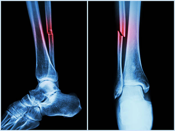
Fracture shaft of fibula bone ( leg bone ) . X-ray of leg ( 2 position : side and front view )

X-ray image of diabetic foot amputation, AP and lateral view.


Vertical light blue banner with ankle and foot joints traumas concept. Realistic bones of foot skeleton of human leg. For advertising, medical publications in social media. Vector illustration stock vector.

Arch of foot pain bones skeleton realistic illustration. Medial view. Anatomy of joints, human leg realistic black and yellow transparente skeleton. For medical orthopedic advertising. Vector illustration stock vector.

Calcaneal spur anatomy. Foot problem, diagnostic and treatment in podiatry clinic. Heel bone outgrowth from calcaneal tuberosity. Ankle pain and swelling. X ray examination of feet vector illustration

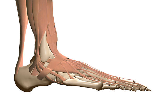
Foot bone and Achilles tendon.
Next