Black female radiologist looking at a patientâs CAT scan to write the report at the clinic - Diagnostics tools concepts
Browse 720+ kidney ct scan stock photos and images available, or search for kidney mri to find more great stock photos and pictures.
Black female radiologist looking at a patientâs CAT scan to write the report at the clinic - Diagnostics tools concepts
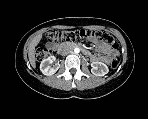
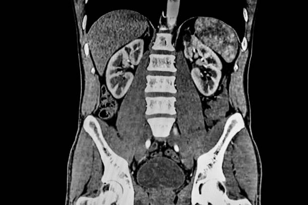
Normal kidneys on computed tomography scan

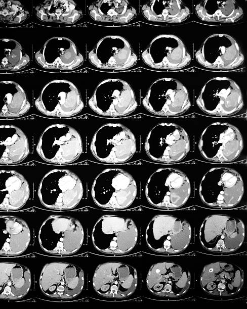
computed tomography of the chest. Medical background. monochrome photos

A medical MRI scan of a female body. SHowing the Kidneys, Liver and spine.
Abdomen cat scan with view of pancreas cyst.

CT Abdomen Cross section View. CT scan (computed tomography) of chest organs.

Asian Female Doctor using a laptop and looking at screens of Magnetic Resonance Imaging.

Kidneys (marked by arrows) on abdominal CT scan

From left to right, axial, coronal and sagittal contrast-enhanced CT of the abdomen and pelvis of a young child demonstrating a large, aggressive abdominal tumor arising from the left kidney. This tumor and the attached kidney were removed and found to be a Wilms tumor. Named after Max Wilms, a German doctor who described the disease in 1899, Wilms tumor or nephroblastoma is the most common abdominal cancer and most common kidney cancer in children. It is often only diagnosed once it is large enough to feel or be seen pushing out of the abdomen. Fortunately, the vast majority of Wilms tumors are favorable histology and have a good chance of being cured with treatment. Treatment depends on the stage (size, location, spread) of tumor and begins with surgery to remove the tumor followed by chemotherapy, and often also radiation therapy.

Pretty young woman doctor working,writing prescription for sick patient...CAT scan of human abdomen on monitor

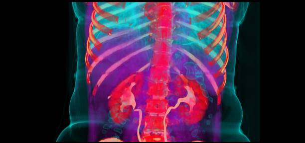
volume rendering CT image shows aorta and renal artery.

MRI imaging of abdomen, focal nodular hyperplasia of the liver

Senior radiologist of Oncology institute is examing MRI and CAT scans of human abdomen on his monitors

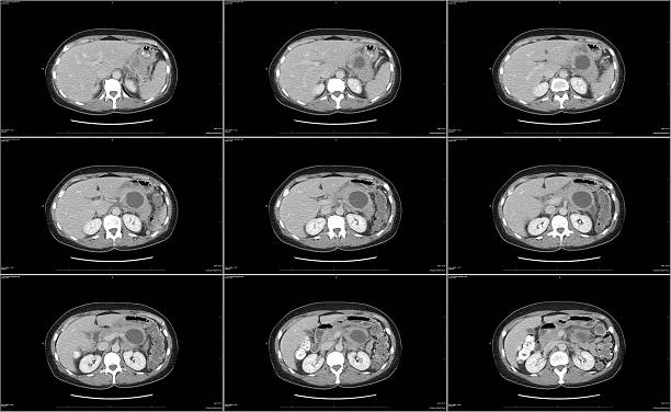
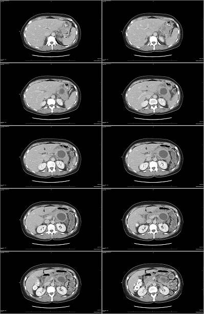
Computational tomography image of the abdominal and pelvic regions.

CT scan of abdomen by mri scanning abdomen.

Senior radiologist of Oncology institute is examing MRI scans of human brain on his monitors

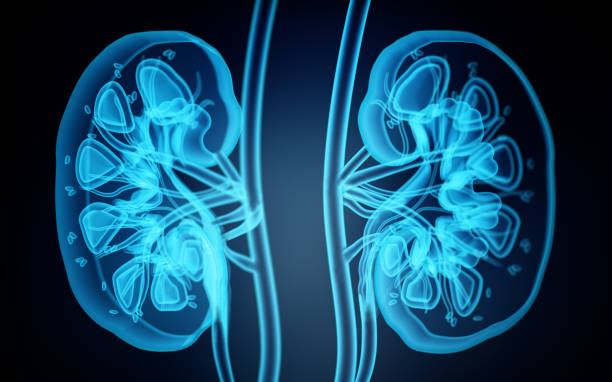
Set of illustrations for kidney cancer disease which include the symptoms, causes, risk factors, and the diagnosis for the illness.

Pretty young woman doctor working,examining patients file.. picture of CAT scan of human abdomn on monitor

CAT scan of human abdomen...man with aneurysm of abdominal aorta

Anatomy of human body on a white background

Human internal organs on whole body computed tomography

Pretty young woman doctor in her office working,checking CAT scan picture of human abdomen on computer monitor

Doctor looks at computed tomography images of abdomen and cyst on kidney

adbominal conputed tomography with injection

Who has illness of appendicitis.

Kidneys (marked by yellow) on abdominal CT scan

Computational tomography image of the abdominal and pelvic regions.
Abdomen cat scan with view of pancreas cyst.
Set is designed with suitable visuals for all medical and healthcare

Focused African American female radiologist looking at a patientâs scans to write the report at the clinic - Diagnostics tools concepts

Yung male doctor with stethoscope watching CAT scan image of human abdomen

Yung male doctor with stethoscope watching CAT scan image of human abdomen
© 2025 iStockphoto LP. The iStock design is a trademark of iStockphoto LP. Browse millions of high-quality stock photos, illustrations, and videos.
Do Not Sell or Share