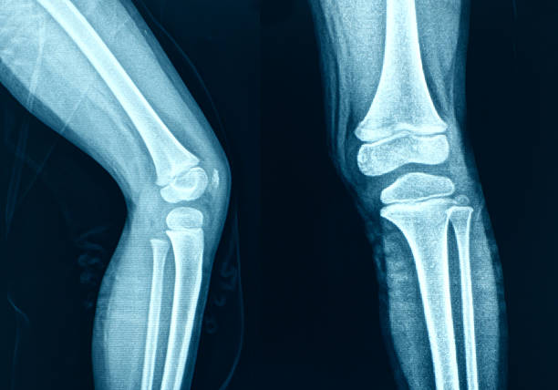
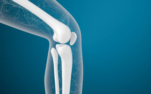
X-ray image of knee
Browse 13,500+ kneecap stock photos and images available, or search for kneecap pain to find more great stock photos and pictures.
knee anatomy. side and front view. Cross section of the joint showing the main parts: femur, fibula, articular capsule, menisci, muscles and ligaments. vector illustration

Healthy and unhealthy dog's knee joint, the medial luxating patella or knee cap dislocation illustration.

Young woman feeling pain in her knee. Woman runner got sports injury running on forest trail. Woman runner hold her sports injured knee.

Chondromalacia patella knee breakdown compared with healthy outline diagram. Labeled educational kneecap tissue damage with cartilage problem and anatomical leg joint structure vector illustration.
Leg bones and knees, 3d rendering. Computer digital drawing.
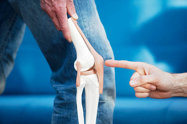
Artificial model of a knee joint. A finger pointing at the patella. XXL size image.
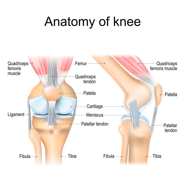
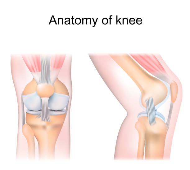
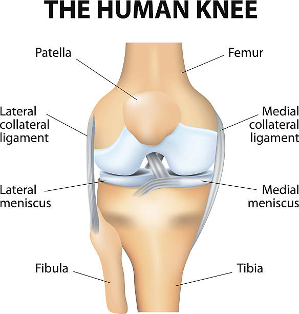
Knee anatomy. Structure of leg joint. Major parts. Vector poster with text label for medical education

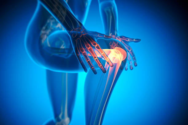
X ray illustration of a running man with pain in knee joint with blue background - 3D illustration
Medical X-Ray illustration of pain in knee joint - 3D illustration

Knee pain while running is often due to runner's knee, IT band syndrome, and knee bursitis.
Knee Joint Icon. Human Bones Joint Symbol for Medical Apps and Websites
Knee bones vector. Human bone and joint icon set. Rheumatology and traumatology, vector design and illustration. Vector illustration

Patellar dislocation. Normal position of kneecap and Patella displaced. Anatomy of the Knee
Human Knee joint anatomy. Vector
Digital medical illustration depicting a vertical patellar fracture. Anterior (front) view.
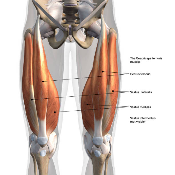
Frontal view of man's isolated leg muscles with labeled names

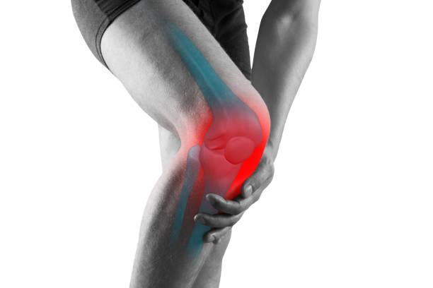
Pain in the Knee Joint - Medical Illustration - 3D Rendering

Human knee joint 3d model vector illustration. Low poly design future technology cure pain treatment. Blue background and red injury man body leg medicine template art

Knee Cartilage Injury - Torn Meniscus - Stock Illustration as EPS 10 File

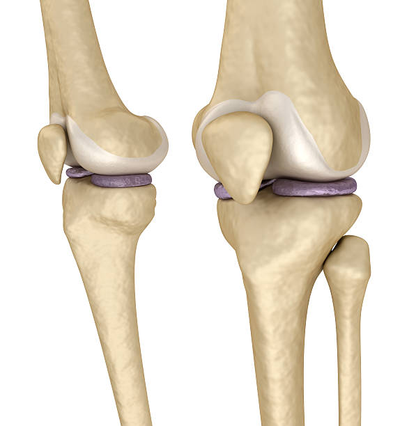
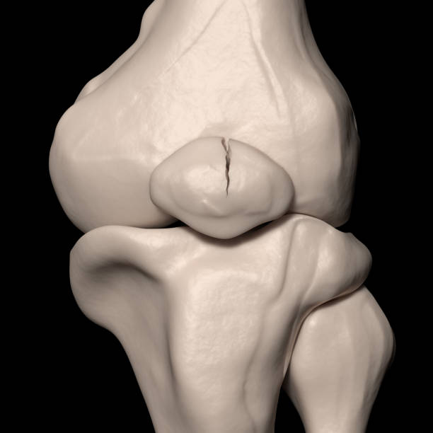
medically accurate illustration of the knee bones

3D illustration of Patella - Part of Human Skeleton.
Human Knee joint anatomical diagram, medical scheme. Educational information template
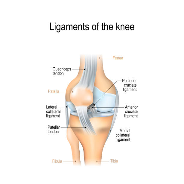
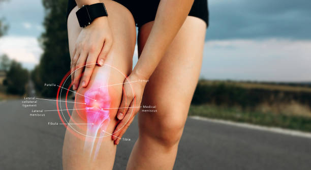
Ligaments of the knee. Anterior and Posterior cruciate ligaments, Patellar and Quadriceps, tendons, Medial and Lateral collateral ligaments. joint anatomy. Vector illustration for biological, medical, science and educational use

X ray illustration of a running man with pain in knee joint with blue background - 3D illustration

Female doctor is rewinding knee bandage to man. Doctor taking care of a patient with trauma of patella

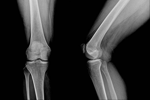
x-ray show knee joint replacement

Woman in the park holding painful knee after sports injury
Knee pain, man with legs ache, chiropractic treatments concept with highlighted skeleton, isolated on white background

Mature Adult, Senior Adult, Knee, Pain, Problems

Doctor traumatologist demonstrating bones of knee joint on artificial model and taking xray picture closeup. X ray diagnosis of rheumatoid arthritis concept
Bone icon set , vector illustration

Senior man runner with knee pain in the park

Knee joint icon. Knee bones graphic sign. Symbol human joint isolated on white background. Vector illustration

Human knee joint medical icon, emblem for orthopedic clinic

Total Knee Replacement. Knee implant. Vector

Knee joint icon. Knee bones graphic sign. Symbol human joint in the circle isolated on white background. Flat design. Vector illustration

Normal position of kneecap and patella displaced. Illustration isolated on white background. Graphic concept for your design

Knee joint icon. Knee bones graphic sign. Symbol human joint in the circle isolated on white background. Flat design. Vector illustration
knee bones pain white background knee injury
© 2025 iStockphoto LP. The iStock design is a trademark of iStockphoto LP. Browse millions of high-quality stock photos, illustrations, and videos.
Do Not Sell or Share