Images
Labeled Hand Bones Pictures, Images and Stock Photos
Browse 130+ labeled hand bones stock photos and images available, or start a new search to explore more stock photos and images.
Most popular

Anatomy of dog paws with forelimb and hindlimb bones vector illustration. Educational labeled skeleton comparison with zoological inside structure scheme. Animal legs inner closeup examination model.
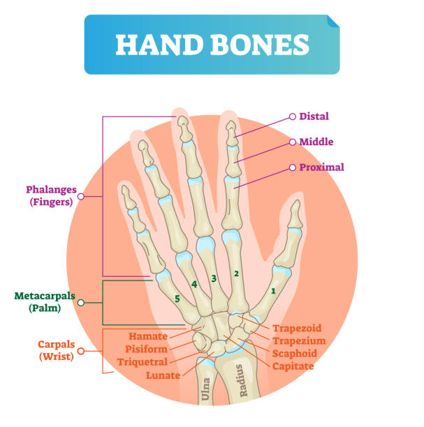
Hand bones vector illustration. Labeled educational human arm structure with phalanges, metacarpals, hamate, pisiform, triquetral and lunate. Palm finger parts scheme.
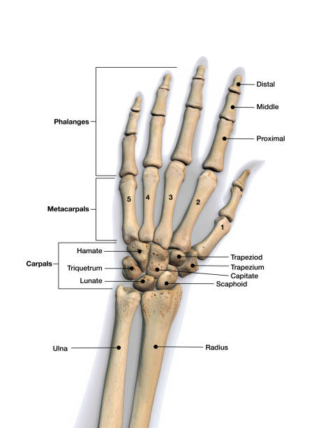
Skeletal bones of wrist and hand with labeling. Dorsal (back) view.
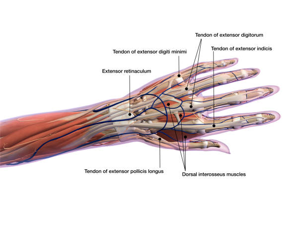
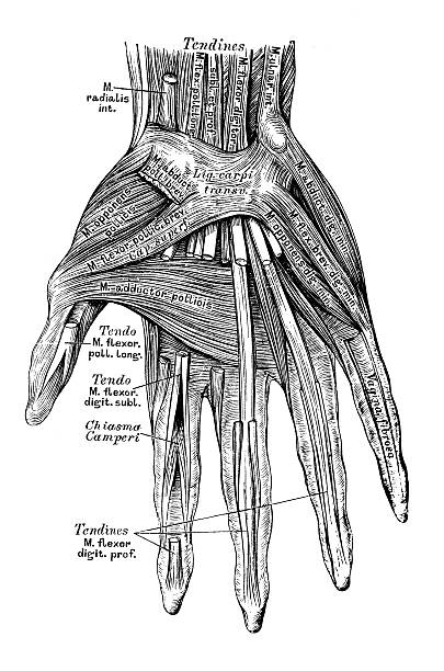
Muscle, tendons and connective ligaments of wrist and hand with labeling. Dorsal (back) view.
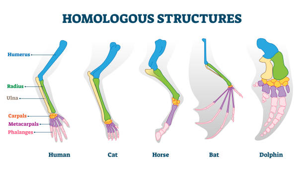
Homologous structures vector illustration. Biological species example scheme. Labeled structural diagram with bone titles. Humerus, ulna and carpals in various creature skeletons from common ancestry.

Wrist cross section educational structure scheme vector illustration. Anatomical labeled arm research with physiological description and explanation. Nerve, musculature, artery and bone location.

Carpal bones with hand palm skeletal structure and anatomy outline diagram. Labeled educational scheme with medical left hand model and isolated hamate, trapezoid or pisiform bone vector illustration.

Human anatomy scientific illustrations with latin/italian labels: wrist joint

Plantigrade, Digitigrade and Unguligrade comparison vector illustration. Educational labeled structure scheme with human, dog and pig legs collection. Bone skeleton parts with location explanation.

Anatomy of dog paw structure with forelimb and hindlimb comparison scheme vector illustration. Educational labeled pads parts description with digital, metacarpal, digital and carpal location examples

Thumb ulnar collateral ligament as finger injury and problem outline diagram. Labeled educational hand xray with bones and cartilage after medical condition and pain explanation vector illustration.

Human anatomy scientific illustrations with latin/italian labels: hand bones

Anatomy of dog paw structure with forelimb and hindlimb comparison scheme vector illustration. Educational labeled pads parts description with digital, metacarpal, digital and carpal location examples

Labeled human anatomy of male lower arm brachioradialis muscles isolated within the skeletal system bones in a frontal view on a white background.

Human anatomy scientific illustrations with latin/italian labels: hand muscles

Labeled human anatomy of male forearm Flexor Carpi Ulnaris muscles isolated within the skeletal system bones in a frontal view on a white background.

Extensor carpi ulnaris muscle for arm and hand wrist movement outline diagram. Labeled educational fusiform muscular system in lateral part of posterior forearm vector illustration. Skeletal bones.

Illustration of a The skeleton of the hand with muscle insertions


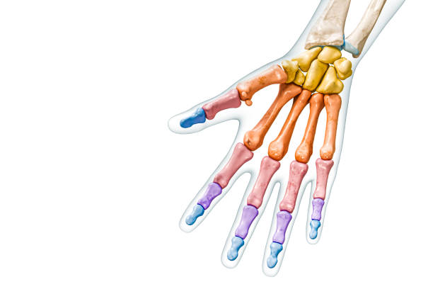
Human hand parts and Bones. Left hand on the white background. Skeletal System and Phalanges fingers. anatomy of human hand and wrist. Labeled

Illustration of a Bones ,ligaments of a hand

Human anatomy scientific illustrations with latin/italian labels: forearm muscles


The wrist is a complex joint that bridges the hand to the forearm. It is actually a collection of multiple bones and joints. The bones comprising the wrist include the distal ends of the radius and ulna, 8 carpal bones, and the proximal portions of the 5 metacarpal bones
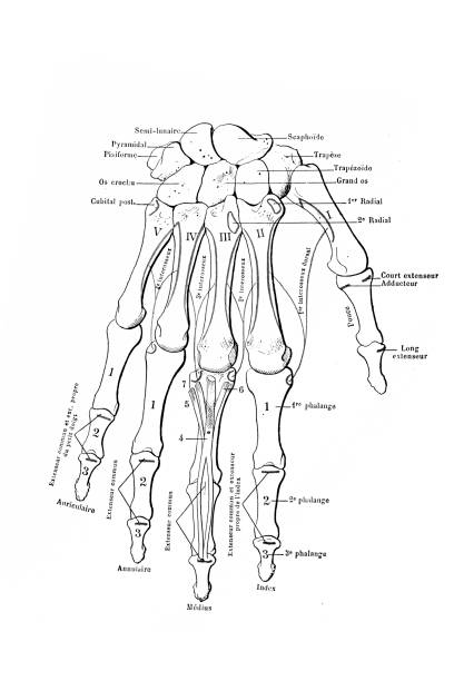
Illustration of a The skeleton of the hand with muscle insertions

Human anatomy scientific illustrations with latin/italian labels: wrist joint

Illustration of a Bones ,ligaments of a hand

Illustration of a Cross section bones of a hand

Illustration of a Cross section bones of a hand

Human anatomy scientific illustrations with latin/italian labels: forearm muscles

Labeled human anatomy of male forearm Extensor Digitorum muscles isolated within the skeletal system bones in a rear view on a white background.

Labeled human anatomy of male lower arm Extensor Digiti Minimi muscles isolated within the skeletal system bones in a rear view on a white background.
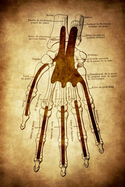
Illustration of a The skeleton of the hand with muscle insertions


Adductor pollicis muscle with hand or palm skeletal system outline diagram. Labeled educational scheme with proximal phalanx of thumb, metacarpals and capitate bones vector illustration. Arm xray.

Human anatomy scientific illustrations with latin/italian labels: hand muscles

Human anatomy scientific illustrations with latin/italian labels: hand muscles
Human anatomy scientific illustrations with latin/italian labels: hand muscles

Human anatomy scientific illustrations with latin/italian labels: hand muscles

Human anatomy scientific illustrations with latin/italian labels: hand muscles

Trichome and bristle types comparison and division groups outline diagram. Labeled educational biological categories with plant hair differences vector illustration. Tapering, glandular and stellate.

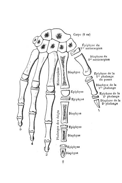
Illustration of a Ossification of the bones of the hand


Process Of Injury Treatment and Bandaging, Infographics For Healthcare Or Medical-related Content. Hand With Wound Being Wrapped and Bandaged With White Bandage. Cartoon Vector Illustration


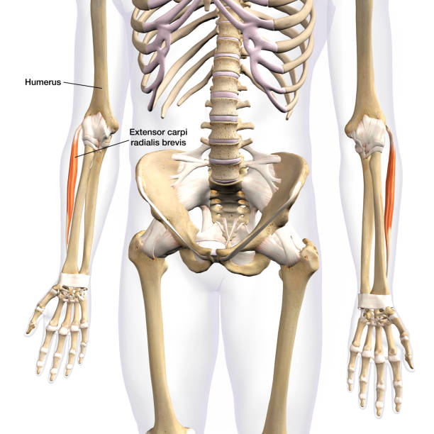
Labeled human anatomy of male lower arm Carpi Radialis Brevis muscles isolated within the skeletal system bones in a frontal view on a white background.

Next