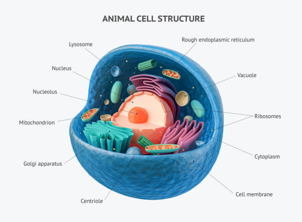
3d rendering of biological animal cell with organelles cross section isolated on white. Animal cell with placed text annotations to all organelles
Browse 100+ labeled mitochondria diagram stock photos and images available, or start a new search to explore more stock photos and images.
3d rendering of biological animal cell with organelles cross section isolated on white. Animal cell with placed text annotations to all organelles

Human metabolism vector banner. Labeled chemical energy educational scheme. Explanation diagram with food carbohydrates, fats and proteins reactions to create ATP and heat. Biological diet infographic

ATP ADP cycle. Adenosine triphosphate (ATP) is a organic chemical that provides energy for cell. intracellular energy transfer. Adenosine diphosphate (ADP) is organic compound for metabolism in cell. Vector diagram for educational, biological, medical and science use. model of molecule adenosine triphosphate, and Adenosine diphosphate

Neuromuscular junction vector illustration scheme. Labeled medical infographic. Motor neuron and muscle cell structure closeup. Diagram with myofibril and muscle fibers.
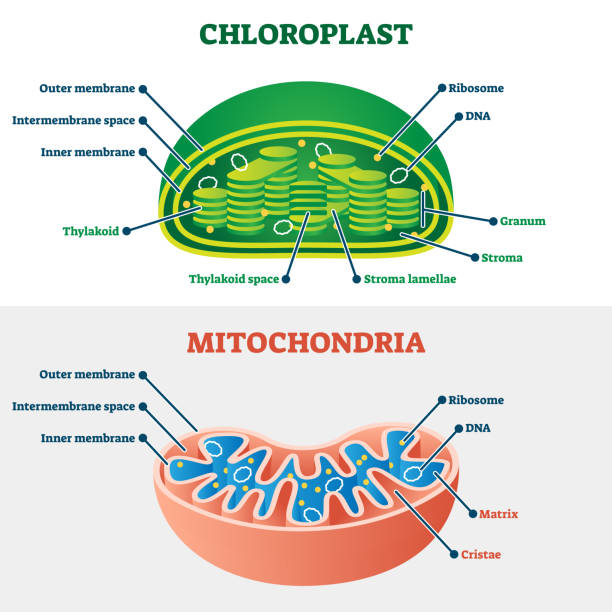
Chloroplast vs mitochondria vector illustration. Labeled educational structure scheme. Biological cell part diagram for school handout. Physiology closeup model with plant energy organelles comparison

Dopamine vector illustration. Labeled diagram with its action and pathways. Scheme with closeup presynaptic axon, terminal, synaptic cleft, dendrite and receiving cells.
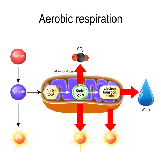
Aerobic respiration. Cellular respiration. Pyruvate enter the mitochondria in order to be oxidized by the Krebs cycle. products of this process are carbon dioxide, water, and energy. Vector diagram for educational, biological, science and medical use
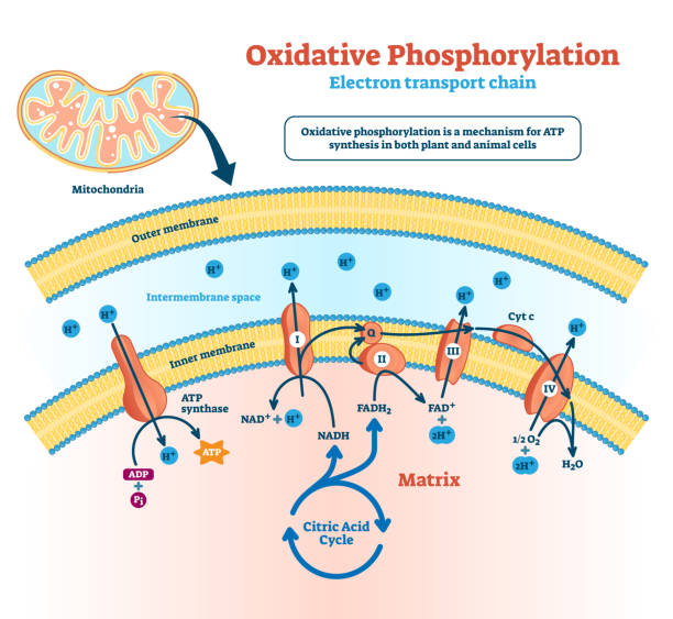
Oxidative phosphorylation vector illustration. Labeled electron transport linked metabolism scheme. Educational diagram with cells use enzymes to oxidize nutrients process in explanation infographics.
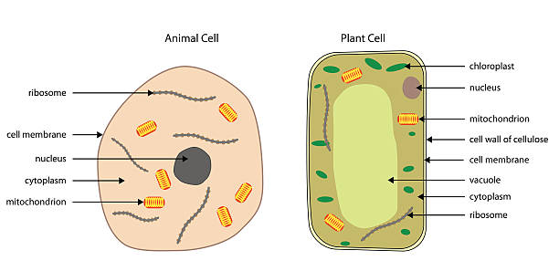
Labelled diagrams of typical animal and plant cells with editable layers.

Cell organelles biological vector illustration diagram. Cross sections of nucleus, cytoplasm liquid, centresome tubes, mitochondria, golgi apparatus, membrane, endoplasmic reticulum and RNA ribosome.

Adipose tissue vector illustration. Labeled medical body fat explanation scheme. Educational diagram with WAT, BAT and beige fat locations and structure. Infographics with inner skin section closeup.
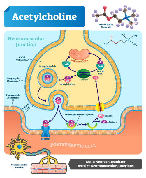
Acetylcholine vector illustration. Labeled scheme with structure of neurotransmitter, neuromuscular junction, synaptic vesicle, axon and cleft. Anatomical closeup diagram

Toxoplasma gondii structure. Vector illustration flat design

Plasmodium is the malaria parasite, is a large genus of parasitic protozoa. Infection with these protozoans is known as malaria, a deadly disease. Diagram of Plasmodium merozoites structure. vector illustration for medical, educational and science use

Aerobic Respiration bio anatomical vector illustration diagram, labeled educational medical scheme. Clean outline style drawing poster. Presentable scientific information close up cross section design

Insulin secretion vector illustration. Biological pancreas function labeled scheme. Full cycle diagram with glucose transporter, metabolism, ATP and potassium channel.

ATP ADP cycle. intracellular energy transfer. Energy absorb and Energy released in a cell. Adenosine triphosphate (ATP) and Adenosine diphosphate (ADP). Vector diagram for education, biological, medical and science use.

Yeast cell vector illustration. Labeled fungus microorganism closeup structure diagram. Biological scheme with educational internal parts titles. Single celled natural fermentation process ingredient.

Synapse vector illustration. Labeled diagram with neuromuscular junction, glandular and other neirons example. Closeup with isolated axon, cleft and dendrite structure.

Fat cell structure vector illustration. Labeled anatomical adipocytes scheme. Cytoplasm, reservoir, golgi apparatus and endoplasmic reticulum educational diagram.

Toxoplasma gondii is an obligate intracellular, parasitic protozoan that causes the disease toxoplasmosis. Diagram of Toxoplasma structure.

Plasmodium is an intracellular, parasites of vertebrates and insects (mosquito) that causes the disease malaria. Diagram of Plasmodium structure. vector illustration for medical, educational and science use

Cell anatomy vector illustration. Labeled educational structure diagram. Isolated microscopic biological scheme with cytoplasm, mitochondria, ribosome and endomlasmic reticulum location explanation.
Cells consist of a protoplasm enclosed within a membrane, which contains many biomolecules such as proteins and nucleic acids.

Structure of the Chondrocytes. Chondrocytes cells in healthy cartilage.

Chloroplast vs mitochondria process educational scheme vector illustration. Labeled photosynthesis and cellular respiration interaction diagram. Graphic with green plant chemical formula or ATP energy

Aerobic respiration with mitochondrion in cell: Glycolysis, Oxidative decarboxylation of pyruvate, Citric acid cycle and Oxidative phosphorylation. Cellular respiration. Krebs cycle

Protist cell anatomy with euglena microorganism structure outline diagram. Labeled educational scheme with green organism parts description vector illustration. Eukaryotic biological inner structure.

Internal structure of leaf diagram illustration

3D Isometric Flat Vector Conceptual Illustration of Animal Cells, Educational Diagram

3D Isometric Flat Vector Illustration of Autophagy, Diagram of the Process

Internal structure of leaf diagram illustration

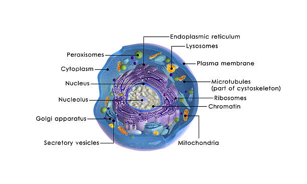
An infographic of eukaryotic cell

Plant cell structure. vector diagram. anatomy of a biological cell with labeled parts. cross section of a plant cell. Illustration for education. Poster

Mucous Neck Cell Structure Illustration. Human Stomach Epithelium Cells Diagram. Gastric Mucous Neck Cell Anatomy. Biology Diagram of Histology of Mucous Neck Cell

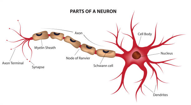
Anatomy of a typical human neuron. Structure neuron

Cell organelles biological anatomy vector illustration diagram of the nucleus, cytoplasm liquid, centresome tubes, mitochondria, golgi apparatus, membrane, endoplasmic reticulum and RNA ribosome.
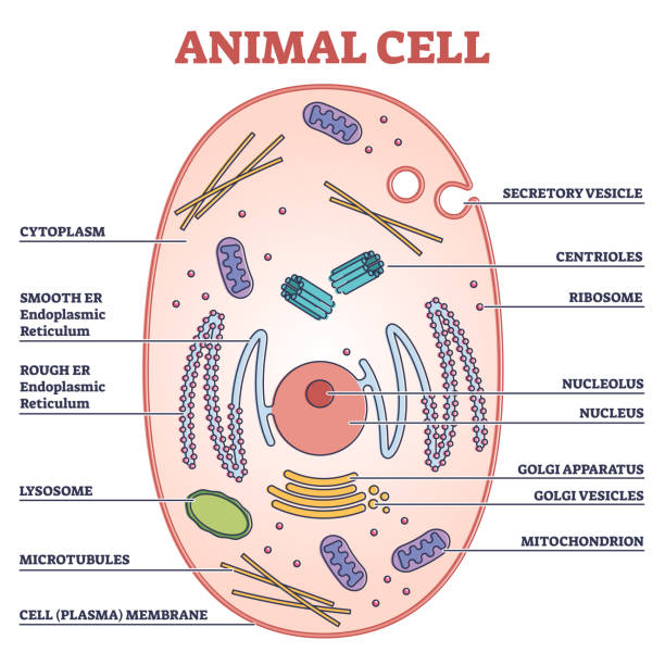
Animal cell with labeled anatomical structure parts in educational outline concept diagram. Biological explanation with inner sections vector illustration. Graphic with microscopic cross section model

Internal structure of leaf diagram illustration

ATP ADP cycle. Adenosine triphosphate ATP and Adenosine diphosphate ADP molecules. Molecular chemical structural formula and model. Cellular energy. Krebs cycle. Aerobic and Anaerobic respiration. Vector illustration

Amoeba labeled vector illustration. Educational single cell animal structure scheme with ectoplasm, nucleus, mitochondrion, pseudopod, lysosome and contractile vacuole.
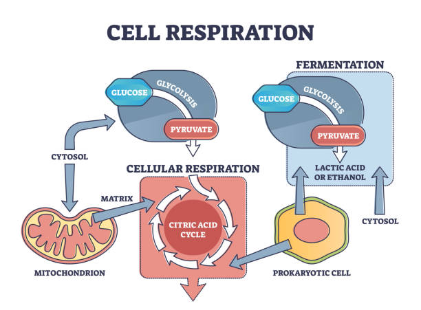
Cell respiration process explanation with biological stages outline diagram. Educational labeled scheme with citric acid, cytosol and glycolysis as molecular chemical cycle info vector illustration.

Energy intake of the cell. Structure of the cell

Chondrocyte vector illustration infographic. Medical labeled diagram structure of chondrocyte in lacunae, interterritorial matrix, elastic, hyaline and fibro cartilage.

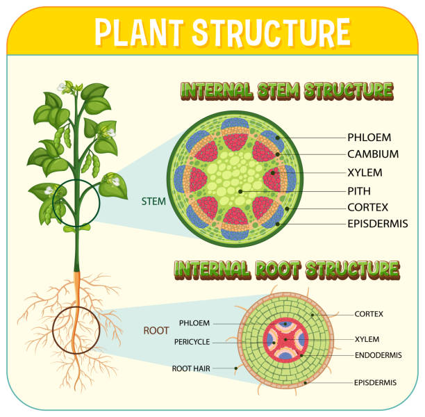
Internal structure of root diagram illustration

Internal structure of leaf diagram illustration

Internal structure of leaf diagram illustration

Internal structure of leaf diagram illustration

Animal cell anatomy infographics with detailed educative diagram and labelled elements realistic vector illustration.
Internal structure of root diagram illustration

Vector illustration of human mucous neck cell with detailed labeled parts nucleus, mitochondria, Golgi apparatus, endoplasmic reticulum, mucin granules, microvilli, and vacuoles.

Modern flat vector illustration of a labeled plant cell structure. Includes organelles such as chloroplast, nucleus, vacuole, mitochondria, and cell wall, clearly marked for educational purposes. Ideal for biology classrooms, science textbooks, school projects, digital education platforms, and academic presentations. Clean design enhances usability for both students and educators in biological sciences.