Structure of kinesin. Kinestin is motor proteins into eukaryotic cells. kinesin walking on a microtubule
Browse 280+ microtubule stock photos and images available, or search for neuron microtubule to find more great stock photos and pictures.
Structure of kinesin. Kinestin is motor proteins into eukaryotic cells. kinesin walking on a microtubule

Brain cells die, neuron diseased, certain areas of brain shrink memory loss or changes in memory for people age 65 and up at risk could affect younger people. Info graphic vector.

The actin cytoskeleton is essential for maintaining the shape and structure of cells, and enabling cell migration, 3D rendering

Fibroblast vector illustration. Microscopic close-up with extracellular, collagen fibrils, elastin, hyaluronic acid. Scheme with microtubule, golgi apparatus, nucleus, mitochondrion and ribosomes.

Microtubules, 3D computer artwork. Microtubules are polymers of the protein tubulin. They are a component of the cytoskeleton, which maintains a cell's shape, allows some cellular mobility and is involved in intracellular transport.The tubular polymers of tubulin can grow as long 50 micrometres and are highly dynamic. In Alzheimer's disease, the transport of tau-protein (belonging to the MAP proteins) stabilizing the microtubules is disturbed and allows phosphate-groups to attach to the tau-protein, destabilizing the microtubuli of brain axons. This leads to the agglutination of the nerve cells resulting in neuronal degeneration.

A cell during anaphase. astral microtubules generate forces that stretch the cell into an oval 3d rendering

Pathological phosphorylation (yellow) of Tau proteins (red-orange) leads to disintegration of microtubuli in the neuron axon an aggregation of the tau proteins. The transport of synaptic vesicles (orange-blue) is interrupted.
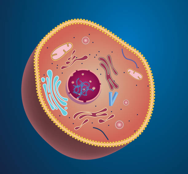
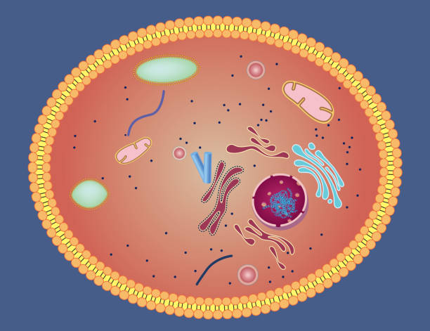
Isolated cell biology pictogram. Cell anatomy structure vector illustration. Cell structure detailed colorful anatomy with description.

Biological anatomy of centrioles

Brain cells die, neuron diseased, certain areas of brain shrink memory loss or changes in memory for people age 65 and up at risk could affect younger people. Info graphic vector.
Synaptic transmission, human nervous system. Concept consciousness. Brain synapses. Transmission synapse, signals, impulses in brain, information transfer at the cellular or atomic level. 3D Rendering

Dopamine vector illustration. Labeled diagram with its action and pathways. Scheme with closeup presynaptic axon, terminal, synaptic cleft, dendrite and receiving cells.

Microtubules, 3D computer artwork. Microtubules are polymers of the protein tubulin. They are a component of the cytoskeleton, which maintains a cell's shape, allows some cellular mobility and is involved in intracellular transport.The tubular polymers of tubulin can grow as long 50 micrometres and are highly dynamic. In Alzheimer's disease, the transport of tau-protein (belonging to the MAP proteins) stabilizing the microtubules is disturbed and allows phosphate-groups to attach to the tau-protein, destabilizing the microtubuli of brain axons. This leads to the agglutination of the nerve cells resulting in neuronal degeneration.
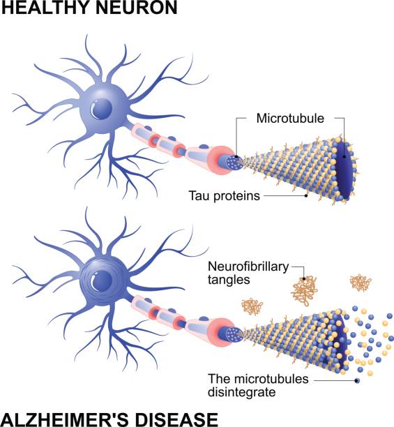
Alzheimer's disease is the change in tau protein that results in the breakdown of microtubules in brain cells. Mechanism of disease. Diagram shows two neurons: healthy cell and neuron with Alzheimer's disease. Tau hypothesis. Neurofibrillary tangles

Synaptic transmission, human nervous system. Concept consciousness. Brain synapses. Transmission synapse, signals, impulses in brain, information transfer at the cellular or atomic level. 3D Rendering
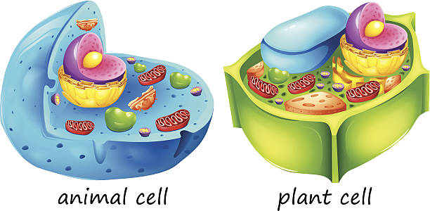
lllustration of the animal and plant cells on a white background

Serototin vector illustration. Labeled diagram with gut brain axis and CNS. Intestinal microbiota influence brain behavior and intestinal cycle. Educational infographic.

Animal cells are generally smaller than plant cells. Animal cells range from 10 to 30 micrometers in length, while plant cells range from 10 and 100 micrometers in length. Animal cells come in various sizes and tend to have round or irregular shapes. Plant cells are more similar in size and are typically rectangular or cube shaped.

Using a transparent effect.
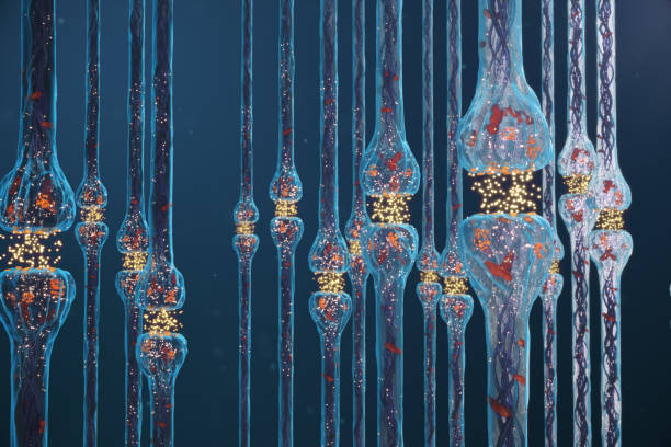
Synaptic transmission, nervous system receptors. Concept consciousness. Brain synapses. Transmission synapse, impulses in brain, information transfer at the cellular or atomic level. 3D Rendering

Intracellular transport. Computer artwork of a vesicles (spheres) being transported along a microtubule by a kinesin motor protein. Kinesins are able to 'walk' along microtubules. Microtubules are polymers of the protein tubulin and are a component of the cytoskeleton.

Nerve cell types and organelles of the cell body Close-up detailed anatomy illustration
Gradient and transparent effect used.
Found only in animal cells, these paired organelles are typically located together near the nucleus in the centrosome, a granular mass that serves as an organizing center for microtubules

3d illustration universal blood , red blood cells with enzyme

Forensic Science in Lab. Forensic scientist working with DNA sample
Gradient and transparent effect used.

Internal anatomy of the prokaryotic cell. Different types of bacteria. Comparison with the eukaryotic cell.

Synaptic transmission, nervous system receptors. Concept consciousness. Brain synapses. Transmission synapse, impulses in brain, information transfer at the cellular or atomic level. 3D Rendering
Gradient and transparent effect used.

3D rendering of the nucleus of a neuron cell

Enzyme ATPases as Kinesins move along microtubule filaments, and are powered by the hydrolysis of adenosine triphosphate or ATP 3d rendering 3d rendering

Mammalian cell forming an X shape, viewed under a fluorescent microscope.

Alzheimer's disease. dementia. Vector Background with neurons and amyloid plaques (tau protein that lead to the disintegration of microtubules in brain cells).

Synaptic transmission, human nervous system. Concept consciousness. Brain synapses. Transmission synapse, signals, impulses in brain, information transfer at the cellular or atomic level, 3D Rendering

Synapse vector illustration. Labeled diagram with neuromuscular junction, glandular and other neirons example. Closeup with isolated axon, cleft and dendrite structure.
Gradient and transparent effect used.

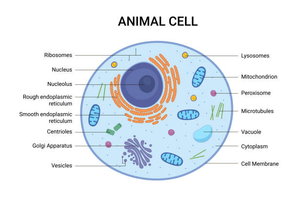
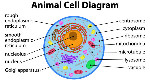
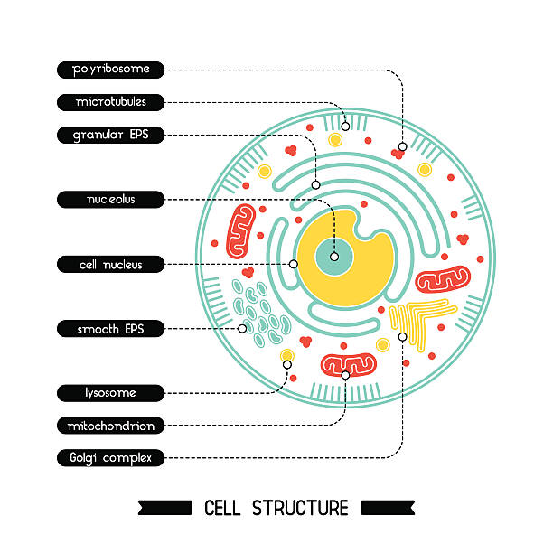
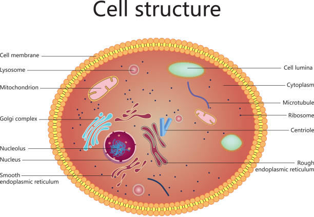
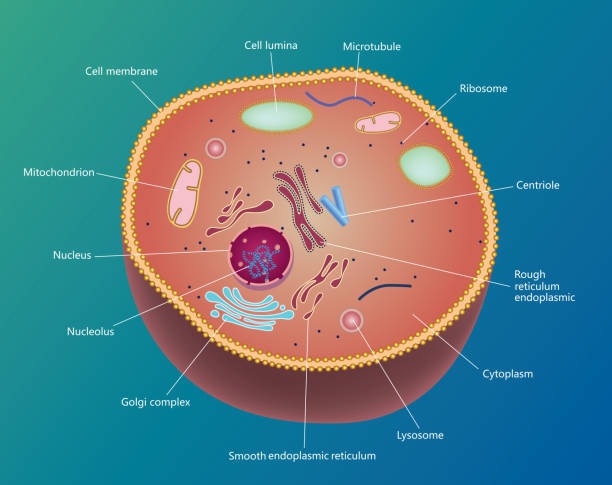
Cell anatomy vector illustration. Labeled educational structure diagram. Isolated microscopic biological scheme with cytoplasm, mitochondria, ribosome and endomlasmic reticulum location explanation.

Transmission electron microscope (TEM) micrograph showing two synapses with clear synaptic vesicles. The postsynaptic element (a dendrite) shows ribosomes and cisternae of rough endoplasmic reticulum

Peroxisome anatomy