Images
Phagocytosis Diagram Pictures, Images and Stock Photos
Browse 170+ phagocytosis diagram stock photos and images available, or start a new search to explore more stock photos and images.
Most popular
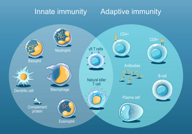
Adaptive and Innate immunity. Cells of The Immune System. Immune response. Immunology infographic. Rapid and slow response. Isometric flat vector Illustration

Phagocytosis. Neutrophil that uses its plasma membrane to engulf a bacterium. From endocytosis to exocytosis. educational scheme. Digestion process in phagocyte. immune system mechanism. vector illustration.

immune system cells. macrophage, t-cell, b-cell and antibodies.
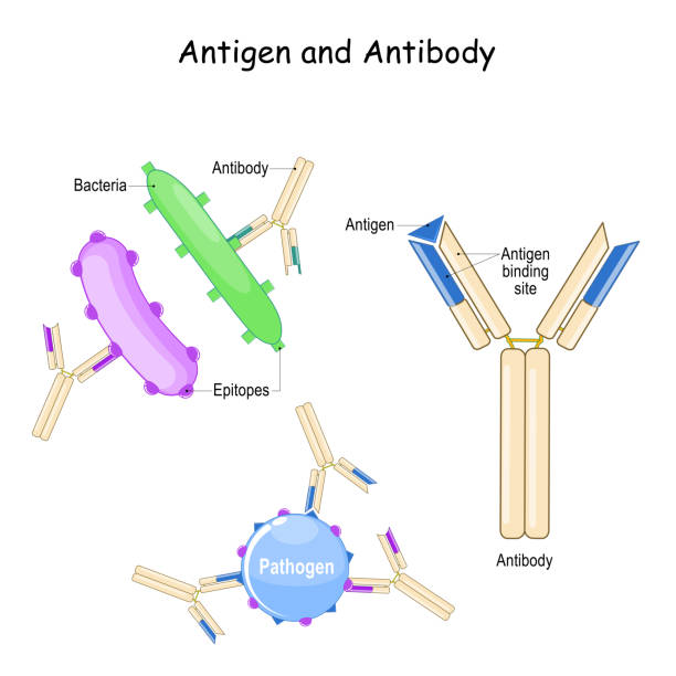
Antibody and Antigen. Humoral immunity and antigen-antibody complex. Two bacteria with different epitopes and antibodies. Biology, Immunology, and Microbiology study"r"n
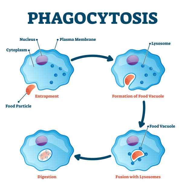
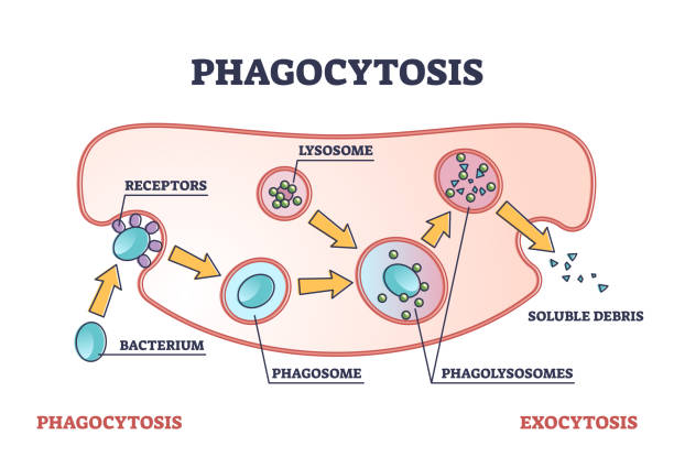
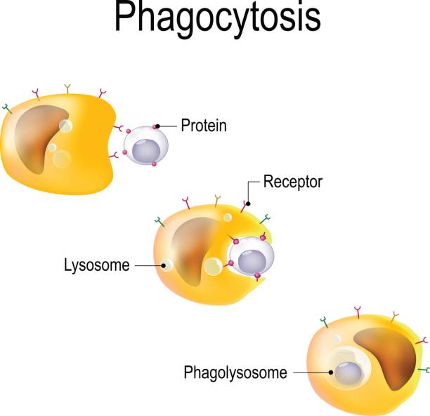
Phagocytosis vector illustration. Labeled endocytosis educational scheme. Cycle with entrapment, vacuole formation, lysosomes fusion and digestion process. Educational immune system mechanism closeup.

Leukocyte adhesion and migration. After activation by chemotactic agents, the leukocytes change shape. The leukocytes then crawl and undergo diapedesis by interacting with platelet-endothelial cell adhesion molecules.

Phagocytosis. leukocyte and virus. Macrophage is a white blood cell that engulfs and digests viruses, microbes, cancer cells etc. Human immune system. Vector illustration for educational, scientific, medical and microbiological use
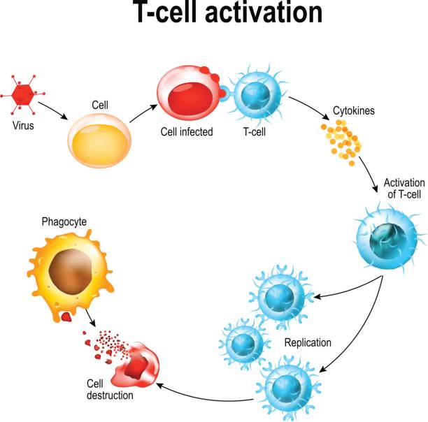
Activation of T-cell leukocytes. T-cell encounters its cognate antigen on the surface of an infected cell. T cells direct and regulate immune responses and attack infected or cancerous cells.

Cytotoxic T cell. T-cell regulate immune responses, release the perforin and granzymes, and attack infected or cancerous cells. Through the action of perforin, granzymes enter the cytoplasm of the target cell, and lead to apoptosis (cell death).

Phagocytosis. Professional phagocytic cells. Neutrophils, macrophages, monocytes, dendritic cells, osteoclasts and eosinophils are immune response to most infections. Vector illustration. Medical poster.
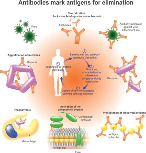
An antibody (Ab), also known as an immunoglobulin is a large, Y-shaped protein produced mainly by plasma cells that is used by the immune system to identify and neutralize pathogens such as bacteria and viruses. Vector graphic.

Activation of B-cell leukocytes: lymphoblast, activation, memory B-leukocyte, virus, plasma cell, antibody, antigen, and naive lymphocyte
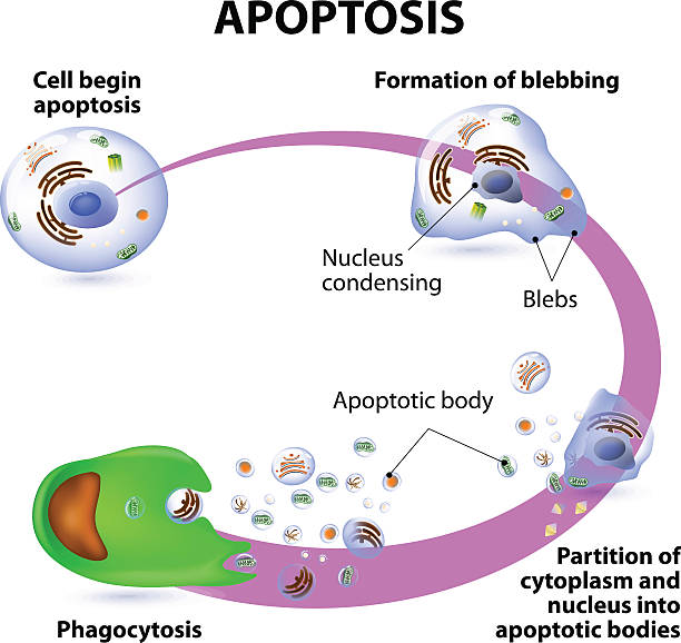
Apoptosis is the process of programmed cell death. Vector diagram
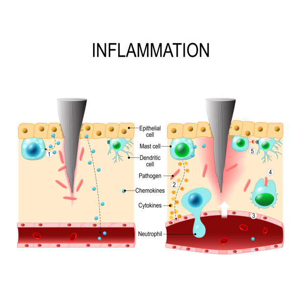
Inflammation is a process by which white blood cells protect of the body from infection with bacteria and viruses. Phagocytes are attack any patogens. Neutrophils migrate from blood vessels to the infected tissue. Immune system. Vector diagram for educational, biological, medical and science use
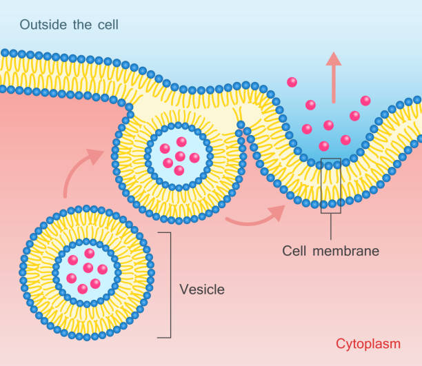
Exocytosis - vesicle transport that carry very large molecules across the cell membrane. Vector illustration
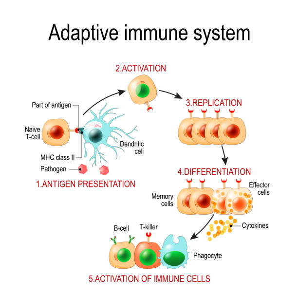
Adaptive immune system from Antigen presentation to activation of other immune cells. specific immune. T-helper and T-killer cells. Memory and Effector cells. Viruse, Lymphocyte, antibody and antigen. Vector diagram for educational, biological, and science use

Apoptosis. Programmed cell death. Caspases. Cellular homeostasis. Cell fragmentation. Apoptotic bodies for Phagocytosis. Vector. Schematic diagram. Detailed poster.

Apoptosis or Necrosis. Difference between necrotic death of a cell, and apoptosis of a cell. Comparison of the premature death of cells and programmed death. Morphological changes. Vector poster. Isometric Flat illustration.

Epstein-Barr virus. life cycle. EBV replication: Entry to the cell, latency and reactivation. human herpesvirus that caused of cancer, and infectious mononucleosis

Hallmarks of aging. Chromosomes with Telomeres before and after division of new and senescent cell. Cell division will cease once telomeres shorten to a critical length. Cellular aging. Vector illustration

autophagy of mitochondria. Diagram of the process of autophagy from Forming a membrane and autophagosome to fuse phagosome and lysosome when contents of the vesicles are degraded and recycled. Autophagy defects linked to various diseases and cancer development. Vector poster
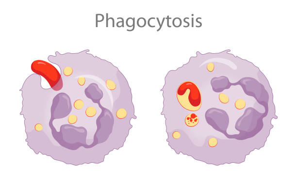
Phagocytes contain membranous sacs called lysosomes that contain various digestive enzymes, microbicidal chemicals, and toxic oxygen radicals. The lysosomes fuse with the phagosomes containing the ingested microbes and the microbes are destroyed

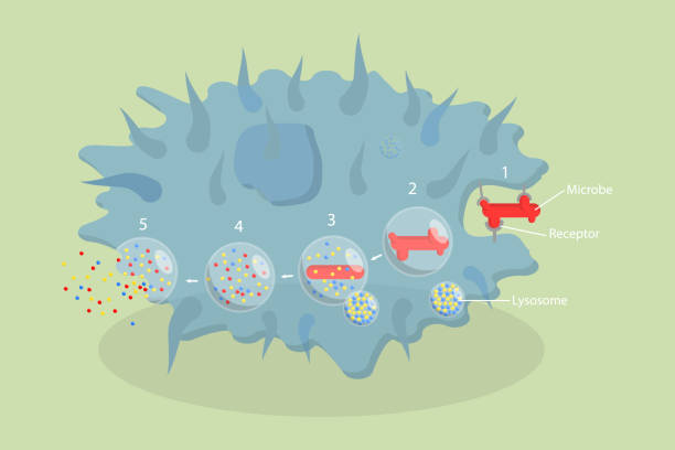
Phagocytosis in 4 steps: Binds with pathogen, pathogen is ingested by a phagocyte, fusion of lysosomes with the phagosome, and Antigen presentation. immune system. Vector diagram for educational, biological, and science use
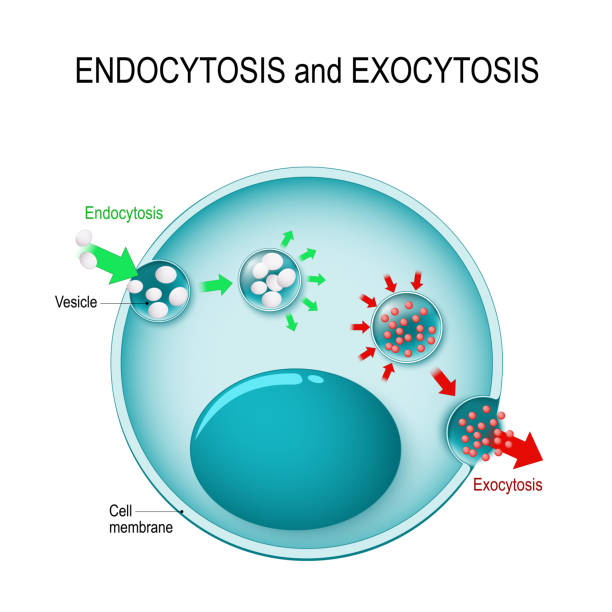
endocytosis and exocytosis in the cell. endocytosis: transports proteins into the cell. exocytosis: transports molecules out of the cytoplasm. vesicles fuses with membrane, contents are secreted into the extracellular environment

humoral immunity. Lymphocyte, antibody and antigen. Vector diagram

B cells Activation. B-cell lymphocytes (white blood cell) that function in the humoral immunity component of the adaptive immune system. leukocyte that secreting antibodies
Phagocytosis as cellular ingesting and eliminating process outline diagram. Cell immunity response and anatomical organism mechanism to eliminate enemies, bacterium and particles vector illustration.

difference between exocytosis and endocytosis. The cell transports molecules into and from the cell. Vector illustration for science and educational use


The Epstein-Barr virus (EBV) replication cycle (Entry to the cell, latency and reactivation). human herpesvirus. the cause of infectious mononucleosis and cancer.
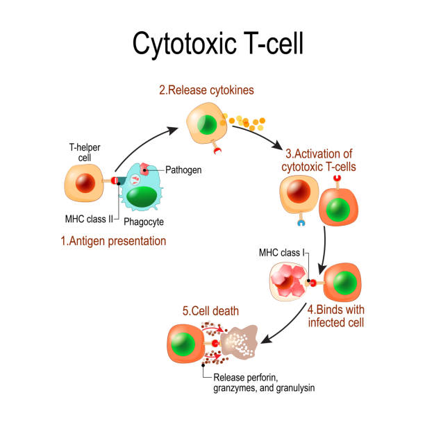
Cytotoxic T cell. T-cell regulate immune responses, release the perforin and granzymes, and attack infected or cancerous cells. Through the action of perforin, granzymes enter the cytoplasm of the target cell, and lead to apoptosis (cell death

Red blood cell life cycle medical vector illustration circulation diagram. Biological anatomy scheme with forming blood cells and transporting through inner organs.


Endocytosis. The transport of macromolecules into a cell in a vesicle. Vector illustration design
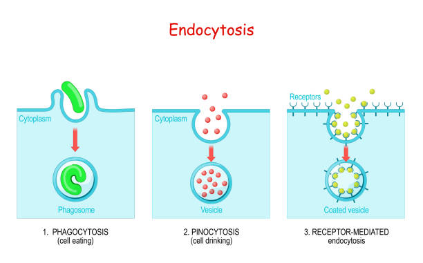
endocytosis. phagocytosis is cell eating, pinocytosis is a cell drinking, receptor-mediated endocytosis - when cells absorb metabolites, hormones, proteins and viruses by receptors on the surface of the cell.
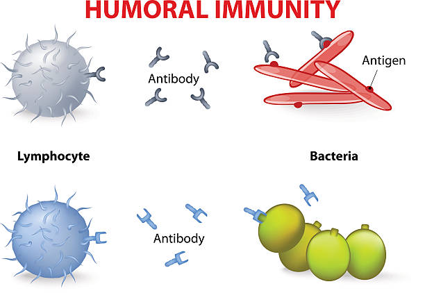
humoral immunity. Lymphocyte, antibody and antigen. Vector diagram
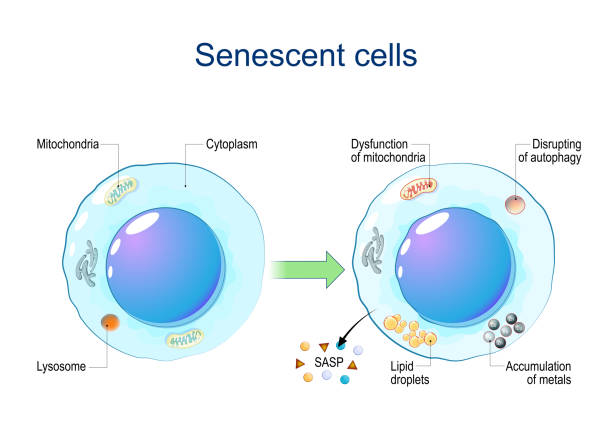
Senescent cells. Cellular senescence from Dysfunction of mitochondria, accumulation of metals, Disrupting of autophagy, Lipid droplets to release of Senescence-associated secretory phenotype SASP and chronic inflammation. DNA damage response. Aging cells. Vector diagram
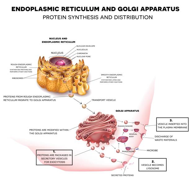
Endoplasmic reticulum and Golgi Apparatus. Protein synthesis and distribution detailed drawing


B cell activation. Antigen presentation. Plasma cells and Antibody production. B cell signaling pathways. immune response. Vector poster
Phagocytosis in immune system or apoptosis. three steps.
3D Isometric Flat Vector Conceptual Illustration of Phagocytosis, Labeled endocytosis Educational Scheme

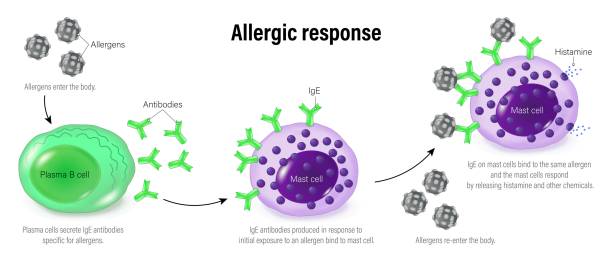
Allergy mechanism diagram. Allergic diseases concept. Immunoglobulin E antibodies in the human immune system. Binding to an allergen and to a receptor on mast cells medical flat vector illustration.

Phagocytosis. macrophage absorption of bacteria. Stages of mechanism of the immune response from entrapment or endocytosis to phagosome formation, degradation and exocytosis. Vector illustration. Medical poster.
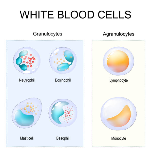
Granulocytes and Agranulocytes. White blood cells. Two types of leukocytes. Close-up of Cells of Immune system Lymphocyte, Monocyte, Neutrophil, Eosinophil, Basophil and Mast cell. Vector illustration
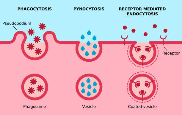
Three major types of endocytosis. Cell eating, cell drinking, receptors coated pit on cell membrane. Vector illustration.

The different types of endocytosis: receptor-mediated endocytosis, pinocytosis (cell drinking) and phagocytosis (cell eating). vesicle, coated vesicle, and phagosome. vector illustration for medical, educational and science use
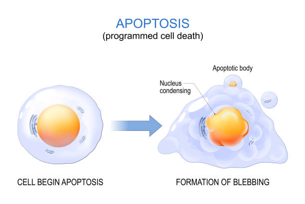
Apoptosis. programmed cell death. aging process in cells. Structural changes of ageing and senescent cells from normal cell to final stage of formation of membrane blebbing. Vector illustration

Images of macrophages.


3D Isometric Flat Vector Conceptual Illustration of Neutrophil, Educational Scheme
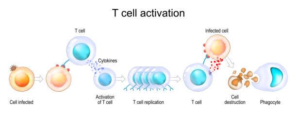
Activation of leukocytes. T-cell encounters its cognate antigen on the surface of an infected cell. T-cells direct and regulate immune responses and attack infected or cancerous cells. Cell-mediated immunity. The Adaptive and Innate immune system. vector poster
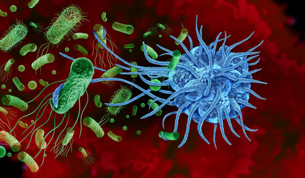
Phagocyte Attacking Bacteria as a white blood cell or leukocyte immune cell that engulfs and digests harmful particles with extended pseudopodia to protect the body.

Allergy mechanism diagram. Allergic diseases concept. Immunoglobulin E antibodies in the human immune system. Binding to an allergen and to a receptor on mast cells medical flat vector illustration.
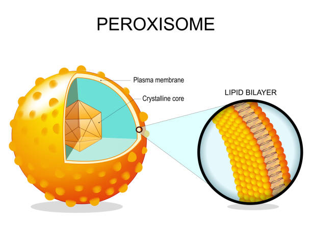
Peroxisome anatomy. Cross section of a cell organelle. Close-up of a Lipid bilayer Plasma membrane, Crystalline core, transport proteins. Vector illustration

Neutrophil vector illustration. Medical educational scheme with labeled capillary, circulation, adherence, deformability, chemotaxis and phagocytosis. Apoptosis and microorganisms icroscopic closeup.


T cell Activation. T lymphocyte, is a white blood cell. cell-mediated immunity
Next