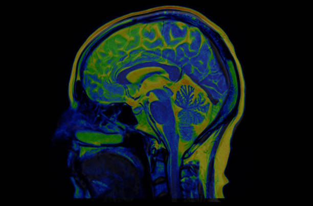
"Spect is a nuclear medicine tomographic imaging technique and it consists of injecting radioactive tracer (for instance, Technetium-99m, 99mTc) into bloodstream, followed by the reading of the radioactivity by the gamma camera.SPECT imaging is performed by using a gamma camera to acquire multiple 2-D images from multiple angles. A computer is then used to apply a tomographic reconstruction algorithm to the multiple projections, yielding a 3-D dataset. This dataset may then be manipulated to show thin slices along any chosen axis of the body, similar to those obtained from other tomographic techniques, such as MRI, CT, and PET. to reconstruct.In the brain, the radioactivity measures the blood flow that indirectly indicates the metabolic activity. Higher metabolic activity is indicated by color orange.Spect can be used to detect area of the brain responsible for generating epilepsy seizures. This is a computer generated image, it is not as sharp as a regular photograph, but this is the sharpest image we can get to date."