Normal Human Skin Split Layers Cube with Muscle, physical structure of skin anatomy Illustration about medical and healthcare diagram, health science biology and dermatology vector.
Browse 620+ subcutaneous layer stock photos and images available, or search for epidermis to find more great stock photos and pictures.
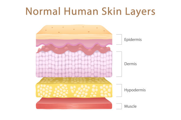
Normal Human Skin Split Layers Cube with Muscle, physical structure of skin anatomy Illustration about medical and healthcare diagram, health science biology and dermatology vector.
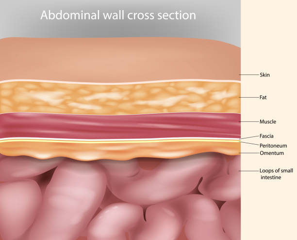
Abdominal wall cross section Anatomy. Abdominal wall layers Medical Illustration
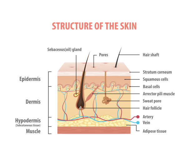
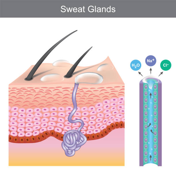
Human skin. Layered epidermis with hair follicle, sweat and sebaceous glands. Healthy skin anatomy medical vector illustration. Dermis and epidermis skin, hypodermis
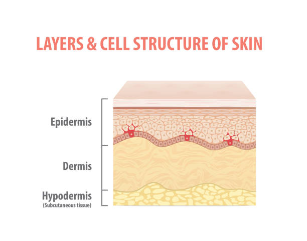
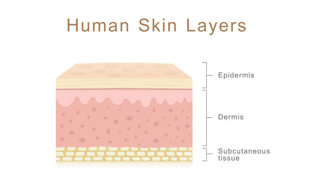
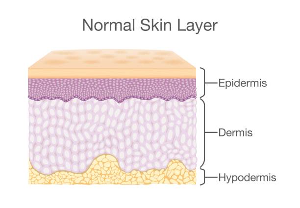
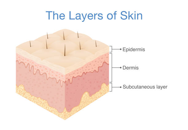
Cross-section illustration of human skin, composed of three primary layers: epidermis, dermis and subcutis.
Human skin layers, healthcare and medical illustration about human skin

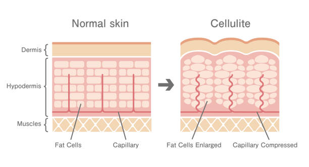
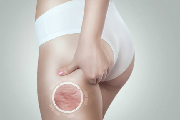
Cellulite formation. Orange peel syndrome. Adiposis edematosa. Cross section of Cellulite and healthy skin. Skin anatomy. Fat tissue of human body. Vector illustration. Epidermis and dermis texture.
Skin is the soft outer covering of vertebrates. Other animal coverings such as the arthropod exoskeleton have different developmental origin, structure and chemical composition.
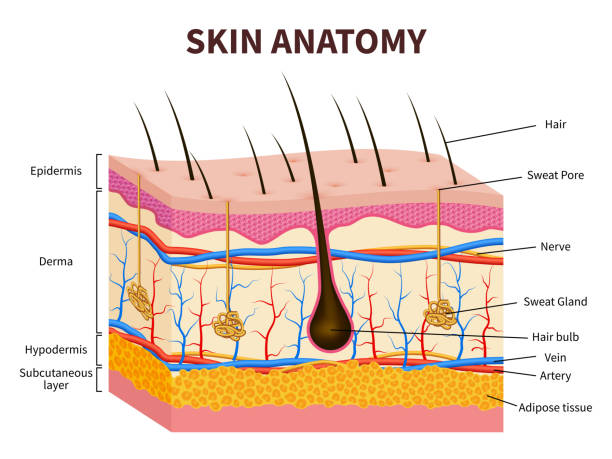
Skin layers, structure anatomy diagram vector illustration. Cartoon human skin infographic anatomical education background, epidermis with hair follicle, layered hypodermis and dermis, sweat pore

Human skin anatomy. Digital illustration.

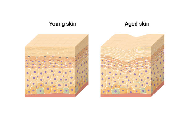
Visual representation of skin changes over a lifetime. Collagen and elastin form the structure of the dermis making it tight and plump. Fibroblasts synthesize collagen and elastin.

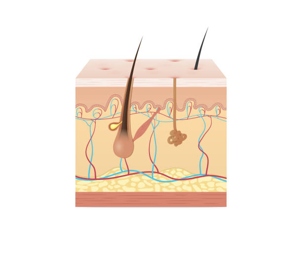
Layers Of Human Skin with hair follicle, sweat and sebaceous glands. Epidermis, dermis, hypodermis and muscle tissue. Vector illustration for your design and medical use
Human skin structure with collagen and elastane fibers, hyaluronic acids, fibroblasts. Schematic illustration. Layers of the human skin. skin and health care concept Vector diagram
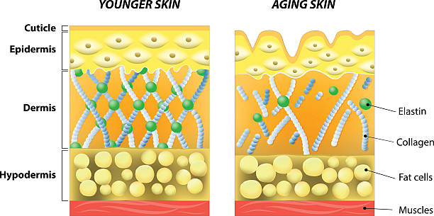
younger skin and aging skin. elastin and collagen. A diagram of younger skin and aging skin showing the decrease in collagen and broken elastin in older skin.

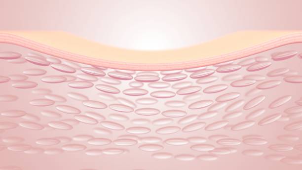
Skin structure 3d render. Abstract skin layer with cells isolated on beige background. Human normal skin epidermis, dermis, subcutaneous fat or hypodermis, healthy skin anatomy. 3D illustration

The human skin is the outer covering of the body. In humans, it is the largest organ of the integumentary system. The skin has multiple layers of ectodermal tissue and guards the underlying muscles, bones, ligaments and internal organs.

Normal Human Skin Layers Isometric Cube with Muscle, physical structure of skin anatomy Illustration about medical and healthcare diagram, health science biology and dermatology vector.

Human skin structure. Vector illustration of epidermis anatomy. Vector illustration of epidermis anatomy.

Visceral and subcutaneous fat around waistline. Location of visceral fat in abdominal cavity. Types of human obesity. Medical scheme. Vector illustration isolated on white background.

Skin Type Sticker. Age related dermis with wrinkles. Layers of skin with uneven epidermis. Design element for scientific books. Cartoon isometric vector illustration isolated on white background

Three main layer of the human skin. Illustration about medical diagram.
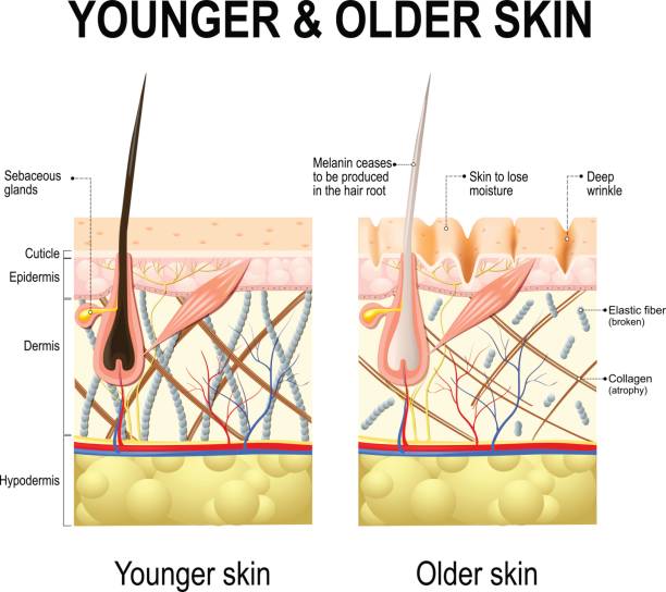
Human skin changes or ageing skin. A diagram of younger and older skin showing the decrease in collagen fibers, atrophy and broken elastin, formed wrinkles, hair becomes gray in the elderly.
Layer of Healthy Human Skin in vector style and components information. Illustration about medical diagram.

Vector illustration of human hair diagram. Piece of human skin and all structure of hair on the white background. Medical Treatment of baldness, epilation concept

The Layer of Human Skin in vector style and components information. Illustration about medical and health.
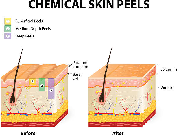
chemical peeling or procedure chemexfoliation. Human skin layers

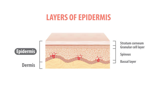
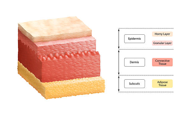
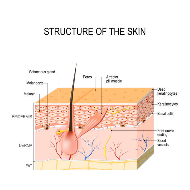
Layers Of Human Skin. Epidermis (horny layer and granular layer), Dermis (connective tissue) and Subcutaneous fat (adipose tissue)

Normal Human Skin Layers Cube with Muscle, physical structure of skin anatomy Illustration about medical and healthcare diagram, health science biology and dermatology vector.

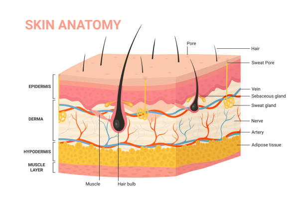
Skin anatomy. Layers: epidermis (with hair follicle, sweat and sebaceous glands), derma and fat (hypodermis). Vector diagram for educational, medical, biological, and scientific use
The Layer of the human skin. Illustration about medical diagram.
Dry skin icon 3d render. Structure of damaged or aging skin with flaking and wrinkles. Epidermis, dermis and subcutaneous tissue skin layers. Isolated skin surface with bubble cells. 3D illustration

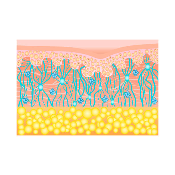
The formation of cellulite. Cellulite occurs in most females and rarely in males. Vector diagram.
Healthy Human Skin. hair follicle, cell structure and layers. Vector illustration for your design and medical use. human anatomy.

Visual representation of skin changes over a lifetime.

Medical infographic illustrations of hair growth cycle. Vector pictures of human biology. Hair human banner, anatomy root follicle