Images
Tunica Adventitia Pictures, Images and Stock Photos
Browse 40+ tunica adventitia stock photos and images available, or search for adventure to find more great stock photos and pictures.
Most popular


Human blood vessel anatomy. Detailed scheme. Editable vector illustration isolated on a light background. Medial, scientifical, healthcare concept. Graphic design.
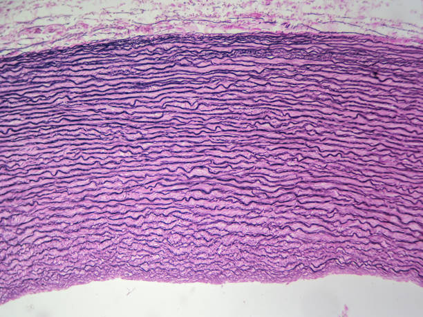
Aorta from primate showing elastic connective tissue (100x)


Chasubles in the interior of the Cathedral of Saint Gerland or San Gerlando in the old town of Agrigento, Sicily, Italy,


Mechanism of a mitochondria.


Chasubles in the interior of the Cathedral of Saint Gerland or San Gerlando in the old town of Agrigento, Sicily, Italy,

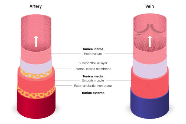
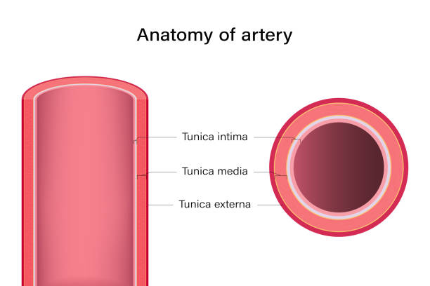
The filtration of substances across an arterial wall and hypertension depends on blood pressure


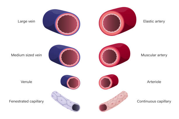
Light micrograph of a longitudinal section of a large vein showing, from the top, the tunica intima with the endothelium and a thin subendothelial connective tissue. A media layer formed by a few bundles of circular cross sectioned myocytes. The majority of the wall is occupied by intermingled bundles of collagen fibres belonging to the tunica adventitia. Two small nerve fiber bundles can be seen in the adventitia.
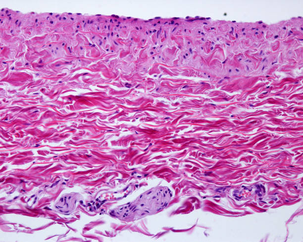
Very low magnification micrograph showing the wall of a large vein. The muscular layer is very thin. Almost the entire wall corresponds to the adventitia layer, rich in bundles of collagen fibers.

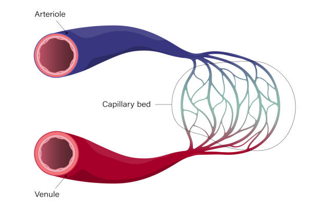
This is the aspect of the blood vessel.


Gaius Plinius Secundus ( AD 23/24 – AD 79 ), called Pliny the Elder was a Roman author, naturalist, natural philosopher, and naval and army commander of the early Roman Empire, and a friend of the emperor Vespasian. Original edition from my own archives Source : Larousse 1899
Chasubles in the interior of the Cathedral of Saint Gerland or San Gerlando in the old town of Agrigento, Sicily, Italy,


Cross section of blood vessel.
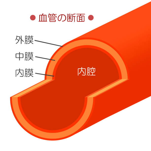
This is the structure of the blood vessel.


Light micrograph of a longitudinal section of a large vein showing, from the top, the tunica intima with the endothelium and a thin subendothelial connective tissue. A media layer formed by a few bundles of circular cross sectioned myocytes. The majority of the wall is occupied by intermingled bundles of collagen fibres belonging to the tunica adventitia. A small blood vessel (belonging to the vasa vasorum system) can be seen in the adventitia.


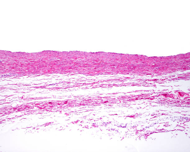
Aorta from primate showing elastic connective tissue (400x)

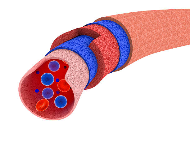
Digital medical illustration: Microscopic cross section of human blood vessel featuring: