A scientist placing a slide under a microscope
Browse 2,700+ under the microscope stock photos and images available, or search for nature under the microscope or human body under the microscope to find more great stock photos and pictures.
A scientist placing a slide under a microscope

A scientist using a pipette with a microtiter plate and a petri dish

Lenses of the fluorescent microscope.

Stem of cotton cross section. Light microscope slide with microsection of plants of the genus Gossypium in the mallow family Malvaceae. Plant anatomy. Biology. Photo.
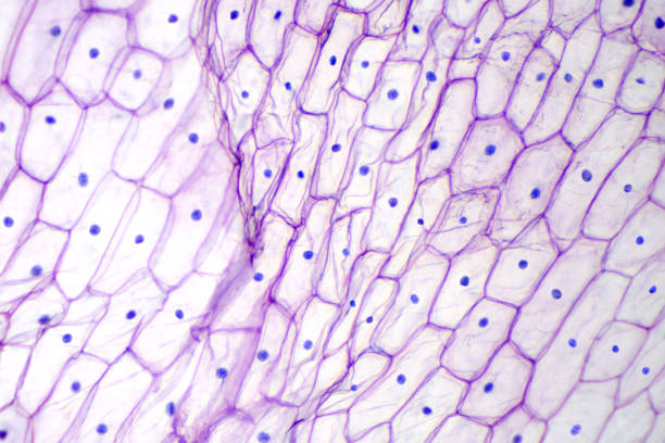
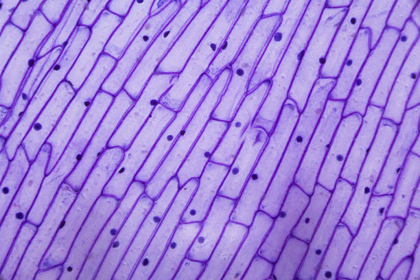
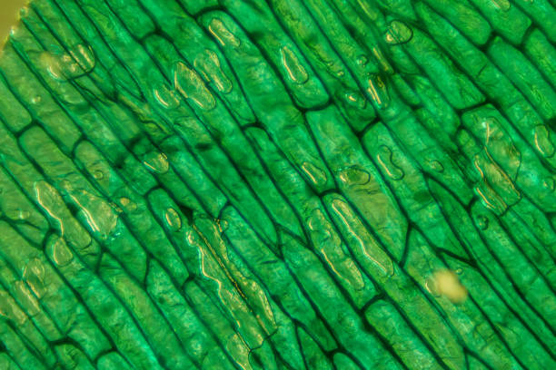
Onion epidermis under light microscope. Purple colored, large epidermal cells of an onion, Allium cepa, in a single layer. Each cell with wall, membrane, cytoplasm, nucleus and large vacuole. Photo.

A scientist placing a slide under a microscope

A scientist placing a slide under a microscope

Spinal fluid smear on the test glass and stain for lab test

Businessman standing under a microscope being examined 3d isometric vector illustration concept for banner, website, landing page, ads, flyer template

Pine mature wood cross section. Light microscope slide with microsection of an evergreen conifer in the genus Pinus. Plant anatomy. Biology. Photo.

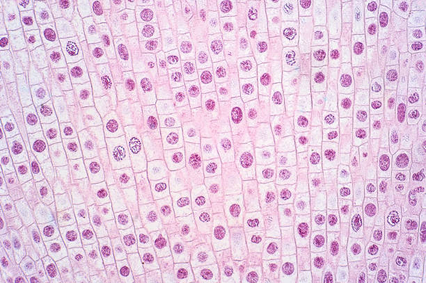
Root tip of Onion and Mitosis cell in the Root tip of Onion under a microscope.

many blured bacteria close up under the microscope. Abstract
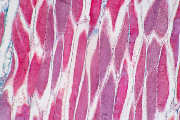
Characteristics of anatomy and Histological sample Striated (Skeletal) muscle of mammal Tissue under the microscope.

The study of biological virus under an electron microscope Virus cells under a microscope. 3D render. Bacteria. Disease. 3D illustration Rendering
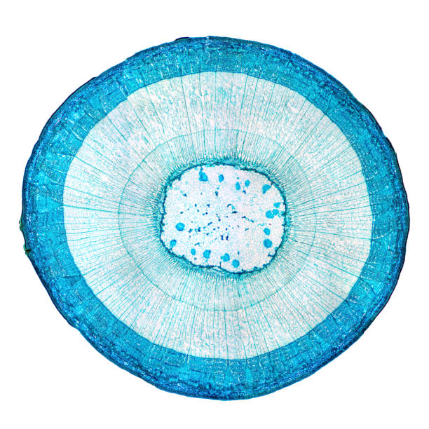
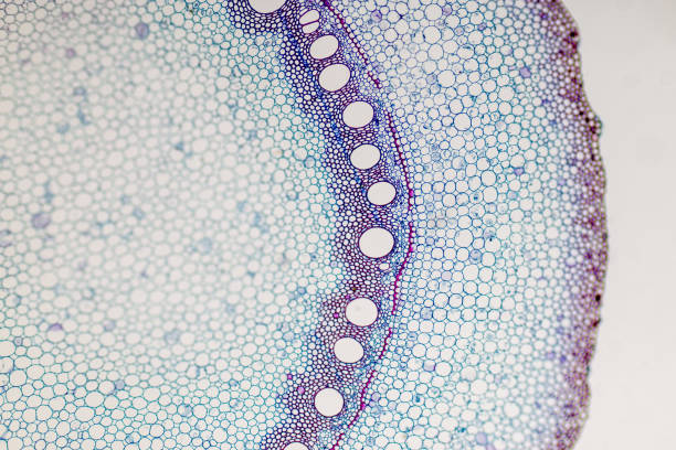
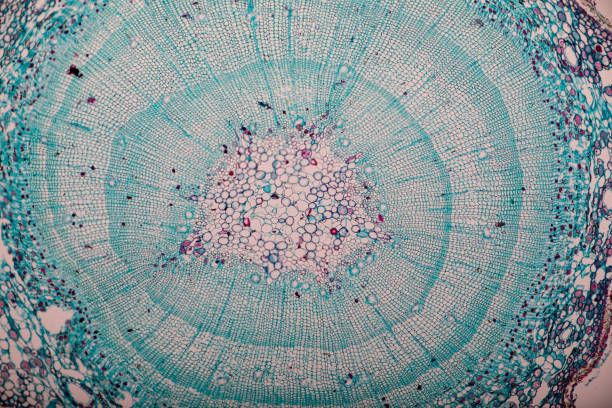
Stem of wood dicotyledon, whole cross section under microscope. Light microscope slide with the microsection of a wood stem with vascular bundles, concentric arranged in a ring. Plant anatomy. Photo.

Host cells with spores (mold) are inside wood under the microscope for education.

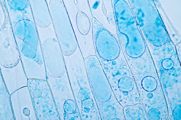
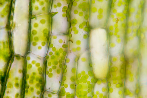
microscopic view of plant cells for botanic education and analysis

Creative ebru background with abstract acrylic painted waves. Beautiful marbling texture. Handmade marble surface. Blue and yellow colors.

Macro increasing of a fungal mold Mucoraceae that grew on canned red beans. Shot through an optical microscope
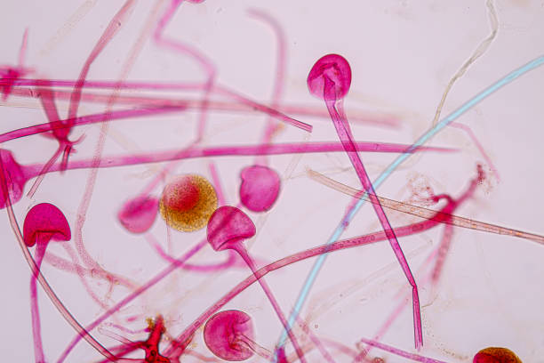
Rhizopus is a genus of common saprophytic fungi on Slide under the microscope .

Scientific futuristic dark background with color gradients, glass in a round eye under a microscope

Magnifying glass with bacteria. White circle on blue background. Medicine concept. Vector illustration. Stock image. EPS 10.

Macro increasing of a fungal mold Mucoraceae that grew on canned red beans. Shot through an optical microscope

microscopic view of plant cells for botanic education and analysis

microscopic view of plant cells for botanic education and analysis

many blured bacteria close up under the microscope. Abstract

The study of plant tissues under the microscope in the laboratory.

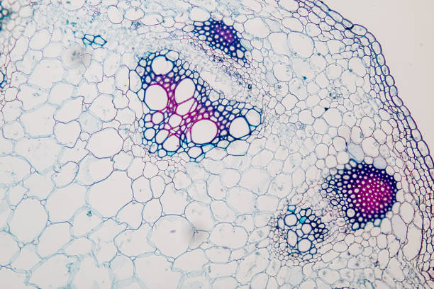
Stem of wood dicotyledon, half cross section under microscope. Light microscope slide with the microsection of a wood stem with vascular bundles, concentric arranged in a ring. Plant anatomy. Photo.

imitation of bacteria and microorganisms, microscopic view, macro abstract background

Characteristics of Kingdom Fungi for education in Microbiology laboratory.