
Images
Urothelium Pictures, Images and Stock Photos
Browse 270+ urothelium stock photos and images available, or start a new search to explore more stock photos and images.
Most popular
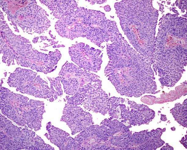
Low grade papillary carcinoma (urothelial carcinoma) formed by papillae with fibrovascular core and coated with transitional epithelium. Cells are relatively uniform without nuclear polymorphism and lack of cellular polarity.
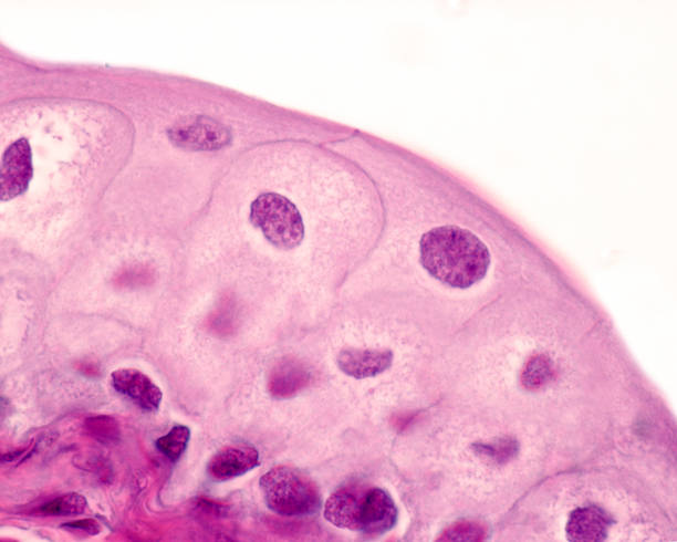
In the urinary tract there is a type of epithelium, the transitional epithelium or urothelium, which is exclusive to this region of the body. It is an epithelium with a stratified appearance, that is, whose cells are arranged in several layers from the base to the light. Above the basal layer, a variable number of cell layers are located depending on the state of distension of the organ, which can be very well evidenced in the urinary bladder. The image pertains to an empty bladder and shows 6 to 7 layers of nuclei, which are reduced to 2-3 in a full bladder. Finally, it is also characteristic of the transitional epithelium that the superficial cells and, sometimes, also in the intermediate layers, are binucleated or show very large nuclei, corresponding to polyploid nuclei.
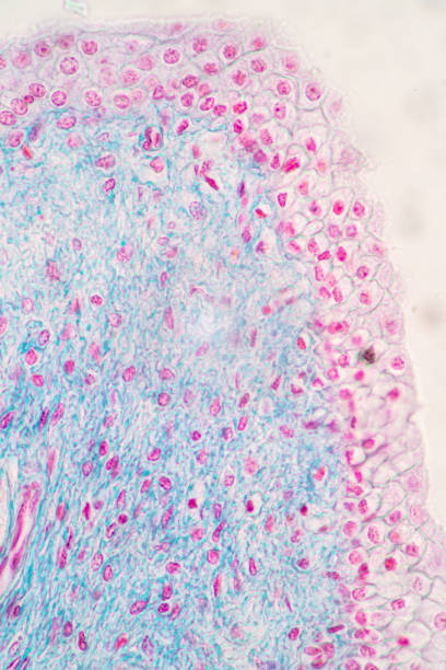
Dog tissue sample. Captured by a scientific microscope and Canon 5D Mark IV
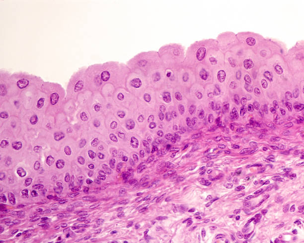
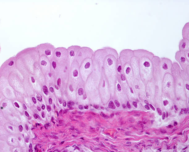
Light micrograph of the mucosa of the urinary bladder lined by transitional epithelium (urothelium) resting on a connective tissue lamina propria. The superficial layer shows large cells with big nuclei, and a rounded apical surface.
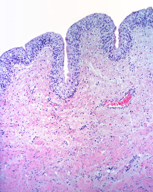
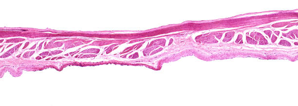
Mucosa of the urinary bladder formed by a transitional epithelium and a lamina propria of connective tissue. Below, the muscular layer formed by smooth muscle fibers begins
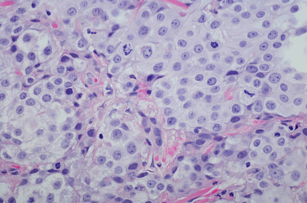
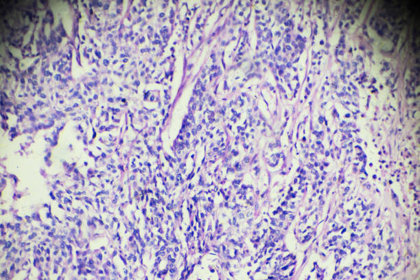
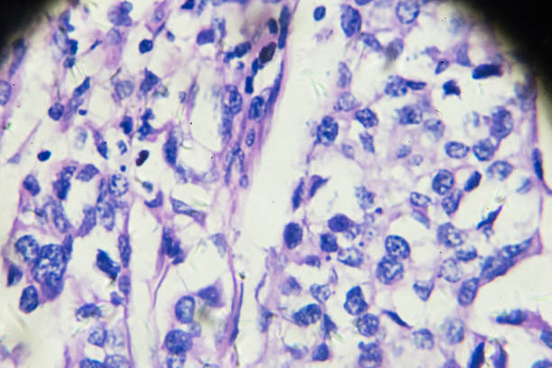
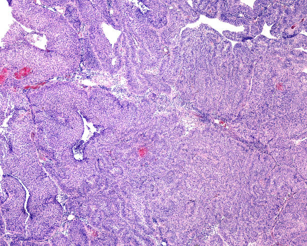
Micrograph of Invasive urothelial carcinoma high grade. Invasive urothelial carcinoma is a type of Transitional cell carcinoma (TCC, also urothelial cell carcinoma or UCC) and is a type of cancer that develops in the urinary system: the kidney, urinary bladder, and accessory organs. It is the most common type of bladder cancer and cancer of the ureter, urethra, and urachus. Invasive urothelial carcinoma originates from tissue lining the inner surface of these hollow organs - transitional epithelium. It can extend from the kidney collecting system to the bladder. H&E Stain
Micrograph of Invasive urothelial carcinoma high grade. Invasive urothelial carcinoma is a type of Transitional cell carcinoma (TCC, also urothelial cell carcinoma or UCC) and is a type of cancer that develops in the urinary system: the kidney, urinary bladder, and accessory organs. It is the most common type of bladder cancer and cancer of the ureter, urethra, and urachus. Invasive urothelial carcinoma originates from tissue lining the inner surface of these hollow organs - transitional epithelium. It can extend from the kidney collecting system to the bladder. H&E Stain


High magnification micrograph of the surface of a transitional epithelium or urothelium. The most important physiological property of this epithelium is, without a doubt, its great impermeability, which prevents the urine collected with so much work by the kidney, from passing through the epithelium and returning back to the body. To do this, these cells have one of the most compact tight junction systems in the entire body. With the light microscope and at high magnifications, these junctions are reflected by a remarkable reinforcement of the intercellular boundaries that can be seen in this image.

In the urinary tract, from the renal pelvis to the urethra, there is a special type of epithelium: it is the transitional epithelium, also called urothelium because it is exclusive to this tract. It is an epithelium with a stratified appearance, with cells whose nuclei are arranged in several layers. However, it has been shown that superficial cells emit processes that reach the basal layer. The surface cells are large and often shaped like an inverted club or pear, as can be seen in the micrograph. Also typical is the domed, globular appearance of the luminal surface of epithelial cells.


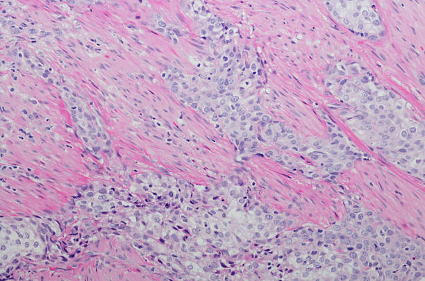
Transitional cell cancer of bladder under microscopy zoom in different areas


Transitional cell cancer of bladder under microscopy zoom in different areas


Transitional cell cancer of bladder under microscopy zoom in different areas


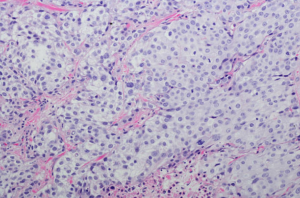
Micrograph of Invasive urothelial carcinoma high grade. Invasive urothelial carcinoma is a type of Transitional cell carcinoma (TCC, also urothelial cell carcinoma or UCC) and is a type of cancer that develops in the urinary system: the kidney, urinary bladder, and accessory organs. It is the most common type of bladder cancer and cancer of the ureter, urethra, and urachus. Invasive urothelial carcinoma originates from tissue lining the inner surface of these hollow organs - transitional epithelium. It can extend from the kidney collecting system to the bladder. H&E Stain
Showing Light micrograph of the Urinary bladder human under the microscope for education in the laboratory.
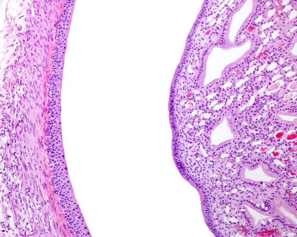
Kidney. Renal papilla and kidney pelvis. Calyx of the renal pelvis(left) lined by transitional epithelium (urothelium). Outside the mucosa, a layer of smooth muscle cells can be seen. On the right, the papilla of the renal pyramid emptying into a minor calyx. The papilla has large papillary ducts of Bellini.


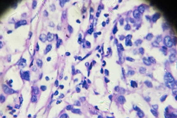
Transitional cell cancer of bladder under microscopy zoom in different areas
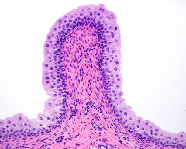
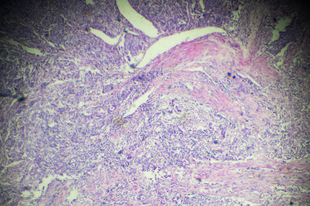
Microscopic image showing a urinary bladder papilloma formed by papillae with fibrovascular core and coated with transitional (urothelial) epithelium. Light microscope micrograph. H&E stain
Next