00:12
Hand Drawn Anatomy of Human Lungs on Chalkboard
Browse 50+ human anatomy diagram organs drawings stock videos and clips available to use in your projects, or start a new search to explore more stock footage and b-roll video clips.

Motion Clip of Hand Drawn Sketch An Anterior View of The Human Skull, A Bony Structure That Forms The Head in Vertebrates.

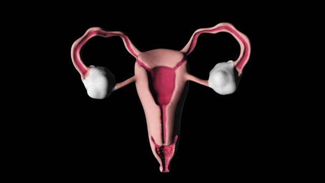
The uterus or womb is a major female hormone-responsive reproductive sex organ of most mammals, including humans.

the sheath of cells and connective tissue which surrounds the root of a hair.

The nervous system is the part of an animal's body that coordinates its voluntary and involuntary actions and transmits signals between different parts of its body.

The nervous system is the part of an animal's body that coordinates its voluntary and involuntary actions and transmits signals between different parts of its body.

Concept of application new technology in future medicine

The nervous system is the part of an animal's body that coordinates its voluntary and involuntary actions and transmits signals between different parts of its body.

The nervous system is the part of an animal's body that coordinates its voluntary and involuntary actions and transmits signals between different parts of its body.

Cardiac muscle (heart muscle) is involuntary striated muscle that is found in the walls and histological foundation of the heart, specifically the myocardium. Cardiac muscle is one of three major types of muscle, the others being skeletal and smooth muscle.

The uterus or womb is a major female hormone-responsive reproductive sex organ of most mammals, including humans.
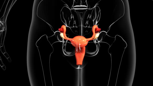
The uterus or womb is a major female hormone-responsive reproductive sex organ of most mammals, including humans.

Animation of blue coloured human body anatomy and internal organs.

Animation of human body anatomy and internal organs isolated on white background.

The uterus or womb is a major female hormone-responsive reproductive sex organ of most mammals, including humans.

Animated anatomy drawing of a human body with internal organs on a white blueprint grid paper with crayons on a cork bulletin board.

The uterus or womb is a major female hormone-responsive reproductive sex organ of most mammals, including humans.

Concept of application new technology in future medicine

The human skull, an intricate and vital component of the human body, serves multiple crucial functions that extend beyond its anatomical structure. Composed of several bones intricately fused together, the skull not only provides protection for the brain and sensory organs but also plays a significant role in facial structure, communication, and cultural symbolism. The human skull consists of two main parts: the cranium and the mandible. The cranium, or braincase, is formed by several bones that encase and protect the brain. These include the frontal, parietal, temporal, occipital, sphenoid, and ethmoid bones, which are joined together by sutures. The mandible, or lower jawbone, articulates with the skull at the temporomandibular joint, allowing for movement during chewing and speech.

Liver anatomy model for medical education purposes. Liver diseases

Human liver anatomy closeup. Realistic anatomy structure of liver organ of hepatic system and organ of digestive gallbladder. Human liver model for medicines pharmacy and education

The thyroid gland is found in the neck, below the thyroid cartilage. The thyroid gland controls how quickly the body uses energy, makes proteins, and controls how sensitive the body is to other hormones.

A close-up view of a detailed anatomical model of the human rectum and anus

Concept of application new technology in future medicine
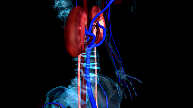
the kidneys are located in the abdominal cavity, more specifically in the paravertebral gutter and lie in a retroperitoneal position at a slightly oblique angle. There are two kidneys. One is on each side of the spine.

the kidneys are located in the abdominal cavity, more specifically in the paravertebral gutter and lie in a retroperitoneal position at a slightly oblique angle. There are two kidneys. One is on each side of the spine.
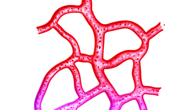
Capillaries are the smallest of a body's blood vessels and are parts of its microcirculation.

Hand Drawn Animation of Human Anatomy Cranial Bones on Chalkboard

4k animation of a human liver contour on a white background. Stock video for presentation about alcohol addiction. Self-drawing liver with alpha channel.

The heart has four chambers, two upper atria, the receiving chambers, and two lower ventricles, the discharging chambers. The atria are connected to the ventricles by the atrioventricular valves and they are separated by the coronary sulcus.

The circulatory system also called the cardiovascular system, is an organ system that permits blood to circulate and transport nutrients (such as amino acids and electrolytes), oxygen, carbon dioxide, hormones, and blood cells to and from cells in the body to nourish it and help to fight diseases, stabilize body temperature and pH, and to maintain homeostasis.

Selfie drawing of human intestines by one line on a white background. Esophagus animation for health and nutrition whiteboard presentation.

The nervous system is the part of an animal's body that coordinates its voluntary and involuntary actions and transmits signals between different parts of its body.

the kidneys are located in the abdominal cavity, more specifically in the paravertebral gutter and lie in a retroperitoneal position at a slightly oblique angle. There are two kidneys. One is on each side of the spine.

the kidneys are located in the abdominal cavity, more specifically in the paravertebral gutter and lie in a retroperitoneal position at a slightly oblique angle. There are two kidneys. One is on each side of the spine.

the kidneys are located in the abdominal cavity, more specifically in the paravertebral gutter and lie in a retroperitoneal position at a slightly oblique angle. There are two kidneys. One is on each side of the spine.

You can use this Video Template for Your Cool promotion. All you need to do is add Your text in your desired editing software.

the kidneys are located in the abdominal cavity, more specifically in the paravertebral gutter and lie in a retroperitoneal position at a slightly oblique angle. There are two kidneys. One is on each side of the spine.