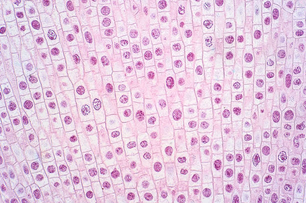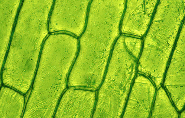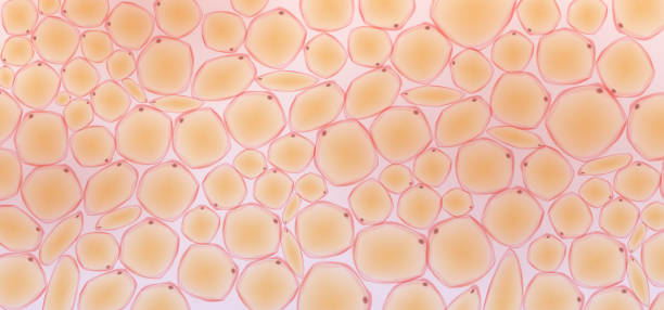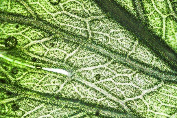
Microscopic image of Microscopic image of nymphaea of aqustio stem
Browse 68,200+ microscope cells stock photos and images available, or search for human cells or dna to find more great stock photos and pictures.
Microscopic image of Microscopic image of nymphaea of aqustio stem
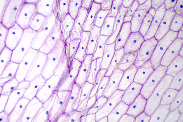
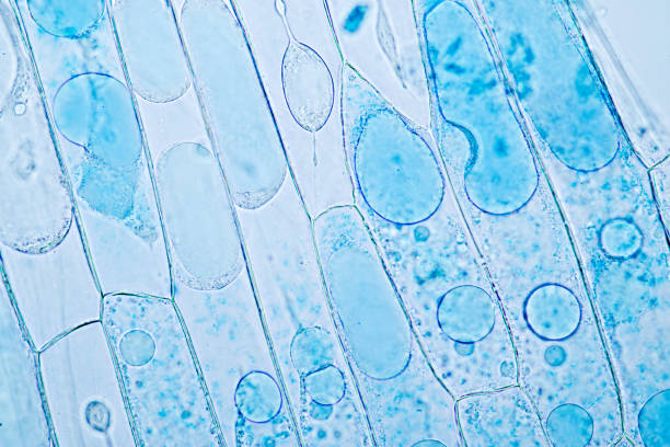
Onion epidermis under light microscope. Purple colored, large epidermal cells of an onion, Allium cepa, in a single layer. Each cell with wall, membrane, cytoplasm, nucleus and large vacuole. Photo.
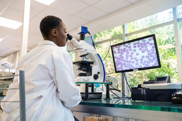
Rear view of a woman pathologist looking at a sample using microscope with magnified image seen on computer screen. Female technician working in a medical laboratory.

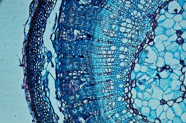
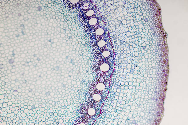
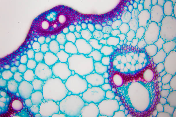
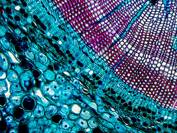
Tilia Stem under Optical Microscope
Lung cancer - adenocarcinoma: Therapies targeting specific genetic alterations such as EGFR, ALK and ROS1 are appropriate for selected cases (photographed and uploaded by US surgical pathologist).
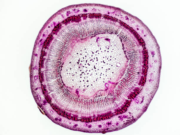
linden stem (Tilia platyphyllos) cross section under the microscope showing phloem, vascular cambium, medullary ray and pith - optical microscope x32 magnification
Cancer Cells under microscope background 3D illustration
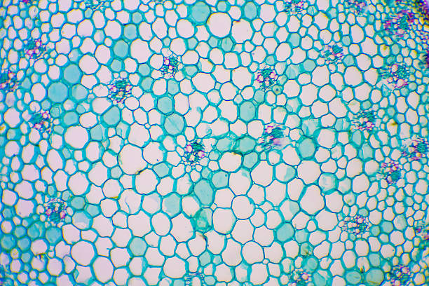
Microscopic image of Microscopic image of nymphaea of aqustio stem

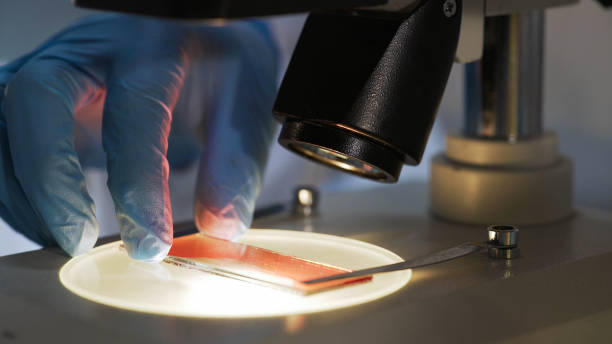
scientist hands with microscope, examining samples and liquid. Medical research with technical equipment

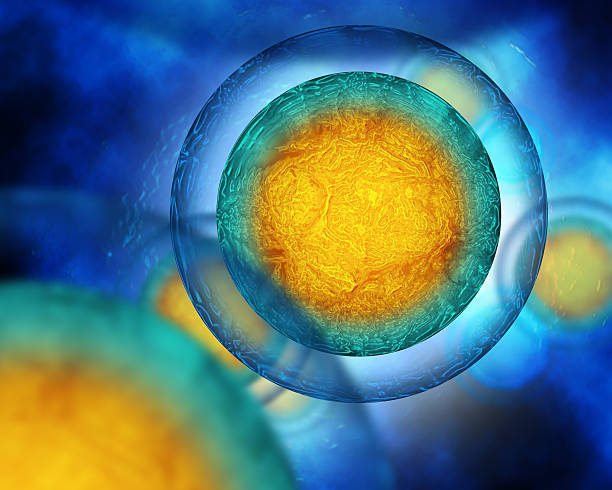
In vitro fertilisation concept. Artificial insemination or fertility treatment macro photography.

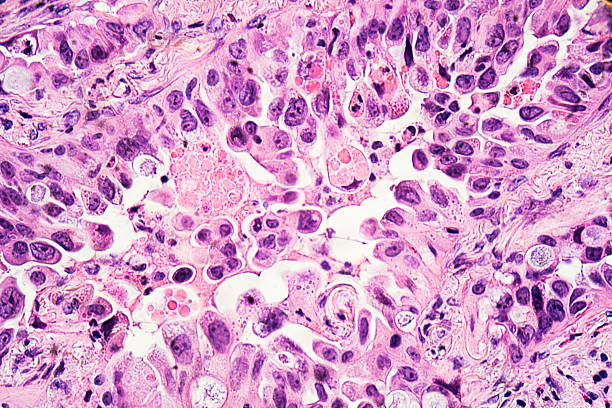
Squamous cell carcinoma or squamous cell cancer (SCC or SqCC) is a cancer of a kind of epithelial cell, the squamous cell. These cells are the main part of the epidermis of the skin, and this cancer is one of the major forms of skin cancer. However, squamous cells also occur in the lining of the digestive tract, lungs, and other areas of the body, and SCC occurs as a form of cancer in diverse tissues, including the lips, mouth, esophagus, urinary bladder, prostate, lung, vagina, and cervix, among others. Despite sharing the name squamous cell carcinoma, the SCCs of different body sites can show tremendous differences in their presenting symptoms, natural history, prognosis, and response to treatment. Micrograph of squamous cell carcinoma of the head and neck
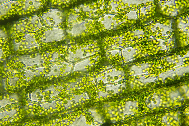
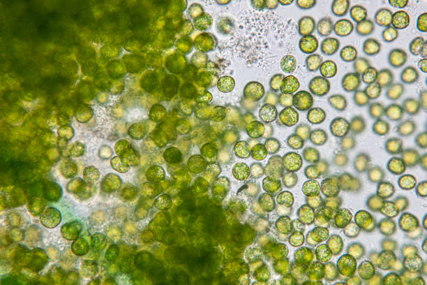
Cells of algae with chloroplast, Microscopic magnification
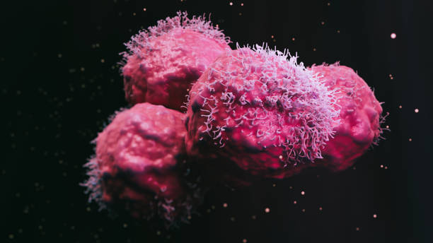
Cancer malignant cells - 3d rendered image, abstract enhanced scanning electron micrograph (SEM) of cancer malignant cells. Visual of overall shape of the cell's surface at a very high magnification. Medical research concept.

Cancer cells vis - 3d rendered image, enhanced scanning electron micrograph (SEM) of cancer cell. Visual of overall shape of the cell's surface at a very high magnification. Medical research concept.
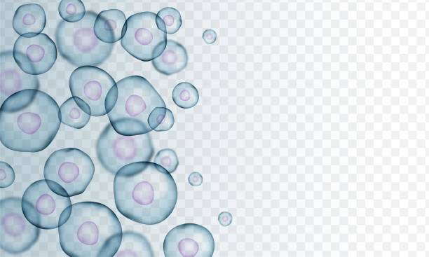
Transparent cell stem background template. Biology research dna nucleus cells vector pattern.
Monkeypox mutation,Mutated fever monkey,variant of smallpox
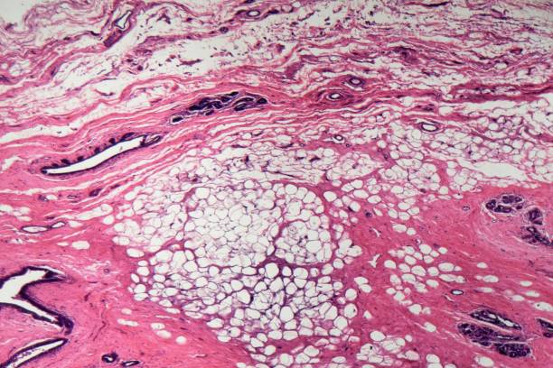
Section of human mammary gland cells under the microscope
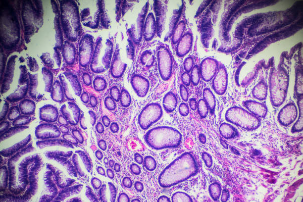
Squamous epithelial cells under microscope view for education histology. Human tissue.
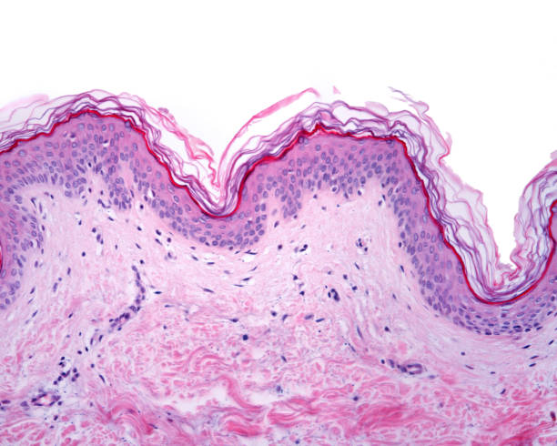
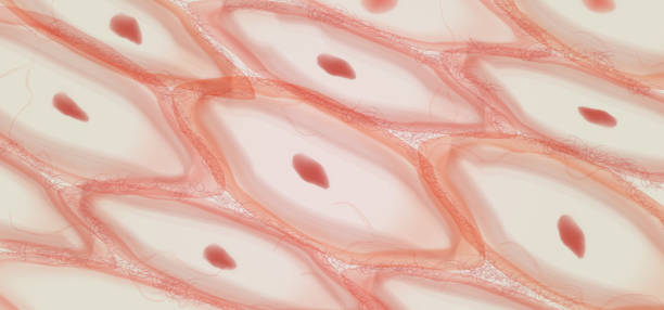
Thin skin showing the epidermis with their different layers resting on dermis.
Pine Stem Cross Section seen on microscope at 200x Magnification. Phase Contrast Optical Microscope
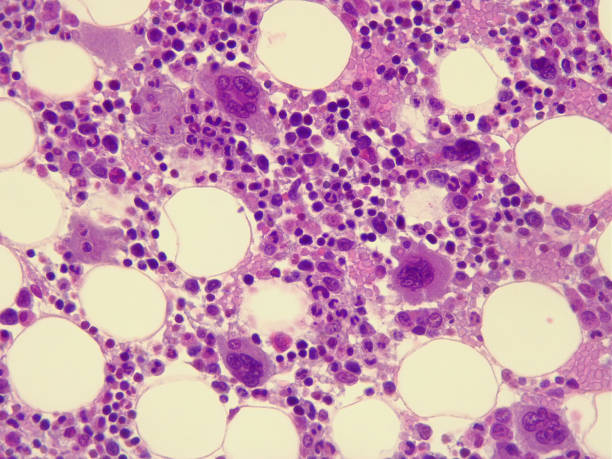
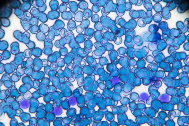
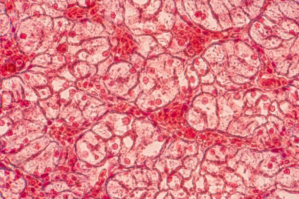
Human bone marrow under the microscope. 400x magnification
Micrograph of myeloma neoplasm bone marrow biopsy. Hematoxylin and eosin staining (H&E)
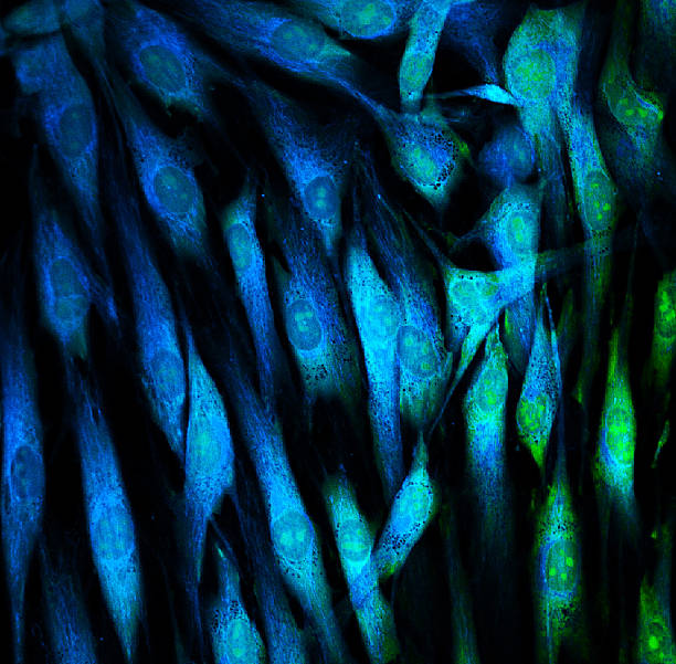
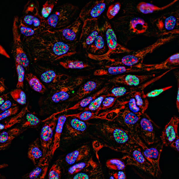
Fibroblasts (skin cells) labeled with fluorescent dyes
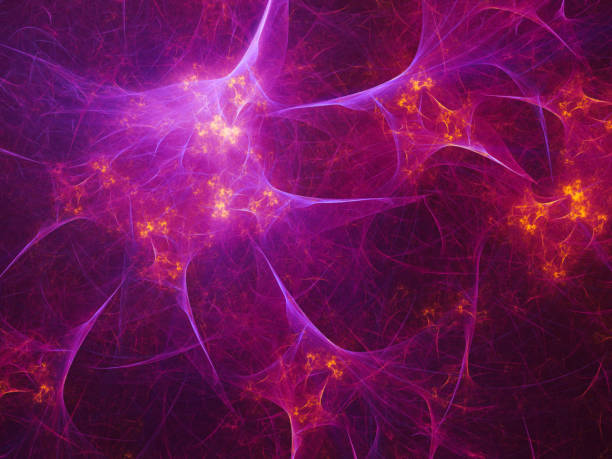
Abstract fractal art background which could suggest a neural network, or the nervous system, or other themes of connectivity and biology.

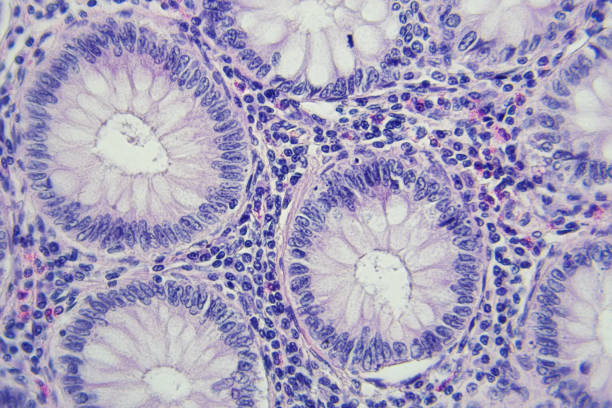
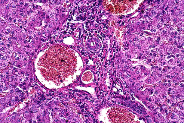
Histological Pancreas human, Liver human, Vermiform appendix human and Kidney Human under the microscope for education.
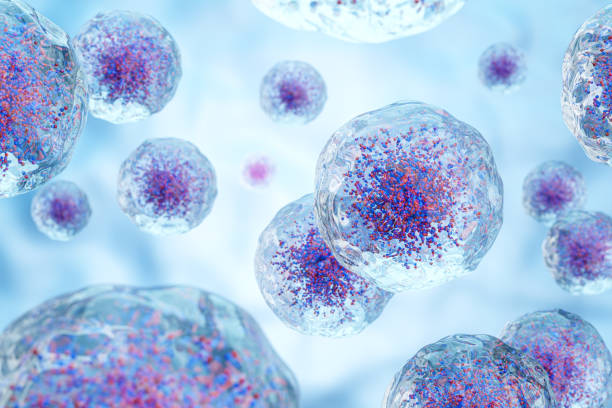
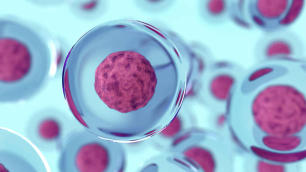
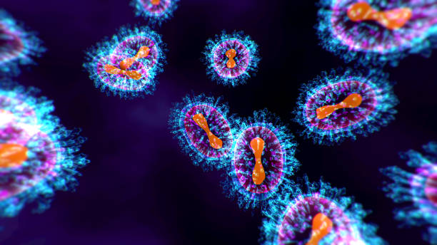
Embryonic stem cells colony under a microscope. Cellular therapy and research of regeneration and disease treatment in seamless 3D illustration. Biology and medicine of human body concept. 4K
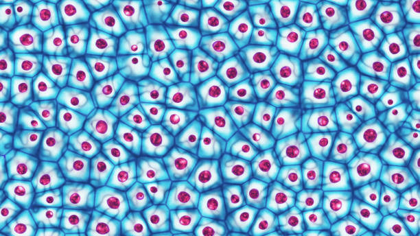
Science background with cells. Illustration contains transparency and blending effects, eps 10
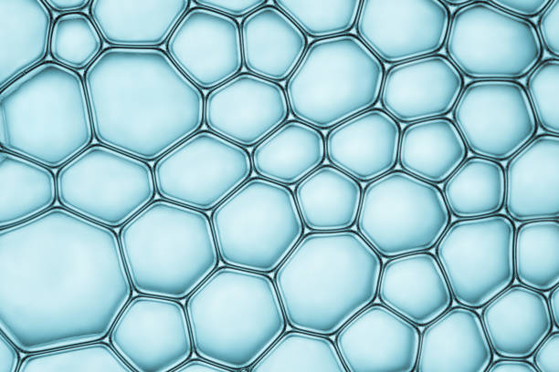
Macro close up of soap bubbles look like scientific image of cell and cell membrane
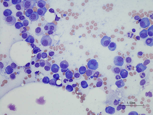
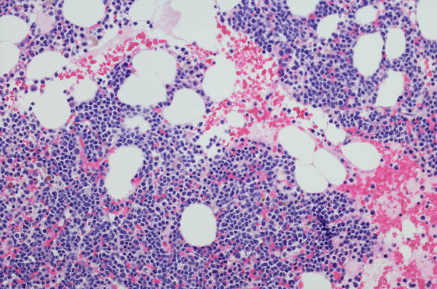
Microscopic photo of a professionally prepared slide demonstrating Plasma cell myeloma from bone marrow aspirate. Wright Giemsa stain.
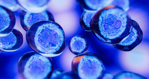
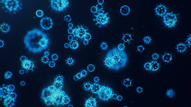
egg cells flowing in a blue background

Microscopic photo of a professionally prepared slide demonstrating invasive keratinizing squamous cell carcinoma of the skin.
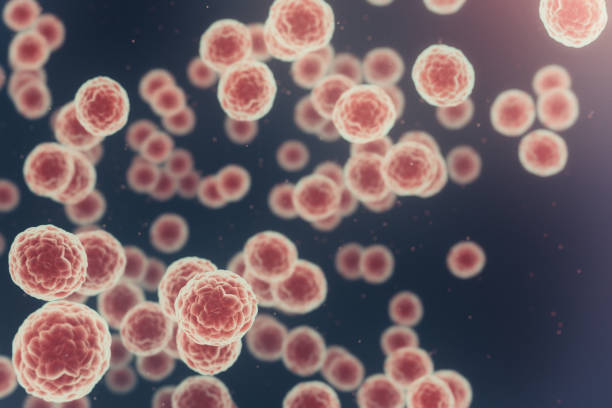
Sick cells 3d conceptual illustration. Cancer cells